![]()
Part 1
The Microalgal Cell with Reference to Mass Cultures
![]()
1
The Microalgal Cell
Robert A. Andersen
Friday Harbor Laboratories, University of Washington, Friday Harbor, WA, USA
Provasoli-Guillard National Center for Marine Algae and Microbiota, Bigelow Laboratory for Ocean Sciences, East Boothbay, ME, USA
Abstract
Microalgae are a diverse collection of microorganisms that conduct oxygen-evolving photosynthesis. Their biochemical diversity includes production of a wide array of carbohydrates, lipids, and proteins that are commercially valuable. Many produce several different morphologies, for example, flagellate, coccoid, and cyst stages. Many species are capable of sexual reproduction, some microalgae apparently having only asexual reproduction (e.g., Chlorella, Nannochloropsis). Algal ultrastructure is also diverse, paralleling their biochemical and physiological diversity. Many genomes of microalgae have been sequenced, and these are providing new insights into algal diversity. Genomic research has corroborated known endosymbiotic events and has revealed unknown, or cryptic, such events. Endosymbiosis has been a major factor in the production of algal diversity, and once it is better understood, this may be a practical means for producing new combinations of traits that have commercial application. The current state of algal taxonomy is summarized.
keywords algae; carbohydrate; chloroplast; endosymbiosis; genome; lipid; morphology; physiology; phytoplankton; protein
1.1 INTRODUCTION
Algae are primarily oxygen-releasing photosynthetic organisms with simple body plans – no roots, stems, or leaves. Algae are usually aquatic organisms. They do not form a single monophyletic group and consequently cannot be easily defined. Although algae as a group are ubiquitous, individual species occupy specific habitats. Some algae are attached to a substrate like plants, some are motile like animals, some are simply suspended in water, some grow loosely on soil, trees, and animals, and some form symbiotic relationships with other organisms (e.g., corals, lichens). The internal cell structure of algae varies greatly. Microalgae lack complex multicellular structures that are found in seaweeds. The cyanobacteria or blue-green algae have a prokaryotic cell structure and closely resemble bacteria. Eukaryotic algal cells have a nucleus and usually one or more chloroplasts; they also have mitochondria, Golgi bodies, endoplasmic reticulum, and other typical eukaryotic organelles. Despite the difficulty in presenting a clear definition for algae, thousands of books, scores of scientific journals, and numerous internet websites are dedicated solely to compiling our knowledge of algae (Lee, 2008; Graham et al., 2009).
1.2 GROSS MORPHOLOGY
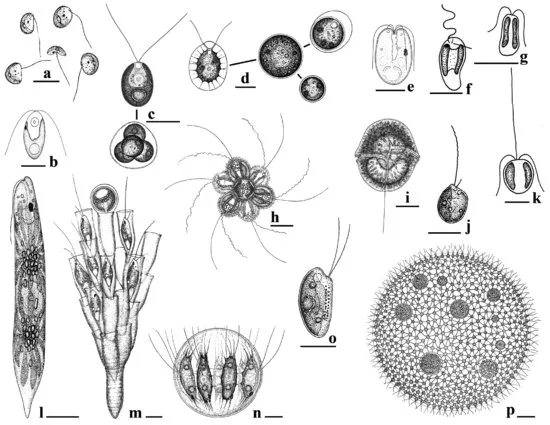
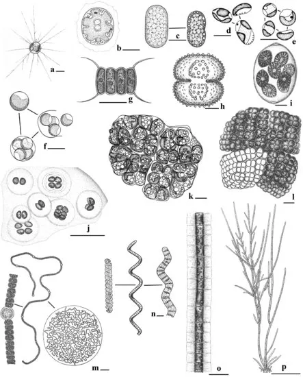
Microalgae appear in a wide variety of shapes and forms. This morphological variation occurs not only among species but also among different life stages of the same species. The common forms are defined with adjectives such as amoeboid, palmelloid (= capsoid), coccoid, filamentous, flagellate, and sarcinoid (Figs. 1.1 and 1.2). Scientists use morphological life forms when generally discussing algae and their stages; there are, however, hundreds of thousands of algal species, and they do not always fit neatly into a few convenient categories. The first algae were morphologically simple organisms; today's simplest morphologies, however, are frequently the result of evolutionary reduction through which the algae are better able to survive because of their simplicity. In the following text, algal forms are treated from simple to complex, and this approach is strictly arbitrary (i.e., it does not reflect “primitive” vs. “advanced”).
Flagellates may be single cells where each cell is an independent organism propelled through water with one or more flagella (e.g., Pedinomonas, Chlamydomonas, Gymnodinium, Ochromonas, Tetraselmis) (Fig. 1.1). Several to many flagellate cells may be joined together to produce a motile colony (e.g., Dinobryon, Synura). Large colonies, such as Volvox (Fig. 1.1p), have hundreds of cells. Most flagellate cells have two flagella, but marine picoflagellates may have only one flagellum (e.g., Micromonas and Pelagomonas) while Pyramimonas may have up to 16 flagella per cell. Haptophyte algae usually have a haptonema positioned between the two flagella (Fig. 1.1k), and the haptonema can be used for attaching to surfaces or collecting particles of food. Many common flagellate algae also produce nonmotile stages, as shown for Chlamydomonas and Haematococcus (Figs. 1.1c and 1.1d). Changing the environmental conditions can induce these alternate stages, and the manipulation of stages can be used to advantage in commercial facilities.
Many microalgae have a nonmotile stage as the dominant life form, and in some cases, no motile cells are ever found in the life cycle (Fig. 1.2). Amoeboid algae (e.g., Chlorarachnion, Chrysamoeba, Rhizochromulina) slowly creep across substrates, including the marine snow particles in oceans (Fig. 1.2a). Amoeboid cells may capture bacteria using pseudopods. Coccoid algae reproduce by autospores or zoospores, that is, mother cells undergo synchronized mitotic divisions and the number of daughter cells is fixed (e.g., 2, 4, 8, 16, 32). Single cells, such as Nannochloropsis are free, but commonly coccoid algae produce colonies (e.g., Chlorella, Oocystis, Scenedesmus) (Figs. 1.2d–g and 1.2i). Some, such as Synechococcus (Fig. 1.2c), exist today as single cells or weakly connected cells, but their ancestors were filamentous algae. Palmelloid algae have cells embedded within a gelatinous matrix; usually the cells are not physically connected to each other and only the gel holds them together. The gelatinous mass may be planktonic or attached to a substrate (Fig. 1.2j). The common flagellate Pavlova (Fig. 1.1f), for example, produces large palmelloid sheets when grown under certain culture conditions. Sarcinoid colonies result from equal cell division in three planes so that a cube is produced (Chlorosarcina; Fig. 1.2l). The oil-producing Botryococcus makes a crudely parenchymatous colony (Fig. 1.2k). Filaments are produced when cells attach end to end and form ribbon-like or chain-like assemblages. In their simplest form, filaments are unbranched and consist of a single row of cells (uniseriate) such as Arthrospira/Spirulina (Fig. 1.2n). Complexity develops with side branches (Fig. 1.2p) and multiple rows of cells (multiseriate). The cyanobacterium Nostoc forms large colonies that consist o...