![]()
Section II
Anatomy of movement in everyday living
Joints, muscles and nerve supply
- Positioning movements: the shoulder and elbow
- Manipulative movements: the forearm, wrist and hand
- Nerve supply of the upper limb
- Support and propulsion: the lower limb
- Nerve supply of the lower limb
- Upright posture and breathing: the trunk
![]()
5
Positioning movements: the shoulder and elbow
Key terms
structure and function of the shoulder and elbow, movement and muscles of the shoulder and elbow
Conceptual overview
This chapter outlines the position, structure and function related to both the shoulder and elbow joints. The musculature relative to the shoulder and elbow joints will also be examined. Each joint is looked at in depth, detailing the different muscle groups and relating these to movement seen in everyday activities.
Introduction
The shoulder forms a foundation from which the whole of the upper limb can move. Acting like the cab of a crane, the shoulder allows the hand to be placed in all directions around the body, in the same way as the jib of a crane places its load. In upper limb function, the hand can be held high above the head, in front, behind, to the side and across the body, and touching the body. The role of the shoulder is to position the hand over this wide area.
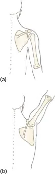
The shoulder not only performs a wide range of movement but also anchors the arm to the trunk, supporting the weight of the upper limb as it moves. The main strut for this purpose is the clavicle, part of the shoulder (pectoral) girdle formed by the clavicle and scapula. When the hand performs precision movements, stability is provided by the joints of the girdle and all the muscles surrounding the shoulder. The shoulder joint is not part of the pectoral girdle but they are mutually dependent in all the movements of the upper limb. Figure 5.1 shows how both the humerus and the scapula both move when the arm is moved towards the vertical.
Movements at the elbow change the functional length of the upper limb, adjusting the distance of the hand from the body. Elbow flexion brings the hand towards the head and body for activities, such as washing, dressing, eating and drinking. Try splinting the elbow in extension to find out how much we depend on elbow flexion for daily activities. The opposite action of elbow extension takes the hand away from the body in reaching and grasping, and also enables the hand to push against resistance, for example sawing wood or pushing a swing door. A person with reduced lower limb function relies on the elbow extensors, together with shoulder muscles, to lift the body weight on the hands to rise from a chair.
Figure 5.1 Posterior view of the scapula and humerus: (a) anatomical position; (b) arm vertical.
Movements of the shoulder, which involve the shoulder girdle and the shoulder joint, will be considered first, followed by the elbow. Upper limb movements depend on the co-operation of the shoulder and the elbow in positioning the hand.
PART I: THE SHOULDER
The shoulder (pectoral) girdle
Position and function
Reflective task
Look at the illustrations of the bones of the pectoral girdle in Appendix I. Use an articulated skeleton to examine: the clavicle linking the sternum and the scapula; the position of the scapula lying over the ribs; and the glenoid fossa of the scapula forming the socket for the head of the humerus.
The bones of the shoulder girdle are the clavicle and the scapula. The clavicle articulates at its medial end with the sternum of the thorax. The scapula is a large, flat triangular bone lying on the ribs, separated by a layer of muscle, in the posterior aspect of the thorax. The scapula is suspended by the muscles attached to its borders and surfaces so that it moves freely on the chest wall. The posterior surface of the scapula has a projecting spine which ends at the acromion process. The lateral end of the clavicle articulates with the acromion process. The head of the scapula lies laterally and has the glenoid fossa, a shallow concavity, for the articulation with the head of the humerus. From the upper part of the head, the coracoid process projects upwards and forwards to lie below the clavicle. The coracoid process provides a base for one of the proximal tendons of the biceps muscle lying on the anterior aspect of the arm.
All movements of the pectoral girdle involve both the clavicle and the scapula together. The movements of the scapula follow the shape of the ribs. The scapula is able to move freely on the thorax, because the muscles between the ribs and the scapula are covered by fascia which allows gliding movements. When the scapula moves on the chest wall, the glenoid fossa is turned to face in different directions, i.e. more directly forwards, backwards, upwards or downwards. This allows the humerus to move further in that particular direction and therefore increases the range of movement at the shoulder joint. If the shoulder girdle becomes fixed, all upper limb activites are restricted and compensation for the reduced range of movement can only be achieved by a shift of the whole body.
In summary, the functions of the shoulder girdle are:
- to anchor the upper limb to the trunk by means of the strut-like clavicle;
- to define the position of the shoulder joint and consequently the direction of the movements of the arm on the trunk;
- to increase the range of movement at the shoulder joint by changes in the angulation of the clavicle and in the position of the scapula on the chest wall.
Joints of the shoulder (pectoral) girdle
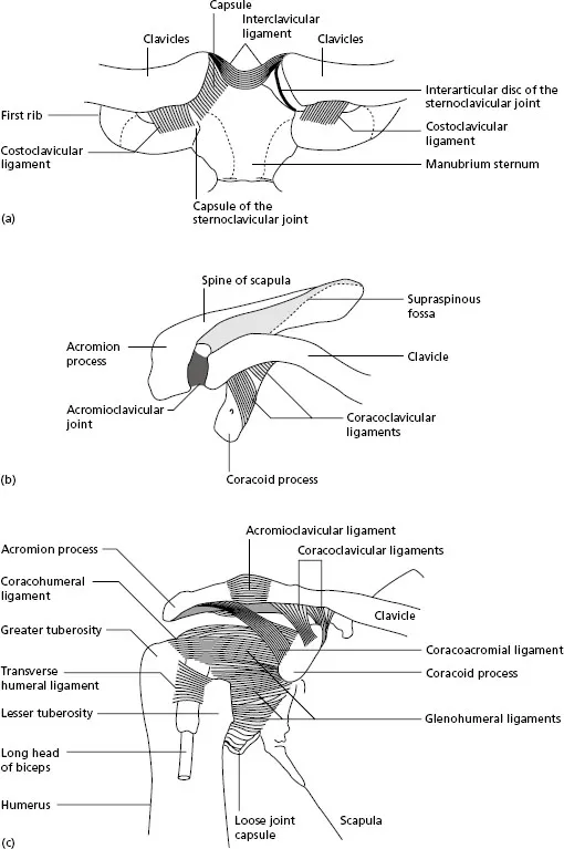
Two articulations are involved in the shoulder girdle. The sternoclavicular joint is a synovial joint between the medial end of the clavicle and the clavicular notch on the manubrium of the sternum. It is divided by an intra-articular disc of fibrocartilage joining the upper end of the clavicle to the first costal cartilge at its sternal end (Figure 5.2a). A strong costoclavicular ligament joins the medial end of the clavicle to the first rib, and the interclavicular ligament joins the medial ends of the right and left clavicles. The disc, together with the ligaments, prevents dislocation of the joint during falls on the outstretched arm or when a heavy load, for example a suitcase, is carried in the hand.
The acromioclavicular joint is a synovial joint that connects the lateral end of the clavicle with the acromion process of the scapula. The capsule is thickened by strong fibres both superiorly and inferiorly. The main factor stabilising the joint is the strong coracoclavicular ligament joining the lateral end of the clavicle to the coracoid process of the scapula (Figure 5.2b).
Reflective task
Palpate the sternoclavicular joint on a partner. Feel the rocking action of the clavicle on the sternum during shrugging the shoulders and folding the arms in front of the body. (A much reduced adjustment takes place at the acromioclavicular joint during these same movements.) Now ask your partner to move the arm in all directions at the shoulder joint. Note that movement at the sternoclavicular joint occurs each time the humerus moves.
Summary of the movements of the shoulder girdle
For the purpose of description, the movements of the shoulder girdle are divided as follows:
- elevation: the scapula moves upwards together with the lateral end of the clavicle. This movement is commonly described as ‘shrugging the shoulders’;
- depression: the scapula and lateral end of the clavicle move down to the resting position;
- protraction: the scapula moves laterally around the chest wall bringing the glenoid fossa to face more directly forwards. The vertebral border of each scapula (see Appendix I ) moves further away from the spine;
- retraction: the scapula moves medially around the chest wall bringing the glenoid fossa to face more directly towards the side. The vertebral border on each scapula moves nearer to the spine;
- lateral rotation: the inferior angle of the scapula moves laterally and the glenoid fossa points upwards;
- medial rotation: the inferior angle of the scapula moves medially and the glenoid fossa returns to the resting position.
These movements of the shoulder girdle increase the range of movement at the shoulder joint. Elevation increases reaching upwards, while depression increases pointing downwards. Protraction takes the hand farther across the body to reach to the opposite side, and retraction takes the hand farther behind the body. Abduction or flexion of the arm, which takes the hand above the head, is increased in range by lateral rotation of the scapula.
Figure 5.2 (a) Sternoclavicular joints, anterior view (left joint with capsule removed); (b) right acromioclavicular joint, superior view; (c) right glenohumeral joint, anterior view.
Reflective task
- Palpate the scapula on a partner whose horizontal arm swings round a wide circle forwards and backwards. Feel the movement of protraction as the arm swings across the front of the body, and retraction as it swings behind the body.
- Lift the arm of a partner through the full range of abduction to reach above the head, then full adduction back to the side. Palpate the scapula during this action. Lateral rotation can be felt as the arm is raised, then medial rotation as the arm is lowered.
The shoulder (glenohumeral) joint
The bony articulation of the shoulder joint occurs between the head of the humerus and the shallow glenoid fossa on the lateral aspect ...