Chest X-Rays for Medical Students
eBook - ePub

Chest X-Rays for Medical Students

Christopher Clarke, Anthony Dux

Share book
  1. English
  2. ePUB (mobile friendly)
  3. Available on iOS & Android
eBook - ePub

Chest X-Rays for Medical Students

Christopher Clarke, Anthony Dux

Book details
Book preview
Table of contents
Citations

About This Book

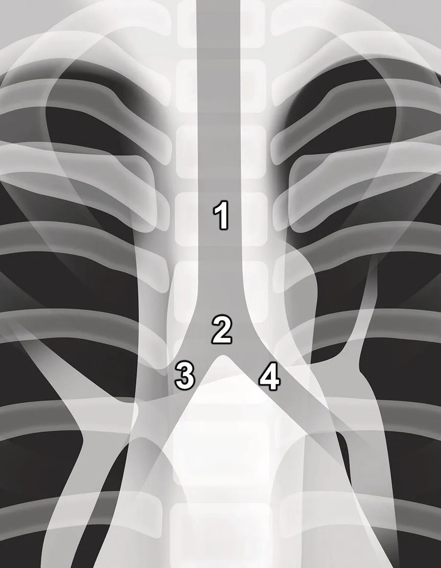
Chest X-rays for Medical Students offers a fresh analytical approach to identifying chest abnormalities, helping medical students, junior doctors, and nurses understand the underlying physics and basic anatomical and pathological details of X-ray images of the chest. The authors provide a memorable framework for analysing and presenting chest radiographs, with each radiograph appearing twice in a side-by-side comparison, one as seen in a clinical setting and the second highlighting the pathology.

This new second edition includes significant revisions, improved annotations of X-rays, expanded pathologies, and numerous additional high-quality images. A comprehensive one-stop guide to learning chest radiograph interpretation, this book:

  • Aligns with the latest Royal College of Radiologists' Undergraduate Radiology Curriculum
  • Offers guidance on how to formulate normal findings
  • Features self-assessment tests, presentation exercises, and varied examples
  • Includes sections on radiograph quality X-ray hazards and precautions

Chest X-rays for Medical Students is an ideal study guide and clinical reference for any medical student, junior doctor, nurse or radiographer.

Frequently asked questions

How do I cancel my subscription?
Simply head over to the account section in settings and click on “Cancel Subscription” - it’s as simple as that. After you cancel, your membership will stay active for the remainder of the time you’ve paid for. Learn more here.
Can/how do I download books?
At the moment all of our mobile-responsive ePub books are available to download via the app. Most of our PDFs are also available to download and we're working on making the final remaining ones downloadable now. Learn more here.
What is the difference between the pricing plans?
Both plans give you full access to the library and all of Perlego’s features. The only differences are the price and subscription period: With the annual plan you’ll save around 30% compared to 12 months on the monthly plan.
What is Perlego?
We are an online textbook subscription service, where you can get access to an entire online library for less than the price of a single book per month. With over 1 million books across 1000+ topics, we’ve got you covered! Learn more here.
Do you support text-to-speech?
Look out for the read-aloud symbol on your next book to see if you can listen to it. The read-aloud tool reads text aloud for you, highlighting the text as it is being read. You can pause it, speed it up and slow it down. Learn more here.
Is Chest X-Rays for Medical Students an online PDF/ePUB?
Yes, you can access Chest X-Rays for Medical Students by Christopher Clarke, Anthony Dux in PDF and/or ePUB format, as well as other popular books in Medicine & Radiology, Radiotherapy & Nuclear Medicine. We have over one million books available in our catalogue for you to explore.

Information

Year
2020
ISBN
9781119504122

II
The ABCDE of chest X‐rays

It is important to use a systematic approach when looking at a chest radiograph. The following ABCDE approach is easy to remember, so when it comes to your exams and you have a moment of panic after being asked to talk about a chest X‐ray, you can stick to these basics even if you don’t have a clue what’s going on!
A is for Airway:
  • Look at the trachea and right and left main bronchi.
B is for Breathing:
  • Look to see if the lungs are uniformly expanded and compare the lung fields.
  • Look around the edges of each lung.
  • Look at the costophrenic angles and the four silhouettes.
C is for Circulation:
  • Look at the cardiac size.
  • Look at the great vessels (pulmonary vessels and aorta).
  • Look at the mediastinum and hila.
D is for Disability:
  • Look for a fracture, especially of the ribs or shoulder girdle.
E is for Everything else (review areas):
  • Look for gas under the diaphragm.
  • Look for surgical emphysema.
  • Look for both breast shadows.
  • Look for foreign bodies and medical interventions.

6
A – Airway

How to review the airway

Start at the top and follow the trachea (1) inferiorly. It should lie in the midline. It divides at the carina (2) to give off the right main bronchus (3) and left main bronchus (4) (Figure 6.1). On a normal radiograph you will often not be able to visualise the more distal airway branches.
Photo depicts the airways of the lung.
Figure 6.1 The airways of the lung.

What to look for

  • Tracheal deviation
p. 24
  • Carinal angle
p. 25

Tracheal deviation

The trachea is considered to be deviated if a portion, anywhere along its length, is completely to the left or right of the midline (the midline being the centre of the vertebral column as indicated by the spinous processes).
Note: Be sure to check that the radiograph is not rotated, as a rotated radiograph can give the impression of tracheal deviation when the trachea is actually central.
If you suspect that the trachea is deviated, look for a possible cause.
  • Deviated towards diseased side (conditions that pull the trachea):
    • lung collapse;
    • pneumonectomy (removal of a lung) or lobectomy (removal of just one lobe);
    • unilateral fibrosis;
    • agenesis of lung (also called lung aplasia – complete absence of a whole lung and its bronchus).
  • Deviated away from diseased side (conditions that push the trachea):
    • tension pneumothorax;
    • massive pleural effusion;
    • mediastinal masses;
    • para‐tracheal masses.
The mos...

Table of contents