Temporary Anchorage Devices in Clinical Orthodontics
eBook - ePub

Temporary Anchorage Devices in Clinical Orthodontics

  1. English
  2. ePUB (mobile friendly)
  3. Available on iOS & Android
eBook - ePub

Temporary Anchorage Devices in Clinical Orthodontics

About this book

Provides the latest information on all aspects of using temporary anchorage devices in clinical orthodontics, from diagnosis and treatment planning to appliances and applications

Written by some of the world's leading experts in orthodontics, Temporary Anchorage Devices in Clinical Orthodontics is a comprehensive, up-to-date reference that covers all aspects of temporary anchorage device (TAD) use in contemporary orthodontics. Taking a real-world approach to the subject, it covers topics ranging from diagnosis and treatment planning to the many applications and management of complications. Case studies demonstrate the concepts, and high-quality clinical photographs support the text throughout.

The book begins with an overview of clinical applications and fundamental principles of TADs. It then goes on to cover biomechanical considerations for controlling target tooth movement with TADs. Biomechanical simulations for various clinical scenarios treated with TADs are addressed next, followed by an examination of histological aspects during the healing process and anatomical considerations with TADs. Other chapters cover: Class II Correction with TADs, Distalization with TADs, TAD-anchored Maxillary Protraction, Maxillary Expansion with TADs, Anterior Open Bite Correction with TADs, TAD-assisted Aligner Therapy, TADs vs. Orthognathic Surgery; Legal Considerations When Using TADs; and much more.

  • Provides evidence-based information on the use of TADs, with a focus on improving outcomes for patients
  • Considers topics ranging from diagnosis and treatment planning to specific clinical applications and appliances
  • Takes a real-world clinical approach, with case studies demonstrating concepts
  • Written by international experts in the field
  • Presents hundreds of high-quality clinical photographs to support the text

Temporary Anchorage Devices in Clinical Orthodontics is an essential resource for orthodontists and orthodontic residents.

Frequently asked questions

Yes, you can cancel anytime from the Subscription tab in your account settings on the Perlego website. Your subscription will stay active until the end of your current billing period. Learn how to cancel your subscription.
At the moment all of our mobile-responsive ePub books are available to download via the app. Most of our PDFs are also available to download and we're working on making the final remaining ones downloadable now. Learn more here.
Perlego offers two plans: Essential and Complete
  • Essential is ideal for learners and professionals who enjoy exploring a wide range of subjects. Access the Essential Library with 800,000+ trusted titles and best-sellers across business, personal growth, and the humanities. Includes unlimited reading time and Standard Read Aloud voice.
  • Complete: Perfect for advanced learners and researchers needing full, unrestricted access. Unlock 1.4M+ books across hundreds of subjects, including academic and specialized titles. The Complete Plan also includes advanced features like Premium Read Aloud and Research Assistant.
Both plans are available with monthly, semester, or annual billing cycles.
We are an online textbook subscription service, where you can get access to an entire online library for less than the price of a single book per month. With over 1 million books across 1000+ topics, we’ve got you covered! Learn more here.
Look out for the read-aloud symbol on your next book to see if you can listen to it. The read-aloud tool reads text aloud for you, highlighting the text as it is being read. You can pause it, speed it up and slow it down. Learn more here.
Yes! You can use the Perlego app on both iOS or Android devices to read anytime, anywhere — even offline. Perfect for commutes or when you’re on the go.
Please note we cannot support devices running on iOS 13 and Android 7 or earlier. Learn more about using the app.
Yes, you can access Temporary Anchorage Devices in Clinical Orthodontics by Jae Hyun Park in PDF and/or ePUB format, as well as other popular books in Medicine & Orthodontics. We have over one million books available in our catalogue for you to explore.

Information

Year
2020
Print ISBN
9781119513476
eBook ISBN
9781119513629
Edition
1
Subtopic
Orthodontics

Section III
Clinical Applications of TADs

33
Three‐dimensional Application of Orthodontic Miniscrews and Their Long‐term Stability

Yoon Jeong Choi and Young‐Chel Park
Department of Orthodontics, Institute of Craniofacial Deformity, Yonsei University College of Dentistry, Seoul, South Korea
In the past, auxiliary appliances such as headgear or intermaxillary elastics were used to move molars because this procedure requires substantial anchorage. Today, orthodontic miniscrews are used to control molar movement because they provide absolute anchorage, which makes it possible to control the molars in three dimensions. This chapter will discuss how miniscrews are used to control transverse, sagittal, and vertical control of molar movement and will examine their long‐term stability.

33.1 Transverse Control – Maxillary Expansion
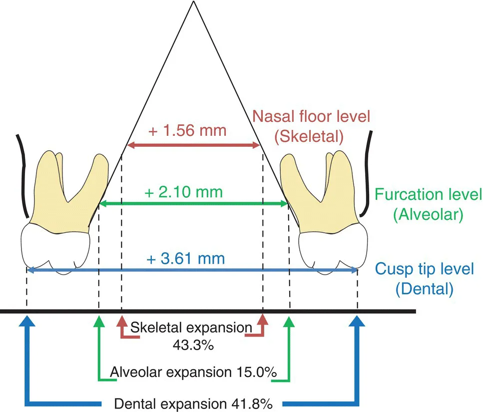
Transverse maxillary deficiency, which is often observed in full‐grown patients, can cause posterior crossbite. Traditionally, surgically assisted rapid palatal expansion (RPE) was the only feasible option for resolving a transverse discrepancy. Since miniscrew‐assisted rapid palatal expansion (MARPE) was first reported in 2010 [1], it has been an effective treatment option for transverse maxillary deficiency. MARPE can expand the maxilla non‐surgically, even in young adults, which quickly made it a favored treatment modality. In this section, the success rate, skeletal and dentoalveolar effects, stability, and changes in the upper airway of MARPE will be covered.
MARPE can expand the maxilla by separating the midpalatal suture (Figure 33.1). The success rate of MARPE for suture opening was reported to be approximately 82–87% [2–4]. The pterygoid plate, which is a part of the sphenoid bone, seems to resist the maxillary expansion as a lateral buttress. When suture opening was successful, the maxilla showed lateral and forward movement (Figure 33.2) [4]. On the coronal section, the maxilla expanded in a pyramidal shape, with a rotational center at the frontonasal suture.
The result of maxillary expansion was stable approximately four years after the treatment [2]. Immediately after the maxillary expansion, the amount of total expansion was composed of 39.1% of skeletal expansion (at the nasal floor level), 7.1% of alveolar expansion (at the furcation level), and 53.8% of dental expansion (at the cusp tip level). As the expansion showed slight but clinically insignificant relapse during retention, the composition of total maxillary expansion was 43.3% of skeletal expansion, 15.0% of alveolar expansion, and 41.8% of dental expansion one year after the expansion (Figure 33.3) [3]. Serial cone‐beam computed tomography (CBCT) images exhibited 1.6 mm apical movement of the buccal alveolar crest of the maxillary first premolar one year after the expansion, while other periodontal measurements showed clinically insignificant changes during the same period. With regard to the nasal airway, the nasal cavity significantly increased in terms of cross‐sectional area and volume, while the nasopharynx did not show clinically significant changes [5].
Previous studies [1–5] have found that non‐surgical MARPE can be effectively used to correct maxillomandibular transverse discrepancy. The maxillary expansion by MARPE remained stable during the long‐term retention and may be helpful for cases of nasal obstruction by increasing the size of the nasal cavity [6]. However, clinicians should monitor periodontal changes carefully because of consequent buccal tipping of the posterior teeth followed by apical movement of the buccal alveolar crest.
Image described by caption.
Figure 33.1 Axial and coronal sections of the maxilla before (top) and after (bottom) MARPE (CBCT images, voxel size 0.3 mm). Note the midpalatal suture is open after the expansion (arrow).
Image described by caption.
Figure 33.2 The midsagittal image before (gray) and immediately after (blue). MARPE superimposed at the anterior cranial base (CBCT images, voxel size 0.3 mm). Note the anterior and inferior movement of the maxilla at the midsagittal section.
Image described by caption and surrounding text.
Figure 33.3 Schematic diagram of the maxillary expansion by MARPE. Eve...

Table of contents

  1. Cover
  2. Table of Contents
  3. List of Contributors
  4. Foreword
  5. Preface
  6. Acknowledgements
  7. About the Editor
  8. Section I: Fundamental Perspectives on TADs
  9. Section II: Three‐dimensional Correction with TADs
  10. Section III: Clinical Applications of TADs
  11. Section IV: Esthetic Control with TADs
  12. Section V: Application of TADs in Surgical Cases
  13. Section VI: Complications with the Use of TADs
  14. Index
  15. End User License Agreement