Laboratory Imaging and Photography: Best Practices for Photomicrography and More is the definitive guide to the production of scientific images. Inside, the reader will find an overview of the theory and practice of laboratory photography, along with useful approaches to choosing equipment, handling samples, and working with microscopic subjects. Drawing from over 150 years of combined experience in the field, the authors outline methods of properly capturing, processing and archiving the images that are essential to scientific research. Also included are chapters on applied close-up photography, artificial light photography and the optics used in today's laboratory environment, with detailed entries on light, confocal and scanning electron microscopy. A lab manual for the digital era, this peerless reference book explains how to record visual data accurately in an industry where a photograph can serve to establish a scientific fact.
Key features include:
Over 200 full-color photographs and illustrations
A condensed history of scientific photography
Tips on using the Adobe Creative Suite for scientific applications
A cheat sheet of best practices
Methods used in computational photography
Frequently asked questions
How do I cancel my subscription?
Simply head over to the account section in settings and click on “Cancel Subscription” - it’s as simple as that. After you cancel, your membership will stay active for the remainder of the time you’ve paid for. Learn more here.
Can/how do I download books?
At the moment all of our mobile-responsive ePub books are available to download via the app. Most of our PDFs are also available to download and we're working on making the final remaining ones downloadable now. Learn more here.
What is the difference between the pricing plans?
Both plans give you full access to the library and all of Perlego’s features. The only differences are the price and subscription period: With the annual plan you’ll save around 30% compared to 12 months on the monthly plan.
What is Perlego?
We are an online textbook subscription service, where you can get access to an entire online library for less than the price of a single book per month. With over 1 million books across 1000+ topics, we’ve got you covered! Learn more here.
Do you support text-to-speech?
Look out for the read-aloud symbol on your next book to see if you can listen to it. The read-aloud tool reads text aloud for you, highlighting the text as it is being read. You can pause it, speed it up and slow it down. Learn more here.
Is Laboratory Imaging & Photography an online PDF/ePUB?
Yes, you can access Laboratory Imaging & Photography by Michael Peres in PDF and/or ePUB format, as well as other popular books in Kunst & Techniken der Fotografie. We have over one million books available in our catalogue for you to explore.
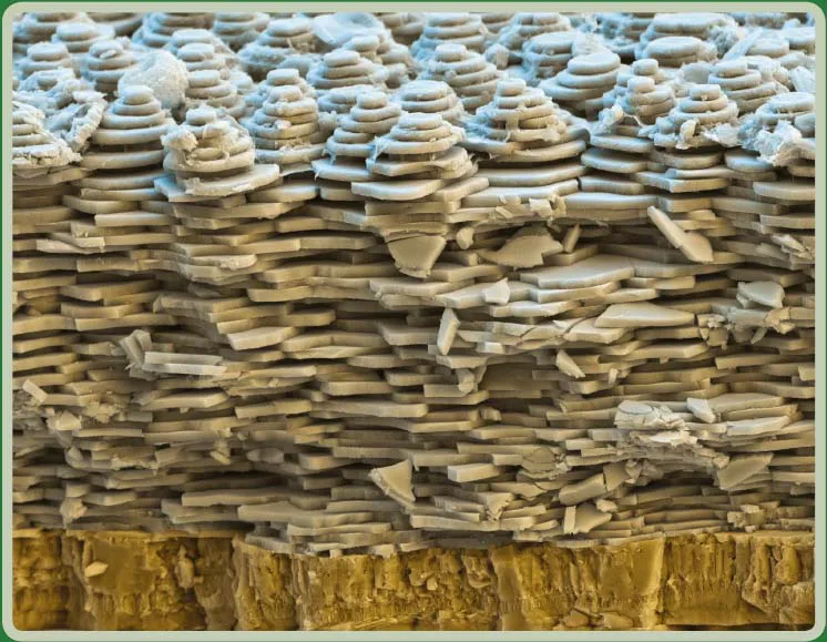
Science or art? This scanning electron photomicrograph features the shell of the white-lipped snail, Cepaea hortensis. The shell is composed of lime. The photograph reveals the layers of the shell, including where the inner wall forms new crystal platelets, and is graphically very interesting. This image is a composite from three detectors, one secondary detector and two backscattered electron-detectors. Compositingand coloring was accomplished using Adobe Photoshop software. The scanning electron microscope used 10 kV with a 13 mm working distance and 2500:1 at 15 ×13 cm. The shell was photographed September 2012 by Nicole Ottawa. Nicole and Oliver Meckes are the owners and photographers of Eye of Science. Image courtesy of Eye of Science, http://www.eyeofscience.com.
A Frame of Reference for the Image in Science
This book has many chapters written to explore various facets of scientific photography. Before delving into the technical chapters, it might be useful to consider the science image as a part of a “bigger conversation”. Beginning with the earliest images produced from a time long gone, people have exhibited an interest in chronicling observations using whatever tools were available. Museums and other cultural heritage institutions have countless examples of these wonderful treasures. Making pictures has always needed and will always require tools, materials, and skills/knowledge. Beginning with the discovery that was needed to make permanent silver-halide photographic images in the early nineteenth century, photography’s technology has experienced a huge transformation to the current digital world we live in. What has not changed during this time of evolution is the motivation as to why people make pictures in the first place. That motivation remains the same. This chapter was written to share some highlights of photography’s evolution specific to science, the challenges and solutions when photographing in science, some key inventors, some important equipment, and some fundamental practices that have spanned more than 150 years.
Today, photographic documentation in science has become a science unto itself. It is difficult to identify an environment or industry not using imaging or images. The idea to use photography in science is by no means new. Making a photograph of an object or event implies a certain importance of that object or the event itself. Many of today’s practices have their origins at the time of photography’s invention and even before. Pictures record history. Pictures can be called photographs or images. Images in science are considered data or facts and they have become an integral part of exploration, discovery, and certainly publishing. Images can record what is not visible to the human visual system and they can make permanent records of transient events to serve as visual notes if needed at a later date. Pictures chronicle things and situations where words might be inadequate and images remember what time will forget. All these outcomes are pretty impressive.
Digital tools have migrated into every discipline and so have the challenges of staying current and relevant for both users and manufacturers alike. With all of the technological advancements, the creation of images that were never possible can easily be produced with the right digital equipment and knowledge. The rapid adoption of the newest technologies is a direct consequence of the simplification of operation of digital tools, the reduction in the costs of all of the necessary equipment, as well as the continual release of easier-to-use software. Simply consider the explosion of smartphones and tablet computing as a microcosm of the digital space that science operates in.
The Science Image: A Point of Departure
Identifying scientific images from other types of photography requires the use of industry-accepted criteria. Before proceeding with that analysis, one characteristic fundamental to any picture is whether a photo itself is a good or a bad picture based on its technical merits. While seemingly an easy task to accomplish, there can be contributing factors that come into play when photographing challenging subjects or in difficult situations. Without doubt, many factors influence outcomes and it has become increasingly easy to operate a modern camera in the automatic mode and get an acceptable result. Sometimes it is actually a rather good result. Making a photographic recording of a subject has never been easier.
Criteria Used to Identify Good Photography:
■Proper or correct exposure: there is detail in both the dark and light areas of the subject.
■Effective isolation of the subject: clear and simple framing and magnification.
■Proper selection and use of shutter speed and aperture based on the sample requirements leading to an adequate range of focus and object free of blur.
■Effective use of light and lighting: subject characteristics are made visible.
■Appropriate sample treatment: the image has a neutral perspective.
■Effective use of focus and/or depth of field (DOF).
■Emphasizing details that are in agreement with the scientific intent.
Additional criteria specific to science photographs:
■Use of a standardized treatment
■Proper use of a scale.
There are a number of other factors that separate good photographs from bad ones that go beyond simply the effective operation of the equipment. These will also play a role in scientific imaging outcomes and include:
■the duration of the event and the photographer’s ability to synchronize the photography with it;
■the frequency of the event and the photographer’s ability to record what is needed to make a useable recording of it;
■access to (the location of) the event.
Making high quality photographs in science can be difficult for these and other reasons. That being said, “making an image is better than making no image.” To be successful, science photographers must be able to innovate and control a multitude of variables that affect the outcome.
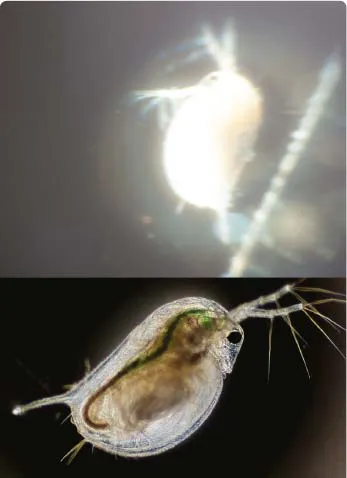
Figure 1.1 These photographs clearly illustrate why the parts of the imaging system matter and skills can make a difference. Both photographs feature a live Daphnia magna (sp). Exposure, sharpness, composition, treatment, and the recording of data are the criteria for a successful photomicrograph. The bottom photograph exhibits proper focus, exposure, isolation, and magnification. The upper photograph is awful.
Science Photographs Require a Scale
Categorizing one thing from another is what people do. People identify things based on experience, criteria, and prior knowledge. On the surface it might be considered easy to identify one thing from another but that is not always the case. Many things do not fit into just one category. Nuances and other factors come into play for identification. Images in particular can operate on many levels and in many environments. Human perception will also play an important role in what is seen, observed, and finally what is believed to have been seen. When a photomicrograph is made, it can operate as a science image or sometimes as art based on a viewer’s point of view. Astronomical photographs are similar in that way. Scientific photography might be described differently depending on who is asked for the definition. It might include references to the equipment that was used, such as in photomicrography, or it might be described by the application that was used, such as in radiography. One absolute requirement for the delineation of a science photograph from other types of photography is the need for the inclusion and proper use of a ruler or other type of scale. Whether a ruler is used for a size comparison or a date or timestamp is included, the inclusion of a scale immediately delineates a science image from other types of photography.
Photographer’s Intent and Subject Matter
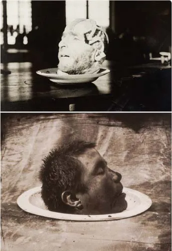
For many, the intent of the photographer might also be used to discriminate scientific photography from other types. Sometimes subtle and not prominent elements play a role in this process. Sometimes the need to identify an image from another type is important while at other times it is not. It is actually quite easy to photograph science subjects in non-scientific ways and it is also possible to evaluate science images—after creation—using a different point of view than when the image was created. The images in Figure 1.3 were produced for very different reasons and at very different times by two different photographers. In both images, the subject might be considered the same. A quick assessment of the images might lead a viewer to conclude they are both medical photographs because of the subject. The image of the Dissected Head on a Soup Plate was made by Dr. Howard Brundage in 1905 and was produced as a medical gross specimen photograph during an autopsy at the State Hospital, Columbus, Ohio. The other image, Head of a Dead Man, was made in 1990 by Joel Peter Witkin as a fine art photograph. While the content in both images, made decades apart, is similar, the intent of the photographers could not be more different. Consequently a subject by itself cannot be used as the sole criterion for the categorization of an image.
Intent can be at the foundation of legal cases as well. In 1993, I participated as an expert witness in a trial where Dr. William Zink, a Florida pediatric orthopedic surgeon, was charged with four counts of child molestation between 1987 and late 1993. Part of the charges included the taking of sexually explicit photographs of the children and other male patients. The physician was ultimately acquitted after a long trial. Fundamental to the case was the need to create a legal definition of a clinical medical photograph. The trial focused a lot of attention on “what discriminated medical photographs featuring children that contained nudity from child pornography.” It took many long weeks of testimony to create a working legal definition for the jury. In the end, the case ultimately came down to trying to determine the physician’s intent. Not specific to this case, it can be difficult if not impossible to measure the intent of an image-maker.
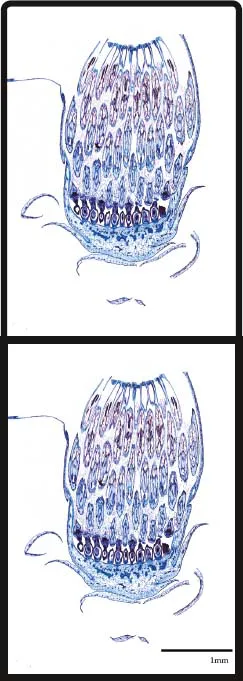
Figure 1.2 These brightfield photomicrographs feature the flower bud of Taraxacum officinale (dandelion) shown in a longitudinal section. It is reproduced without a scale (top) and with a scale (bottom). The inclusion of a scale clearly implies a science image. Without the scale, a viewer might see the image’s characteristics such as its shape, color, and design before assigning the image a science image.
A Picture is worth a thousand words
The need for and the importance of scientific illustrations has a well-documented history. It is easy to find historically important drawings from the seventeenth and eighteenth centuries and sometimes earlier. Leonardo da Vinci, Michelangelo, and early astronomers produced countless drawings of science and in some cases paintings of important discoveries that chronicled— to the best of their abilities and that of the materials of the time—the current scientific frontiers. Initially created as scientific illustrations, these drawings have now become valuable pieces of art over time. Mug shots, early medical photography as well as numerous other types of scientific c photographs from the late eighteenth century have become collectibles and treasured items in today’s society. While they were originally created to chronicle science explorations and for the documentation of things, in modern times they have taken on different roles.
Figure 1.3 Which is the medical photograph and which is the fine art photograph? often the boundaries of distinction are not clearly defined. The subject in both photographs is a human head. The top photograph was made by Dr. Howard Brundage in 1905. The lower photograph was made by Joel Peter witkin in 1990. Upper imag...