An inspirational bible for monochrome photography - this second edition almost doubles the content of its predecessor showing you the path from visualization to print
eBook - ePub
Way Beyond Monochrome 2e
Advanced Techniques for Traditional Black & White Photography including digital negatives and hybrid printing
- 542 pages
- English
- ePUB (mobile friendly)
- Available on iOS & Android
eBook - ePub
Way Beyond Monochrome 2e
Advanced Techniques for Traditional Black & White Photography including digital negatives and hybrid printing
About this book
Trusted by 375,005 students
Access to over 1.5 million titles for a fair monthly price.
Study more efficiently using our study tools.
Information
Part 1
The Basics

© 1996 by Hisun Wong, all rights reserved
From Visualization to Print
Eye and Brain
Now you see it, now you don’t
Photography is a form of visual communication and a category of modern visual art, which simply means that photographs are made to be seen by a group of people other than the artist himself. Successful artists, by intent or by instinct, make use of the fundamentals of human visual perception to improve their works of art. The human reaction to an image is a complex mix of physics, emotion and experience. However, understanding the limits of human vision allows the photographer to distinguish between essential and irrelevant technical accomplishment.
Three essential components are required to make human vision possible. There must be a sufficient amount of light, a light-gathering device to receive and arrange the light into structured optical information, and a processor to sort and administer this information to make it available for further decision and action. In the human visual system, eye and brain work closely together to gather, arrange and process the light around us.

© 1936 by Dorothea Lange, Library of Congress, Prints & Photographs Division, FSA/OWI Collection, [LC-USF34-9058-C]
Electromagnetic Spectrum and Light
Modern humans are constantly exposed to a wide range of electromagnetic radiation (fig.1), but we hardly ever think about it, because our daily lives are filled with radio and television signals, radar, microwaves and the occasional x-ray exposure at the doctor’s office. Low-frequency radiation, such as in radio and television signals, carries little energy and has no effect on the human body. It cannot be seen or felt. Higher frequencies, such as infrared radiation, can be felt by the skin as warmth, and even higher frequencies, such as UV and x-rays, carry sufficient energy to be harmful to humans with prolonged exposures. The highest frequencies, such as gamma radiation and cosmic rays, are packed with energy and would put an end to life on earth, if it were not for the planet’s sensitive atmosphere and its strong magnetic field to protect us. However, most electromagnetic radiation bombards us constantly without ever being detected by any of our senses. There is only a tiny range of frequencies, with a wavelength from roughly 400-700 nm, to which our eyes are sensitive. It is the visible part of the electromagnetic spectrum, better known as ‘light’. Within this range, the human eye sees changes in wavelength as a change of hue.
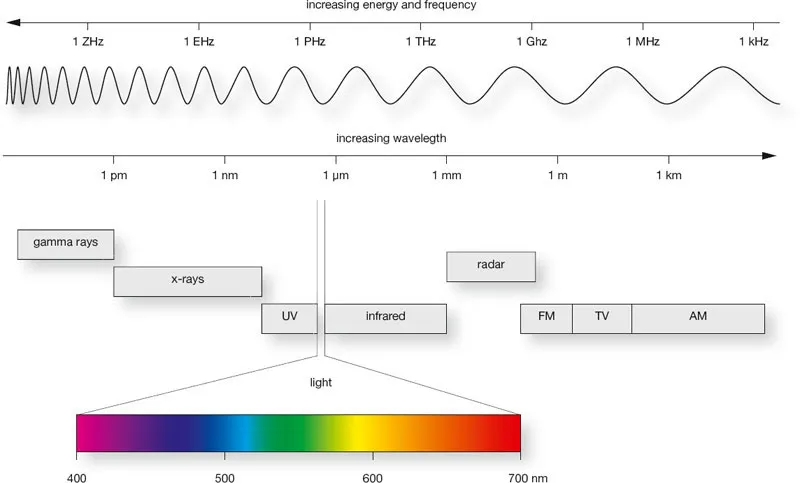
fig.1 Modern humans are constantly exposed to a wide range of electromagnetic radiation, but our eyes are sensitive to only a tiny range of these frequencies. They are the visible part of the spectrum, better known as ‘light’.
The Anatomy of Human Vision
Before we get into the human visual system as a whole, it makes sense to initially understand the optical performance and visual functionality of eye and brain individually. What may come across as a small lesson in human anatomy is actually an essential introduction to the basic phenomena of human vision.
The Human Eye
The human eye is often compared to a photographic camera, because the eye, a sophisticated organ capable of focusing an image onto a light-sensitive surface, is very similar to lens, camera and film (fig.2a), but with some significant differences in operation. The eye is a light-tight hollow sphere (sclera), containing an optical system (cornea and lens), which focuses the incoming light onto a light-sensitive surface (retina) to create an upside-down and reversed image. The amount of incoming light is controlled by the iris, which adjusts the aperture (pupil) as needed. The retinal image is converted into electrical impulses by millions of light-sensitive receptors and transmitted to the brain via the optical nerve.
Sharp focusing is controlled by the ring-shaped ciliary muscle, which surrounds the lens and is able to change its curvature. The muscle contracts to bulge the lens, allowing us to focus on nearby objects, and it relaxes or expands to flatten the lens for far-distance viewing. Changing the optical power of the lens, to maintain clear focus as the viewing distance changes, is a process known as accommodation. As we get older, the lens loses its flexibility, and it becomes increasingly more difficult to focus on close objects.
At infinity focus, the average lens has a focal length of roughly 17 mm. When fully open and adapted to low light levels, the pupil has a diameter of about 8 mm, which the iris can quickly reduce to about 2 mm in order to compensate for very bright conditions and to protect the retina from irreversible damage. In photographic terms, this is equivalent to an f/stop range from f/2 to f/8, covering a subject brightness range of 4 stops or a 16:1 ratio.
The retina is lined with light-sensitive receptors of two types, called rods and cones, which are only responsive to dim and bright light, respectively. At any given time, rods and cones provide a static sensitivity range of about 6 stops. However, rods and cones are able to dynamically alter their sensitivity by regulating the amount of a light-sensitive dye they contain. This enables the retina to adapt to a light-intensity range of 1,000,000:1 and adds 20 stops of dynamic sensitivity to its static range.
Fully building up the light-sensitive dye takes about 8 minutes in cones and up to 30 minutes in rods, which is a process called dark-adaptation. This explains why our vision improves only slowly, when we move from a bright to a dimly lit room. In the reverse process, rods and cones rapidly dispose of the dye, in order to safely adapt to a brighter environment. This is referred to as light adaptation and is typically completed within 5 minutes.
All rods are of a similar design, highly specialized for low-light sensitivity. However, cones come in three different varieties, and each kind produces a slightly different type of dye, making it sensitive to a different wavelength of light. This enables color vision, very similar to the way red, green and blue color receptors enable color imaging in digital camera sensors.
In summary, rods give us sensitive night vision (scotopic) and cones add colorful day vision (photopic) to our sense of sight (fig.2b). Combining the static and dynamic sensitivity range of the retina, and adding the light-regulating support of the iris, provides the human eye with an enormous sensitivity range of 1,000,000,000:1 or almost 30 f/stops, as long as we give it the time to adapt to the dimmest and brightest lighting conditions possible.
Data Sheet of the Human Eye
focal length at infinity | 17 mm |
comfortable min focus distance | 250 mm |
typical aperture range | f/2 - f/8 |
dynamic contrast range | 1,000 : 1 |
max sensitivity range | 1,000,000,000 : 1 |
standard visual angle | 1 arc minute |
min optical resolution | 30 lp/degree |
min reading resolution | 7 lp/mm |
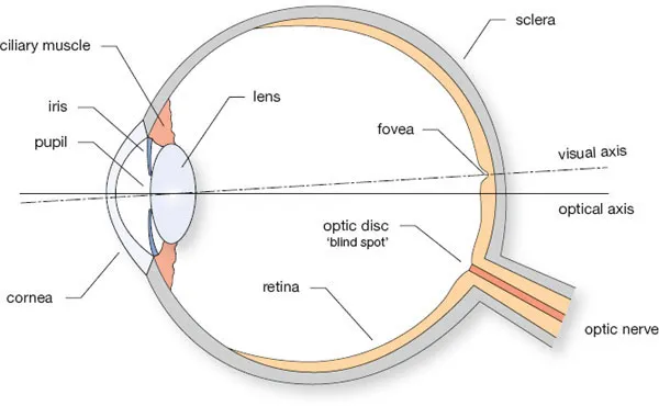
fig.2a anatomy of the human eye
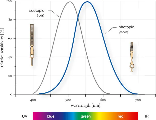
fig.2b spectral sensitivity of the human eye
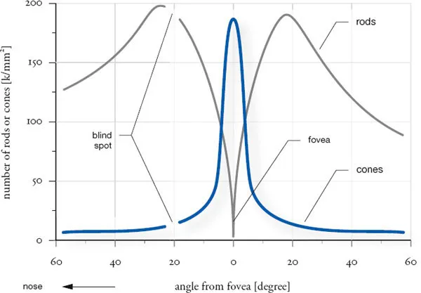
fig.2c population of rods and cones across the retina
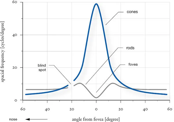
fig.2d visual acuity across retina
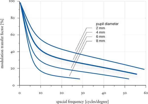
fig.2e visual acuity of the human eye
fig.2 The human eye is often compared to a photographic camera, because the eye, a sophisticated organ capable of focusing an image onto a light-sensitive surface, is very similar to lens, camera and film, but with some significant differences in operation.
There are millions of rods and cones distributed across the retina, but unlike the light-sensitive particles of a silver-gelatin emulsion, rods and cones are not distributed uniformly (fig.2c). Rods predominantly populate the outer surface area of the retina, whereas cones are primarily found around the center. Furthermore, there are two small areas on the retina that are quite different from the rest, and they deserve some special attention.
Close to the center of the retina is a small indentation, called the fovea. Its center, the fovea centralis, which is also the center of human vision, is only 1 mm in diameter. The fovea contains almost exclusively cones and very few rods. In fact, nowhere else on the retina are cones so densely populated as in the fovea. Here, the distance between cones is as small as 2.5 µm, and because of this, humans have excellent visual acuity in bright light. However, peak performance is limited to a relatively small angle of view, only a few degrees, concentrated around the fovea (fig.2d). Everything outside this narrow field of view blends into our relatively fuzzy peripheral vision. Nevertheless, about 50% of the optical impulses, sent to the brain, come from the fovea, and therefore, we can assume an optical resolution of the human eye of at least 30-60 line pairs per degree. The optical resolution of the eye also depends on the diameter of the pupil or, consequently, on illumination levels. Similar to a photographic lens, overall optical performance increases with decreasing aperture until diffraction takes over. Fig.2e shows how a wide-open pupil (8 mm) is limited to 30 lp/degree, a normal pupil opening (4 mm) achieves about 60 lp/mm, and a very small pupil (2 mm) can resolve up to 90 lp/mm. For the purpose of viewing photographs, we can assume an optical resolution of the human eye of 30-90 lp/mm, which is equivalent to viewing angles of 20-60 arc minutes and covers the range from standard to critical viewing conditions.
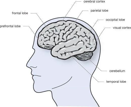
fig.3 The optical information, collected by our eyes, travels along the optical nerve to several areas of the brain for subsequent processing.
About 20° from the center of the fovea is the optical disc. This is the location where the optical nerve is attached to the eye. The optical disc is entirely free of rods or cones, and this complete lack of light receptors is the reason why the optical disc is also referred to as the ‘blind spot’. Amazingly, the blind spot does not disturb human vision at all, because the brain makes use of surrounding optical impulses in order to fill in for the missing image information.
The Human Brain
Comparing the human eye to a camera and lens does not fully appreciate the sophisticated functionality of this complex organ, but it sufficiently illustrates the eye’s contribution to the human visual system. A similar association is often made by comparing the human brain to an electronic computer. The speed with which our brain processes visual input is about the only realistic comparison we can obtain from this analogy, because the brain is much more than just a pile of electronic circuitry.
The eye focuses an upside-down and reversed image onto the retina, where rods and cones convert the optical sensation into electrical signals, which travel along the optical nerve to several areas of the brain for subsequent processing. At first, the visual cortex, which is an area in the occipital lobe of the brain at the back of our head, differentiates between light and shadow, making out borders and edges and combining them into simple shapes. With support of the cerebral cortex in the parietal lobe, the new data is compared with previously memorized information and used to quickly recognize familiar faces and objects, while separating them from the background. But, visual processing does not stop there, because the information is now passed to the temporal lobe, where the meaning of what we have seen is interpreted, and faces and objects are given a name. In the frontal lobe, feelings are added, and finally, in the prefrontal lobe, we order our thoughts and decide what to do next, based on what we have seen.
This is a very simplified overview of the brain’s function as part of the human visual system. What actually happens in our heads is far more complex, and much of the brain’s functionality is still a mystery to modern science. All we know for sure is that whatever our brain does, it does it very, very quickly.
The Human Visual System
The human eye is a camera, and the brain is a fast computer. While this grossly oversimplified statement roughly explains the contribution of both organs to human vision, it cannot illustrate the complexity and sophistication of the human visual system. What we believe to ‘see’ is a combination of the images created by our eyes and the brain’s interpretation of them. In addition, the brain constantly supports the eye to optimize its optical performance and get the most visual information possible. Here are two examples:
The eye is able to recognize minute detail far beyond its inherent optical resolution of 1 arc minute. We can easily distinguish a thin wire against a bright sky down to 1 second of arc, but visual angles alone cannot explain why we can see the dim light of a star, thousa...
Table of contents
- Cover
- Halftitle
- Title
- Copyright
- Contents
- Foreword to the First Edition
- Foreword to the Second Edition
- Preface and Acknowledgments
- Introduction
- Part 1: The Basics
- Part 2: The Science
- Part 3: Odds and Ends
- Appendix
- Glossary
- Bibliography
- Index
Frequently asked questions
Yes, you can cancel anytime from the Subscription tab in your account settings on the Perlego website. Your subscription will stay active until the end of your current billing period. Learn how to cancel your subscription
No, books cannot be downloaded as external files, such as PDFs, for use outside of Perlego. However, you can download books within the Perlego app for offline reading on mobile or tablet. Learn how to download books offline
Perlego offers two plans: Essential and Complete
- Essential is ideal for learners and professionals who enjoy exploring a wide range of subjects. Access the Essential Library with 800,000+ trusted titles and best-sellers across business, personal growth, and the humanities. Includes unlimited reading time and Standard Read Aloud voice.
- Complete: Perfect for advanced learners and researchers needing full, unrestricted access. Unlock 1.5M+ books across hundreds of subjects, including academic and specialized titles. The Complete Plan also includes advanced features like Premium Read Aloud and Research Assistant.
We are an online textbook subscription service, where you can get access to an entire online library for less than the price of a single book per month. With over 1.5 million books across 990+ topics, we’ve got you covered! Learn about our mission
Look out for the read-aloud symbol on your next book to see if you can listen to it. The read-aloud tool reads text aloud for you, highlighting the text as it is being read. You can pause it, speed it up and slow it down. Learn more about Read Aloud
Yes! You can use the Perlego app on both iOS and Android devices to read anytime, anywhere — even offline. Perfect for commutes or when you’re on the go.
Please note we cannot support devices running on iOS 13 and Android 7 or earlier. Learn more about using the app
Please note we cannot support devices running on iOS 13 and Android 7 or earlier. Learn more about using the app
Yes, you can access Way Beyond Monochrome 2e by Ralph Lambrecht,Chris Woodhouse in PDF and/or ePUB format, as well as other popular books in Media & Performing Arts & Digital Media. We have over 1.5 million books available in our catalogue for you to explore.