Responsible for managing the body's waste and regulating the balance of water and electrolytes, the kidneys and renal system, in a sense, make up the body's plumbing network. When any part of the system fails or does not function properly, the body may be subject to unnatural concentrations of certain substances, causing illness or even death. This volume examines the various components of the renal, or urinary, system, and the consequences of dysfunction and disease.

- English
- ePUB (mobile friendly)
- Available on iOS & Android
eBook - ePub
The Kidneys and the Renal System
About this book
Trusted by 375,005 students
Access to over 1.5 million titles for a fair monthly price.
Study more efficiently using our study tools.
Information
Subtopic
PhysiologyIndex
Biological Sciences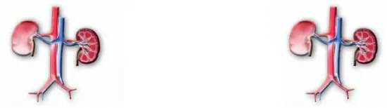
CHAPTER 1
ANATOMY OF THE KIDNEYS AND RENAL SYSTEM
In many respects the human renal, or urinary, system resembles the renal systems of other mammalian species. However, it has its own unique structural and functional characteristics. Structurally, the human renal system consists of the kidneys, where urine is produced, and the ureters, bladder, and urethra for the passage, storage, and voiding (or elimination from the body) of urine. The eliminatory function of the renal system is often described by the terms excretory and urinary. The kidneys, however, both secrete and actively retain within the body certain substances that are as critical to survival as those that are eliminated.
The most important functional components of the renal system are the two kidneys, which control the electrolyte (e.g., salt) composition of the blood and eliminate dissolved waste products and excess amounts of other substances from the blood. The latter substances are excreted in the urine, which passes from the kidneys to the bladder by way of two thin muscular tubes called the ureters. The bladder is a sac that holds the urine until it is eliminated through the urethra.
THE KIDNEYS
The human kidneys are reddish brown paired organs and are about 10 cm (about 4 inches) in length. They are distinguished by their beanlike shape, being concave (curved inward) along the length of one side and convex (curved outward) along the length of the opposite side. Their function is to maintain water balance and to filter metabolic wastes from the blood. (A brief overview of the anatomy of the kidneys is provided here. A detailed discussion can be found in the sections that follow.)
The human kidneys in situ in a male. Encyclopædia Britannica, Inc.
In embryonic development, the kidneys consist of two series of specialized tubules that empty into two collecting ducts, known as the Wolffian ducts. When fully developed, the kidneys contain numerous sophisticated functional units, called nephrons, that filter wastes from the blood and reabsorb water and nutrients. The nephron filtration process results in the final urine product that is ultimately expelled from the body. Each fully developed kidney contains about 1 million to 1.25 million nephrons that filter the entire five-quart water content of the blood every 45 minutes—an equivalent of 160 quarts a day. Of this, only 1½ quarts are excreted. The remainder is reabsorbed by the nephrons.
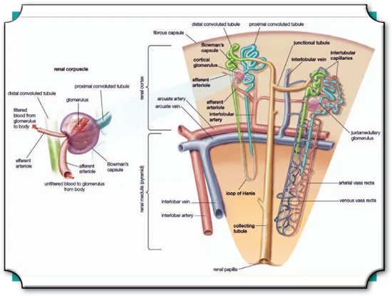
The kidneys can be divided into two major sections: a somewhat granular outer section called the cortex and a smoother inner section called the medulla. The cortex contains clusters of blood vessels, known as glomeruli, and a series of intricately folded, very fine tubes, known as convoluted tubules. These structures form the upper half of each nephron unit. The lower half of the nephron unit, which consists of the loops of Henle and the collecting tubules, lies in the medulla. The long loops of Henle and the long straight collecting tubules give the tissue of the medulla its smooth, somewhat striated appearance.
The urine that is produced by each nephron passes through the collecting tubule in the medulla and is gathered into a cup-shaped cavity called the renal pelvis. The renal pelvis forms the upper end of the ureter, and the urine gathered there ultimately passes through the ureter to the bladder.
ANATOMICAL LOCATION OF THE KIDNEYS
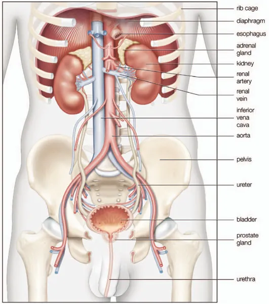
The kidneys are located high in the abdominal cavity and against its back wall. They are found on either side of the backbone (the vertebral, or spinal, column), between the levels of the 12th thoracic and 3rd lumbar vertebrae, and outside the peritoneum (the membrane that lines the abdomen).
The long axes of the kidneys are aligned with that of the body, but the upper end of each kidney (the pole) is tilted slightly inward toward the vertebral column. Situated in the middle of each kidney on the side facing the vertebral column (the medial concave border) is a deep vertical cleft, called the hilus, which leads to a cavity within the kidney known as the renal (kidney) sinus. The hilus is the point of entry and exit of the renal arteries, veins, lymphatic vessels, and nerves and of the enlarged upper extension of the ureters.
THE RENAL CAPSULE
A thin membranous sheath known as the renal capsule covers the outer surface of each kidney. The capsule is composed of tough fibres, chiefly collagen and elastin (fibrous proteins), that help to support the kidney mass and protect the vital tissue from injury. The number of elastic and smooth muscle fibres found in the capsule tends to increase with the individual’s age.
The capsule receives its blood supply ultimately from the interlobar arteries, small vessels that branch off from the main renal arteries. These vessels travel through the cortex of the kidney and terminate in the capsule.
The maximum thickness of the membrane is usually 2–3 mm (0.08–0.12 inch). The capsule surrounds the outer walls and enters into the hollow region of the kidney sinus. The sinus contains the major ducts that transport urine and the arteries and veins that supply the tissue with nutrients and oxygen. The capsule connects to these structures within the sinus and lines the sinus wall.
In a healthy person, the capsule is light reddish-purple in colour, translucent, smooth, and glistening. It can usually be easily stripped from the rest of the kidney’s tissue. A diseased kidney frequently sends fibrous connections from the main body of tissue to the capsule, which makes the capsule adhere more strongly.
RENAL VESSELS AND NERVES
The renal arteries arise, one on each side, from a large vessel known as the abdominal aorta. The renal arteries split off from the abdominal aorta at a point a little above the small of the back (opposite the upper border of the second lumbar vertebra). Close to the renal hilus each artery gives off small branches to the adrenal gland and ureter and then further branches into divisions that extend toward the front (anterior) of the renal tissues as well as behind (or posterior to) these tissues. The large veins carrying blood from the kidneys usually lie in front of the corresponding arteries and join the inferior vena cava (a large vein that travels up from the lower region of the body to the right side of the heart) almost at right angles. The left vein is longer than the right vein because the inferior vena cava lies closer to the right kidney.
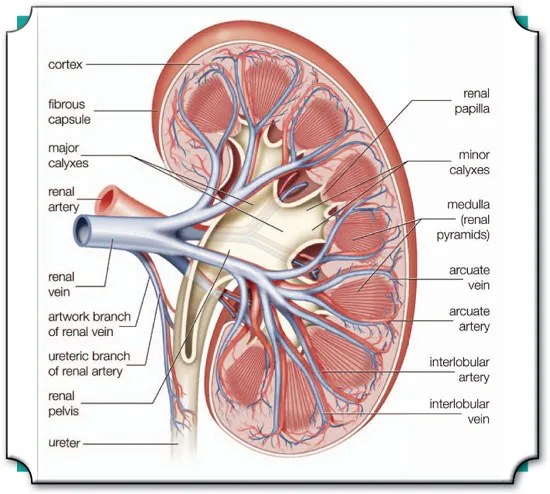
Cross section of the right kidney showing the major blood vessels. Encyclopædia Britannica, Inc.
The kidneys are supplied with sympathetic and parasympathetic nerves of the autonomic nervous system (the part of the nervous system that controls and regulates internal organs without conscious effort). The renal nerves contain both afferent and efferent fibres (afferent fibres carry nerve impulses to the central nervous system, whereas efferent fibres carry impulses away from the central nervous system).
INTERNAL CONFIGURATION: THE RENAL PYRAMID
The renal pyramids are any of the triangular sections of tissue that constitute the medulla, or inner substance, of the kidney. The pyramids consist mainly of tubules that transport urine from the cortex, or outer, part of the kidney, where urine is produced, to the calyxes, or cup-shaped cavities in which urine collects before it passes through the ureter to the bladder. The renal pyramids are visible in cross sections of kidney tissue as comparatively dark cones in the substance of the renal medulla. The bases of the cones face outward, toward the external surface of the kidney, and the apexes project, either singly or in groups, into the renal sinus.
The point of each pyramid, called the papilla, projects into a small cuplike cavity called a minor calyx. Each kidney has about 12 minor calyxes, which combine to form several major calyxes. The major calyxes then converge into the renal pelvis at the upper end of the ureter. Each group of pyramids that projects into a papilla, together with the portion of cortex that arches over the group, is called a renal lobe.
The surface of the papilla has a sievelike appearance because of the many small openings from which urine droplets pass. Each opening represents a tubule called the duct of Bellini, into which collecting tubules within the pyramid converge. Muscle fibres lead from the calyx to the papilla. As the muscle fibres of the calyx contract, urine flows through the ducts of Bellini into the calyx. The urine then flows to the bladder by way of the renal pelvis and a duct known as the ureter.
Between the pyramids are major arteries known as the interlobar arteries. Each interlobar artery branches over the base of the pyramid. Smaller arteries and capillaries divide off from the interlobar arteries to supply each pyramid and the cortex with a rich network of blood vessels. Blockage of an interlobar artery can cause degeneration of a renal pyramid.
Some animals, such as rats and rabbits, have a kidney composed of only one renal pyramid. In humans each kidney has a dozen or more pyramids.
INTERNAL CONFIGURATION: THE RENAL PELVIS
The renal pelvis is shaped somewhat like a funnel that is curved to one side. It is almost completely enclosed in the deep indentation on the concave side of the kidney, which forms the kidney sinus. The large end of the pelvis contains the cuplike major calyxes that collect urine before it flows on into the urinary bladder.
Like the ureter, the renal pelvis is lined with a moist mucous-membrane layer that is only a few cells thick. The membrane is attached to a thicker coating of smooth muscle fibres, which, in turn, is surrounded by a layer of connective tissue. The mucous membrane of the pelvis is somewhat folded so that there is some room for tissue expansion when urine distends the pelvis. The muscle fibres are arranged in a longitudinal and a circular layer. Contractions of the muscle layers occur in periodic waves known as peristaltic movements. The peristaltic waves help to push urine from the pelvis into the ureter and bladder. The lining of the pelvis and of the ureter is impermeable to the normal substances found in urine. Thus, the walls of these structures do not absorb fluids.
MINUTE STRUCTURE: THE NEPHRON AND LOOP OF HENLE
The structural units of the kidneys that actually produce urine are the nephrons. The most primitive nephrons are found in the kidneys (pronephros) of primitive fish, amphibian larvae, and embryos of more advanced vertebrates. The nephrons found in the kidneys (mesonephros) of amphibians and most fish, and in the late embryonic development of more advanced vertebrates, are only slightly more advanced in structure. The most advanced nephrons occur in the adult kidneys, or metanephros, of land vertebrates, such as reptiles, birds, and mammals.
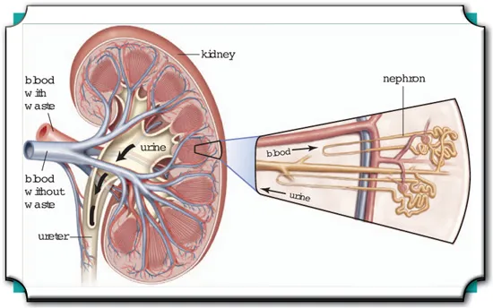
Each kidney has approximately one million nephrons, which filter water and other substances out of the blood to produce urine. Encyclopædia Britannica, Inc.
Each nephron is a long tubule that is closed, expanded, and folded into a double-walled cuplike structure at one end. This structure, called the renal corpuscular capsule, or Bowman’s capsule, encloses a cluster of capillaries (microscopic blood vessels) called the glomerulus. The capsule and glomerulus together constitute a renal corpuscle, also called a malpighian body. Blood flows into and away from the glomerulus through small arteries (arterioles) that enter and exit the glomerulus through the open end of the capsule. This opening is called the vascular pole of the corpuscle.
Each nephron of the kidney contains blood vessels and a special tubule. As the filtrate flows through the tubule of the nephron, it becomes increasingly concentrated into urine. Waste products are transferred from the blood into the filtrate, while nutrients are absorbed from the filtrate into the blood. Encyclopædia Britannica, Inc.
The tubules of the nephrons are 30–55 mm (1.2–2.2 inches) long. The corpuscle and the initial portion of each tubule, called the proximal convoluted tubule, lie in the renal cortex. The tubule descends into a renal pyramid, makes a U-shaped turn, and returns to the cortex at a point near its point of entry into the medulla. This section of the tubule, consisting of the two parallel lengths and the bend between them, is called the loop of Henle or the nephronic loop. After its reentrance into the cortex, the tubule returns to the vascular pole (the opening in the cuplike structure of the capsule) of its own nephron. The final portion of the tubule, the distal convoluted tubule, leads from the vascular pole of the corpuscle to a collecting tubule, by way of a short junctional tubule. Several of the collecting tubules join together to form a somewhat wider tubule, which carries the urine to a renal papilla and the renal pelvis.
The principal function of the loop of Henle appears to be the recovery of water and sodium chloride from the urine. This function allows production of urine that is far more concentrated than blood, limiting the amount of water needed as intake for survival. Many species that live in arid environments such as deserts have highly efficient loops of Henle.
The liquid entering the loop is the solution of salt, urea, and other substances passed along by the proximal convoluted tubule, from which most of the dissolved components needed by the body—particularly glucose, amino acids, and sodium bicarbonate—have been reabsorbed into the blood. The first segment of the loop, the descending limb, is permeable to water, and the liquid reaching the bend of the loop is much richer than the blood plasma in salt and urea. As the liquid returns through the ascending limb, sodium chloride diffuses out of the tubule into the surrounding tissue, where its concentration is lower. In the third segment of the loop, the tubule wall can, if necessary, effect further removal of salt, even against the concentration gradient, in an active-transport process requiring the expenditure of energy. In a healthy person the reabsorption of salt from the urine exactly maintains the bodily requirement: during periods of low salt intake, virtually none is allowed to escape in the urine, but, in periods of high salt intake, the excess is excreted.
Although all nephrons in the kidney have the same general disposition, there are regional differences, particularly in the length of the loops of Henle. Glomeruli that li...
Table of contents
- Cover Page
- Title Page
- Copyright Page
- Contents
- Introduction
- Chapter 1: Anatomy of the Kidneys and Renal System
- Chapter 2: Development and Function of the Kidneys and Renal System
- Chapter 3: The Physiology of Urinary Excretion
- Chapter 4: Renal Disorders of Fluid Regulation and Urinary Function
- Chapter 5: Kidney Failure and Inflammatory and Malignant Renal Diseases and Disorders
- Chapter 6: Evaluation and Treatment of Renal Diseases and Disorders
- Conclusion
- Glossary
- Bibliography
- Index
Frequently asked questions
Yes, you can cancel anytime from the Subscription tab in your account settings on the Perlego website. Your subscription will stay active until the end of your current billing period. Learn how to cancel your subscription
No, books cannot be downloaded as external files, such as PDFs, for use outside of Perlego. However, you can download books within the Perlego app for offline reading on mobile or tablet. Learn how to download books offline
Perlego offers two plans: Essential and Complete
- Essential is ideal for learners and professionals who enjoy exploring a wide range of subjects. Access the Essential Library with 800,000+ trusted titles and best-sellers across business, personal growth, and the humanities. Includes unlimited reading time and Standard Read Aloud voice.
- Complete: Perfect for advanced learners and researchers needing full, unrestricted access. Unlock 1.5M+ books across hundreds of subjects, including academic and specialized titles. The Complete Plan also includes advanced features like Premium Read Aloud and Research Assistant.
We are an online textbook subscription service, where you can get access to an entire online library for less than the price of a single book per month. With over 1.5 million books across 990+ topics, we’ve got you covered! Learn about our mission
Look out for the read-aloud symbol on your next book to see if you can listen to it. The read-aloud tool reads text aloud for you, highlighting the text as it is being read. You can pause it, speed it up and slow it down. Learn more about Read Aloud
Yes! You can use the Perlego app on both iOS and Android devices to read anytime, anywhere — even offline. Perfect for commutes or when you’re on the go.
Please note we cannot support devices running on iOS 13 and Android 7 or earlier. Learn more about using the app
Please note we cannot support devices running on iOS 13 and Android 7 or earlier. Learn more about using the app
Yes, you can access The Kidneys and the Renal System by Britannica Educational Publishing, Kara Rogers in PDF and/or ePUB format, as well as other popular books in Biological Sciences & Physiology. We have over 1.5 million books available in our catalogue for you to explore.