Although much is now known about the biology of reproduction, a great deal remains to be discovered about the diseases and disorders that can damage the reproductive system. Ethical concerns surrounding the issues of birth control and abortion have also provoked much discussion in recent times. This comprehensive narrative details both the biological process of procreation and the often divisive debates on how best to approach matters related to the human body.
Trusted by 375,005 students
Access to over 1.5 million titles for a fair monthly price.
The human reproductive system consists of a network of organs and signaling molecules that interact and communicate to give rise to the human ability to produce and bear live offspring. It is distinguished from all other organ systems of the human body by the fact that it is composed of two anatomically different organ plans, one for the female and one for the male. It is also a slow-developing system, with the reproductive organs obtaining full maturity at some point during adolescence, the transitional phase of growth and development between childhood and adulthood.
Provided all organs are present, normally constructed, and functioning properly, the seven essential features of human reproduction are (1) liberation of an ovum, or egg, at a specific time in the reproductive cycle; (2) internal fertilization of the ovum by spermatozoa, or sperm cells; (3) transport of the fertilized ovum to the uterus, or womb; (4) implantation of the blastocyst, the early embryo developed from the fertilized ovum, in the wall of the uterus; (5) formation of a placenta and maintenance of the unborn child during the entire period of gestation; (6) birth of the child and expulsion of the placenta; and (7) suckling and care of the child, with an eventual return of the maternal organs to virtually their original state.
For this biological process to be carried out, certain organs and structures are required in both the male and the female. The source of the ova (the female germ cells) is the female ovary; that of sperm (the male germ cells) is the testis. In females, the two ovaries are situated in the pelvic cavity. In males, the two testes are enveloped in a sac of skin, the scrotum, lying below and outside the abdomen. Besides producing the germ cells, or gametes, the ovaries and testes are the source of hormones that cause full development of secondary sexual characteristics and the proper functioning of the reproductive tracts. These tracts comprise the fallopian tubes, the uterus, the vagina, and associated structures in females and the penis, the sperm channels (epididymis, ductus deferens, and ejaculatory ducts), and other related structures and glands in males. The function of the fallopian tube is to convey an ovum, which is fertilized in the tube, to the uterus, where gestation (development before birth) takes place. The function of the male ducts is to convey sperm from the testis, to store them, and, when ejaculation occurs, to eject them with secretions from the male glands through the penis.
At copulation, or sexual intercourse, the erect penis is inserted into the vagina, and sperm contained in the seminal fluid (semen) are ejaculated into the female genital tract. Sperm then pass from the vagina through the uterus to the fallopian tube to fertilize the ovum in the outer part of the tube. Females exhibit a periodicity in the activity of their ovaries and uterus, which starts at puberty and ends at menopause. The periodicity is manifested by menstruation at intervals of about 28 days. Important changes occur in the ovaries and uterus during each reproductive, or menstrual, cycle. Periodicity, and subsequently menstruation, is suppressed during pregnancy and lactation.
THE MALE REPRODUCTIVE SYSTEM
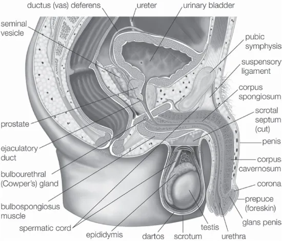
The male gonads are the testes, the source of sperm and of male sex hormones called androgens. The other genital organs are the epididymides; the ductus, or vasa, deferentia; the seminal vesicles; the ejaculatory ducts; and the penis; as well as certain accessory structures, such as the prostate and the bulbourethral (Cowper) glands. The principal functions of these structures are to transport the sperm from the testes to the exterior, to allow their maturation on the way, and to provide certain secretions that help form the semen.
Organs of the male reproductive system. Encyclopædia Britannica, Inc.
EXTERNAL GENITALIA
The two testes, or testicles, which usually complete their descent into the scrotum from their point of origin on the back wall of the abdomen in the seventh month after conception, are suspended in the scrotum by the spermatic cords. Each testis is enclosed in a fibrous sac, the tunica albuginea. The sac is lined internally by the tunica vasculosa, containing a network of blood vessels, and is covered by the tunica vaginalis, which is a continuation of the membrane that lines the abdomen and pelvis. The tunica albuginea has extensions into each testis that act as partial partitions to divide the testis into lobules.
Each lobule contains one or more convoluted, narrow tubes, known as seminiferous tubules, where sperm are formed. The tubules, if straightened, would extend about about 28 inches (70 cm). The multistage process of sperm formation, which takes about 60 days, occurs in the lining of the tubules, starting with the spermatogonia, or primitive sperm cells, in the outermost layer of the lining. Spermatozoa (sperm) leaving the tubules are incapable of independent motion, but they undergo a further maturation process in the ducts of the male reproductive tract. The process may be continued when, after ejaculation, they pass through the female tract. Maturation of the sperm in the female tract is called capacitation. Each sperm is a slender elongated structure with a head, a neck, a middle piece, and a tail. The head contains the cell nucleus. When the sperm is fully mature, it is propelled by the lashing movements of the tail.
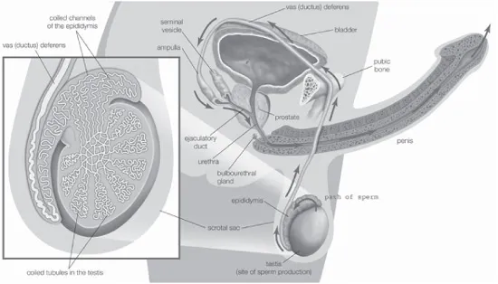
Structures involved in the production and transport of semen. Encyclopædia Britannica, Inc.
Each testis is supplied with blood by the testicular arteries, which arise from the front of the aorta just below the origin of the renal (kidney) arteries. Each artery crosses the rear abdominal wall, enters the spermatic cord, passes through the inguinal canal, and enters the upper end of each testis at the back. The veins leaving the testis and epididymis form a network, which ascends into the spermatic cord. The lymph vessels, which also pass through the spermatic cord, drain to the lateral and preaortic lymph nodes. Nerve fibres to the testis accompany the vessels, passing through the renal and aortic nerve plexuses, or networks.
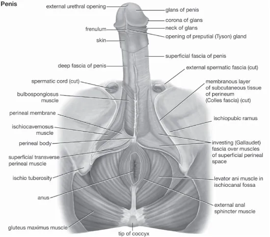
THE PENIS
The penis, the male organ of copulation, is partly inside and partly outside the body. The inner part, attached to the bony margins of the pubic arch (that part of the pelvis directly in front and at the base of the trunk), is called the root of the penis. The second, or outer, portion is free, pendulous, and enveloped all over in skin. It is known as the body of the penis. The organ is composed chiefly of cavernous or erectile tissue that becomes engorged with blood to produce considerable enlargement and erection. The penis is traversed by a tube, the urethra, which serves as a passage both for urine and for semen.
The body of the penis, sometimes referred to as the shaft, is cylindrical in shape when flaccid but when erect is somewhat triangular in cross section, with the angles rounded. This condition arises because the right corpus cavernosum and the left corpus cavernosum, the masses of erectile tissue, lie close together in the dorsal part of the penis, while a single body, the corpus spongiosum, which contains the urethra, lies in a midline groove on the undersurface of the corpora cavernosa. The dorsal surface of the penis is that which faces upward and backward during erection.
The human penis. Encyclopædia Britannica, Inc.
The slender corpus spongiosum reaches beyond the extremities of the erectile corpora cavernosa and at its outer end is enlarged considerably to form a soft, conical, sensitive structure called the glans penis. The base of the glans has a projecting margin, the corona, and the groove where the corona overhangs the corpora cavernosa is referred to as the neck of the penis. The glans is traversed by the urethra, which ends in a vertical, slitlike, external opening. The skin over the penis is thin and loosely adherent and at the neck is folded forward over the glans for a variable distance to form the prepuce or foreskin. A median fold, the frenulum of the prepuce, passes to the undersurface of the glans to reach a point just behind the urethral opening. The prepuce can usually be readily drawn back to expose the glans.
The root of the penis comprises two crura, or projections, and the bulb of the penis. The crura and the bulb are attached respectively to the edges of the pubic arch and to the perineal membrane (the fibrous membrane that forms a floor of the trunk). Each crus is an elongated structure covered by the ischiocavernosus muscle, and each extends forward, converging toward the other, to become continuous with one of the corpora cavernosa. The oval bulb of the penis lies between the two crura and is covered by the bulbospongiosus muscle. It is continuous with the corpus spongiosum. The urethra enters it on the flattened deep aspect that lies against the perineal membrane, traverses its substances, and continues into the corpus spongiosum.
The two corpora cavernosa are close to one another, separated only by a partition in the fibrous sheath that encloses them. The erectile tissue of the corpora is divided by numerous small fibrous bands into many cavernous spaces, relatively empty when the penis is flaccid but engorged with blood during erection. The structure of the tissue of the corpus spongiosum is similar to that of the corpora cavernosa, but there is more smooth muscle and elastic tissue. A deep fascia, or sheet of connective tissue, surrounding the structures in the body of the penis is prolonged to form the suspensory ligament, which anchors the penis to the pelvic bones at the midpoint of the pubic arch.
The penis has a rich blood supply from the internal pudendal artery, a branch of the internal iliac artery, which supplies blood to the pelvic structures and organs, the buttocks, and the inside of the thighs. Erection is brought about by distension of the cavernous spaces with blood, which is prevented from draining away by compression of the veins in the area.
The penis is amply supplied with sensory and autonomic (involuntary) nerves. Of the autonomic nerve fibres the sympathetic fibres cause constriction of blood vessels, and the parasympathetic fibres cause their dilation. It is usually stated that ejaculation is brought about by the sympathetic system, which at the same time inhibits the desire to urinate and prevents the semen from entering the bladder.
THE SCROTUM
The scrotum is a pouch of skin lying below the pubic symphysis and just in front of the upper parts of the thighs. It contains the testes and lowest parts of the spermatic cord. A scrotal septum or partition divides the pouch into two compartments and arises from a ridge, or raphe, visible on the outside of the scrotum. The raphe turns forward onto the undersurface of the penis and is continued back onto the perineum (the area between the legs and as far back as the anus). This arrangement indicates the bilateral origin of the scrotum from two genital swellings that lie one on each side of the base of the phallus, the precursor of the penis or clitoris in the embryo. The swellings are also referred to as the labioscrotal swellings because in females they remain separate to form the labia majora and in males they unite to form the scrotum.
The skin of the scrotum is thin, pigmented, devoid of fatty tissue, and more or less folded and wrinkled. There are some scattered hairs and sebaceous glands on its surface. Below the skin is a layer of involuntary muscle, the dartos, which can alter the appearance of the scrotum. On exposure of the scrotum to cold air or cold water, the dartos contracts and gives the scrotum a shortened, corrugated appearance, whereas warmth causes the scrotum to become smoother, flaccid, and less closely tucked in around the testes. Beneath the dartos muscle are layers of fascia continuous with those forming the coverings of each of the two spermatic cords, which suspend the testes within the scrotum and contain each ductus deferens, the testicular blood and lymph vessels; the artery to the cremaster muscle (which draws the testes upward); the artery to each ductus deferens; the genital branch of the genitofemoral nerve; and the testicular network of nerves.
The scrotum is supplied with blood by the external pudendal branches of the femoral artery, which is the chief artery of the thigh, and by the scrotal branches of the internal pudendal artery. The veins follow the arteries. The lymphatic drainage is to the lymph nodes in the groin.
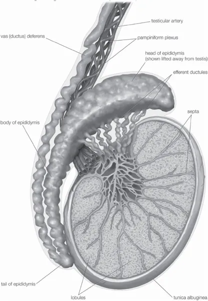
THE TESTES
The testes are the two sperm-producing organs in males. They also produce androgens, the male hormones. In humans the testes occur as a pair of oval-shaped organs and are contained within the scrotal sac.
In humans each testis weighs about 0.875 ounce (25 grams) and is 1.6–2.0 inches (4–5 cm) long and 0.8–1.2 inches (2–3 cm) in diameter. Each is divided by partitions of fibrous tissue from the tunica albuginea into 200 to 400 wedge-shaped sections, or lobes. Within each lobe are 3 to 10 seminiferous tubules. The partitions between the lobes and the seminiferous tubules both converge in one area near the anal side of each testis to form what is called the mediastinum testis.
Human male testis, epididymis, and ductus deferens. Encyclopædia Britannica, Inc.
The testes contain germ cells that differentiate into mature spermatozoa, supporting cells called Sertoli cells, and testosterone-producing cells called Leydig (interstitial) cells. The germ cells migrate to the fetal testes from the embryonic yolk sac. The Sertoli cells, which are interspersed between the germinal epithelial cells within the seminiferous tubules, are analogous to the granulosa cells in the ovary, and the Leydig cells, which are located beneath the tunica albuginea, in the septal walls, and between the tubules, are analogous to the hormone-secreting interstitial cells of the ovary. The Leydig cells are irregularly shaped; commonly have more than one nucleus; and frequently contain fat droplets, pigment granules, and crystalline structures. They vary greatly in number and appearance among the various animal species. Leydig cells are surrounded by numerous blood and lymphatic vessels, as well as by nerve fibres.
The embryonic differentiation of the primitive, indifferent gonad into either the testes or the ovaries is determined by the presence or absence of genes carried on the Y chromosome. Testosterone and its potent derivative, dihydrotestosterone, play key roles in the formation of male genitalia in the fetus during the first trimester of gestation but do not play a role in the actual formation of the testes. The testes are formed in the abdominal cavity and descend into the scrotum during the seventh month of gestation, when they are stimulated by androgens. About 2 percent of newborn boys have an undescended testis at birth, but this condition often corrects itself by the age of three months. The production of testosterone by the fetal testes is stimulated by human chorionic gonadotropin, a hormone secreted by the placenta. Within a few weeks following birth, testosterone secretion ceases, and the cells within the testes remain undeveloped during early childhood. During adolescence, gonadotropic hormones from the pituitary gland at the base of the brain stimulate the development of tissue, and the testes become capable of producing sperm and androgens.
The seminiferous tubules constitute about 90 percent of the testicular mass. In the young male the tubules are simple and composed of undeveloped sperm-producing cells (spermatogonia) and the Sertoli cells. In the older male the tubules become branched, and spermatogonia are changed into the fertile sperm cells after a series of transformations called spermatogenesis. The Sertoli cells found in both young and adult males mechanically support and protect the spermatogonia.
Each seminiferous tubule of the adult testis has a central lumen, or cavity, which is connected to the epididymis and spermatic duct (ductus deferens). Sperm cells originate as spermatogonia along the walls of the seminiferous tubules. The spermatogonia mature into spermatocytes, which mature into spermatids that mature into spermatozoa as they move into the central lumen of the seminiferous tubule. The sperm migrate, by short contractions of the tubule, to the mediastinum testis and are then transported through a complex network of canals (rete testis and efferent ductules) to the epididymis for temporary storage. The sperm move through the epididymis and the spermatic duct to be stored in the seminal vesicles for eventual ejaculation with the seminal fluid. Healthy men produce about one million sperm daily.
In animals that breed seasonally, such as sheep and goats, the testes regress completely during the nonbreeding season and the spermatogonia return to the state found in the young, sexually immature males. Frequently in these animals the testes are drawn back into the body cavity except in the breeding season, when they again descend and mature, a process known as recrudescence.
The principal androgen produced by the testes is testosterone. Testosterone produced locally in the testes and follicle-stimulating hormone (FSH) produced distally in the pituitary gland stimulate the process of spermatogenesis. Testosterone production and spermatogenesis slowly decrease in older men, whereas in women ovarian function ceases abruptly at the time of menopause.
STRUCTURES OF THE SPERM CANAL
The epididymis, ductus deferens (or vas deferens), and ejaculatory ...
Table of contents
Cover Page
Title Page
Copyright Page
Contents
Introduction
Chapter 1: Anatomy of the Human Reproductive System
Chapter 2: Hormones and Reproductive Maturity
Chapter 3: Reproduction and Pregnancy
Chapter 4: Disorders of Reproductive Development and Function
Chapter 5: Infections and Cancers of the Reproductive System
Chapter 6: Infertility and Disorders of Pregnancy
Chapter 7: Issues Concerning Birth Control and Abortion
Conclusion
Glossary
Bibliography
Index
Frequently asked questions
Yes, you can cancel anytime from the Subscription tab in your account settings on the Perlego website. Your subscription will stay active until the end of your current billing period. Learn how to cancel your subscription
No, books cannot be downloaded as external files, such as PDFs, for use outside of Perlego. However, you can download books within the Perlego app for offline reading on mobile or tablet. Learn how to download books offline
Perlego offers two plans: Essential and Complete
Essential is ideal for learners and professionals who enjoy exploring a wide range of subjects. Access the Essential Library with 800,000+ trusted titles and best-sellers across business, personal growth, and the humanities. Includes unlimited reading time and Standard Read Aloud voice.
Complete: Perfect for advanced learners and researchers needing full, unrestricted access. Unlock 1.5M+ books across hundreds of subjects, including academic and specialized titles. The Complete Plan also includes advanced features like Premium Read Aloud and Research Assistant.
Both plans are available with monthly, semester, or annual billing cycles.
We are an online textbook subscription service, where you can get access to an entire online library for less than the price of a single book per month. With over 1.5 million books across 990+ topics, we’ve got you covered! Learn about our mission
Look out for the read-aloud symbol on your next book to see if you can listen to it. The read-aloud tool reads text aloud for you, highlighting the text as it is being read. You can pause it, speed it up and slow it down. Learn more about Read Aloud
Yes! You can use the Perlego app on both iOS and Android devices to read anytime, anywhere — even offline. Perfect for commutes or when you’re on the go. Please note we cannot support devices running on iOS 13 and Android 7 or earlier. Learn more about using the app
Yes, you can access The Reproductive System by Britannica Educational Publishing, Kara Rogers in PDF and/or ePUB format, as well as other popular books in Biological Sciences & Physiology. We have over 1.5 million books available in our catalogue for you to explore.