So automatic and mechanical is breathing for most of us that we often fail to consider the complexities of respiration. Engaging the lungs, airways, and more, the intake of oxygen and release of carbon dioxide are only the most apparent aspects of a much longer routine. Although vulnerable to various infections and other disorders, the respiratory system by and large continues to function in order to sustain us. This book explores each element involved in this subconscious process and the factors that perpetuate human life.
- English
- ePUB (mobile friendly)
- Available on iOS & Android
eBook - ePub
The Respiratory System
About this book
Trusted by 375,005 students
Access to over 1.5 million titles for a fair monthly price.
Study more efficiently using our study tools.
Information
Subtopic
PhysiologyIndex
Biological Sciences
CHAPTER 1
ANATOMY AND FUNCTION OF THE HUMAN RESPIRATORY SYSTEM
Our respiratory system provides us with the fundamental ability to breathe: to inhale and exhale air from our lungs. Breathing, or respiration, is fundamental to survival, and though we possess the ability to consciously control the rate of our breathing, it is otherwise an automatic process, occurring without our having to think about it. Yet, as simple as it is for us to inhale and exhale, supporting this process are a number of complex actions that occur within our bodies. These actions encompass not only muscular movements but also cellular and chemical processes.
The respiratory system consists of two divisions: upper airways and lower airways. The transition between these two divisions is located where the pathways of the respiratory and digestive systems cross, just at the top of the larynx (or voice box). The upper airway system comprises the nose and the paranasal cavities (or sinuses), the pharynx (or throat), and part of the oral cavity. The lower airway system consists of the larynx, the trachea, the stem bronchi, and all the airways that branch extensively within the lungs, such as the intrapulmonary bronchi, the bronchioles, and the alveolar ducts.
THE DESIGN OF THE RESPIRATORY SYSTEM
The human gas–exchanging organ, the lung, is located in the thorax (or chest), where its delicate tissues are protected by the bony and muscular thoracic cage. The lung provides the body with a continuous flow of oxygen and clears the blood of the gaseous waste product, carbon dioxide. Atmospheric air is pumped in and out regularly through a system of pipes, called conducting airways, which connect the gas–exchange region inside the body with the environment outside the body.
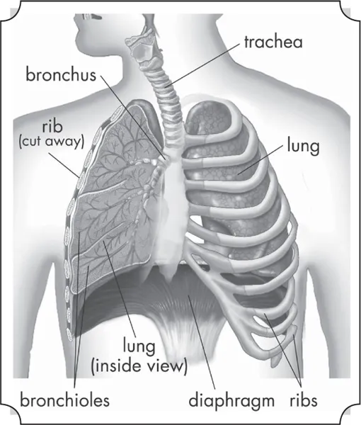
The lungs serve as the gas-exchanging organ for the process of respiration. Encyclopædia Britannica, Inc.
For respiration, the collaboration of other organ systems is essential. The diaphragm, as the main respiratory muscle, and the intercostal muscles of the chest wall play an essential role by generating, under the control of the central nervous system, the pumping action on the lung. The muscles expand and contract the internal space of the thorax, whose bony framework is formed by the ribs and the thoracic vertebrae. Other elements fundamental to the process of respiration include the blood, which acts as a carrier of gases, and the circulatory system (i.e., the heart and the blood vessels), which pumps blood from the heart to the lungs and the rest of the body.
MORPHOLOGY OF THE UPPER AIRWAYS
The nose, sinuses, and pharynx of the upper airways serve the vital role of filtering and warming air as it enters the respiratory tract. The filtering process is vital to clearing inhaled air of dust and other debris, and it protects against the passage into the lungs of potentially infectious foreign agents. The oral cavity, through which air may be inhaled or exhaled, is sometimes also considered a part of the upper airways. In addition to fulfilling a fundamental role in respiration, the structures of the upper respiratory tract also have other important functions, such as enabling the sensation of smell.
THE NOSE
The nose is the external protuberance of an internal space, the nasal cavity. It is subdivided into a left and right canal by a thin medial cartilaginous and bony wall, the nasal septum. Each canal opens to the face by a nostril and into the pharynx by the choana. The floor of the nasal cavity is formed by the palate, which also forms the roof of the oral cavity. The complex shape of the nasal cavity results from projections of bony ridges, the superior, middle, and inferior turbinate bones (or conchae), from the lateral wall. The passageways thus formed below each ridge are called the superior, middle, and inferior nasal meatuses.
On each side, the intranasal space communicates with a series of neighbouring air-filled cavities within the skull (the paranasal sinuses) and also, via the nasolacrimal duct, with the lacrimal apparatus in the corner of the eye. The duct drains the lacrimal fluid into the nasal cavity. This fact explains why nasal respiration can be rapidly impaired or even impeded during weeping: the lacrimal fluid is not only overflowing into tears, it is also flooding the nasal cavity.
The paranasal sinuses are sets of paired single or multiple cavities of variable size. Most of their development takes place after birth, and they reach their final size around age 20. The sinuses are located in four different skull bones: the maxilla, frontal, ethmoid, and sphenoid bones. Correspondingly, they are called the maxillary sinus, which is the largest cavity; the frontal sinus; the ethmoid sinuses; and the sphenoid sinus, which is located in the upper posterior wall of the nasal cavity. The sinuses have two principal functions: because they are filled with air, they help keep the weight of the skull within reasonable limits, and they serve as resonance chambers for the human voice.
The nasal cavity with its adjacent spaces is lined by a respiratory mucosa. Typically, the mucosa of the nose contains mucus-secreting glands and venous plexuses. Its top cell layer, the epithelium, consists principally of two cell types, ciliated and secreting cells. This structural design reflects the particular ancillary functions of the nose and of the upper airways in general with respect to respiration. They clean, moisten, and warm the inspired air, preparing it for intimate contact with the delicate tissues of the gas-exchange area. During expiration through the nose, the air is dried and cooled, a process that saves water and energy.
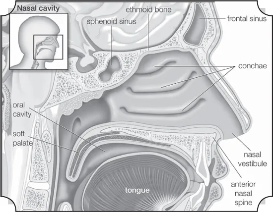
Sagittal view of the human nasal cavity. Encyclopædia Britannica, Inc.
Two regions of the nasal cavity have a different lining. The vestibule, at the entrance of the nose, is lined by skin that bears short thick hairs called vibrissae. In the roof of the nose, the olfactory organ with its sensory epithelium checks the quality of the inspired air. About two dozen olfactory nerves convey the sensation of smell from the olfactory cells through the bony roof of the nasal cavity to the central nervous system.
THE PHARYNX
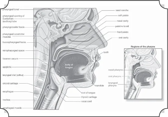
Sagittal section of the pharynx. Encyclopædia Britannica, Inc.
For the anatomical description, the pharynx can be divided into three floors. The upper floor, the nasopharynx, is primarily a passageway for air and secretions from the nose to the oral pharynx. It is also connected to the tympanic cavity of the middle ear through the auditory tubes that open on both lateral walls. The act of swallowing briefly opens the normally collapsed auditory tubes and allows the middle ears to be aerated and pressure differences to be equalized. In the posterior wall of the nasopharynx is located a lymphatic organ, the pharyngeal tonsil. When it is enlarged (as in tonsil hypertrophy), it may interfere with nasal respiration and alter the resonance pattern of the voice.
The middle floor of the pharynx connects anteriorly to the mouth and is therefore called the oral pharynx or oropharynx. It is delimited from the nasopharynx by the soft palate, which roofs the posterior part of the oral cavity.
The lower floor of the pharynx is called the hypopharynx. Its anterior wall is formed by the posterior part of the tongue. Lying directly above the larynx, it represents the site where the pathways of air and food cross each other: air from the nasal cavity flows into the larynx, and food from the oral cavity is routed to the esophagus directly behind the larynx. The epiglottis, a cartilaginous, leaf-shaped flap, functions as a lid to the larynx and, during the act of swallowing, controls the traffic of air and food.
MORPHOLOGY OF THE LOWER AIRWAYS
The major structures of the lower airways include the larynx, trachea, and lungs. The first two of these provide a canal for the passage of air to the lungs, while the lungs themselves receive the air and facilitate the process of gas exchange. The lungs reside within the thoracic cavity (chest cavity), which is the second–largest hollow space of the body. The cavity is enclosed by the ribs, the vertebral column, and the sternum (or breastbone) and is separated from the abdominal cavity (the body’s largest hollow space) by a muscular and membranous partition, the diaphragm. Also residing within the thoracic cavity is the tracheobronchial tree: the heart, the vessels transporting blood between the heart and the lungs, the great arteries bringing blood from the heart out into general circulation, and the major veins into which the blood is collected for transport back to the heart.
The chest cavity is lined with a serous membrane, so called because it exudes a thin fluid, or serum. This portion of the chest membrane is called the parietal pleura. The membrane continues over the lung, where it is called the visceral pleura, and over part of the esophagus, the heart, and the great vessels, as the mediastinal pleura, the mediastinum being the space and the tissues and structures between the two lungs. Because the atmospheric pressure between the parietal pleura and the visceral pleura is less than that of the outer atmosphere, the two surfaces tend to touch, friction between the two during the respiratory movements of the lung being eliminated by the lubricating actions of the serous fluid. The pleural cavity is the space, when it occurs, between the parietal and the visceral pleura.
THE LARYNX
The larynx is an organ of complex structure that serves a dual function: as an air canal to the lungs and a controller of its access, and as the organ of phonation. Sound is produced by forcing air through a sagittal slit formed by the vocal cords, the glottis. This causes not only the vocal cords but also the column of air above them to vibrate. As evidenced by trained singers, this function can be closely controlled and finely tuned. Control is achieved by a number of muscles innervated by the laryngeal nerves. For the precise function of the muscular apparatus, the muscles must be anchored to a stabilizing framework.
The laryngeal skeleton consists of almost a dozen pieces of cartilage, most of them minute, interconnected by ligaments and membranes. The largest cartilage of the larynx, the thyroid cartilage, is made of two plates fused anteriorly in the midline. At the upper end of the fusion line is an incision, the thyroid notch; below it is a forward projection, the laryngeal prominence. Both of these structures are easily felt through the skin. The angle between the two cartilage plates is sharper and the prominence more marked in men than in women, which has given this structure the common name of Adam’s apple.
Behind the shieldlike thyroid cartilage, the vocal cords span the laryngeal lumen. They correspond to elastic ligaments attached anteriorly in the angle of the thyroid shield and posteriorly to a pair of small pyramidal pieces of cartilage, the arytenoid cartilages. The vocal ligaments are part of a tube, resembling an organ pipe, made of elastic tissue. Just above the vocal cords, the epiglottis is also attached to the back of the thyroid plate by its stalk. The cricoid, another large cartilaginous piece of the laryngeal skeleton, has a signet-ring shape. The broad plate of the ring lies in the posterior wall of the larynx and the narrow arch in the anterior wall. The cricoid is located below the thyroid cartilage, to which it is joined in an articulation reinforced by ligaments. The transverse axis of the joint allows a hingelike rotation between the two cartilages. This movement tilts the cricoid plate with respect to the shield of the thyroid cartilage and hence alters the distance between them. Because the arytenoid cartilages rest upright on the cricoid plate, they follow its tilting movement. This mechanism plays an important role in altering length and tension of the vocal cords. The arytenoid cartilages articulate with the cricoid plate and hence are able to rotate and slide to close and open the glottis.
Viewed frontally, the lumen of the laryngeal tube has an hourglass shape, with its narrowest width at the glottis. Just above the vocal cords there is an additional pair of mucosal folds called the false vocal cords or the vestibular folds. Like the true vocal cords, they are also formed by the free end of a fibroelastic membrane. Between the vestibular folds and the vocal cords, the laryngeal space enlarges and forms lateral pockets extending upward. This space is called the ventricle of the larynx. Because the gap between the vestibular folds is always larger than the gap between the vocal cords, the latter can easily be seen from above with the laryngoscope, an instrument designed for visual inspection of the interior of the larynx.
The muscular apparatus of the larynx comprises two functionally distinct groups. The intrinsic muscles act directly or indirectly on the shape, length, and tension of the vocal cords. The extrinsic muscles act on the larynx as a whole, moving it upward (e.g., during high-pitched phonation or swallowing) or downward. The intrinsic muscles attach to the skeletal components of the larynx itself. The extrinsic muscles join the laryngeal skeleton cranially to the hyoid bone or to the pharynx and caudally to the sternum.
THE TRACHEA AND THE STEM BRONCHI
Below the larynx lies the trachea, a tube about 10 to 12 cm (4 to 5 inches) long and 2 cm (0.8 inch) wide. Its wall is stiffened by 16 to 20 characteristic horseshoe-shaped, incomplete cartilage rings that open toward the back and are embedded in a dense connective tissue. The dorsal wall contains a strong layer of transverse smooth muscle fibres that spans the gap of the cartilage. The interior of the trachea is lined by the typical respiratory epithelium. The mucosal layer contains mucous glands.
At its lower end, the trachea divides in an inverted Y into the two stem (or main) bronchi, one each for the left and right lung. The right main bronchus has a larger diameter, is oriented more vertically, and is shorter than the left main bronchus. The practical consequence of this arrangement is that foreign bodies passing beyond the larynx will usually slip into the right lung. The structure of the stem bronchi closely matches that of the trachea.
STRUCTURAL DESIGN OF THE AIRWAY TREE
The hierarchy of the dividing airways, and partly also of the blood vessels penetrating the lung, largely determines the internal lung structure. Functionally, the intrapulmonary airway system can be subdivided into three zones: a proximal, purely conducting zone; a peripheral, purely gas-exchanging zone; and a transitional zone in between, where both functions grade into one another. From a morphological point of view, however, it makes sense to distinguish the relatively thick-walled, purely airconducting tubes from those branches of the airway tree structurally designed to permit gas exchange.
The structural design of the airway tree is functionally important because the branching pattern plays a role in determining air flow and particle deposition. In modeling the human airway tree, it is generally agreed that the airways branch according to the rules of irregular dichotomy. Regular dichotomy means that each branch of a treelike structure gives rise to two daughter branches of identical dimensions. In irregular dichotomy, however, the daughter branches may differ greatly in length and diameter. The models calculate the average path from the trachea to the lung periphery as consisting of about 24 to 25 generations of branches. Individual paths, however, may range from 11 to 30 generations. The transition between the conductive and the respiratory portions of an airway lies on average at the end of the 16th generation, if the trachea is counted as generation zero.
The conducting airways comprise the trachea, the two stem bronchi, the bronchi, and the bronchioles. Their function is to further warm, moisten, and clean the inspired air and distribute it to the gas-exchanging zone of the lung. They are lined by the typical respiratory epithelium with ciliated cells and numerous interspersed mucus-secreting goblet cells. Ciliated cells are present far down in the airway tree, their height decreasing with the narrowing of the tubes, as does the frequency of goblet cells. In bronchioles the goblet cells are completely replaced by another type of secretory cells named Clara cells. The epithelium is covered by a layer of low-viscosity fluid, within which the cilia exert a synchronized, rhythmic beat directed outward. In larger airways, this fluid layer is topped by a blanket of mucus of high viscosity. The mucus layer is dragged along by the ciliary action and carries the intercepted particles toward the pharynx, where they are swallowed. This design can be compared to a conveyor b...
Table of contents
- Cover Page
- Title Page
- Copyright Page
- Contents
- Introduction
- Chapter 1: Anatomy and Function of the Human Respiratory System
- Chapter 2: Control and Mechanics of Breathing
- Chapter 3: Gas Exchange and Respiratory Adaptation
- Chapter 4: Infectious Diseases of the Respiratory System
- Chapter 5: Diseases and Disorders of the Respiratory System
- Chapter 6: Allergic and Occupational Lung Diseases and Acute Respiratory Conditions
- Chapter 7: Approaches to Respiratory Evaluation and Treatment
- Glossary
- Bibliography
- Index
Frequently asked questions
Yes, you can cancel anytime from the Subscription tab in your account settings on the Perlego website. Your subscription will stay active until the end of your current billing period. Learn how to cancel your subscription
No, books cannot be downloaded as external files, such as PDFs, for use outside of Perlego. However, you can download books within the Perlego app for offline reading on mobile or tablet. Learn how to download books offline
Perlego offers two plans: Essential and Complete
- Essential is ideal for learners and professionals who enjoy exploring a wide range of subjects. Access the Essential Library with 800,000+ trusted titles and best-sellers across business, personal growth, and the humanities. Includes unlimited reading time and Standard Read Aloud voice.
- Complete: Perfect for advanced learners and researchers needing full, unrestricted access. Unlock 1.5M+ books across hundreds of subjects, including academic and specialized titles. The Complete Plan also includes advanced features like Premium Read Aloud and Research Assistant.
We are an online textbook subscription service, where you can get access to an entire online library for less than the price of a single book per month. With over 1.5 million books across 990+ topics, we’ve got you covered! Learn about our mission
Look out for the read-aloud symbol on your next book to see if you can listen to it. The read-aloud tool reads text aloud for you, highlighting the text as it is being read. You can pause it, speed it up and slow it down. Learn more about Read Aloud
Yes! You can use the Perlego app on both iOS and Android devices to read anytime, anywhere — even offline. Perfect for commutes or when you’re on the go.
Please note we cannot support devices running on iOS 13 and Android 7 or earlier. Learn more about using the app
Please note we cannot support devices running on iOS 13 and Android 7 or earlier. Learn more about using the app
Yes, you can access The Respiratory System by Britannica Educational Publishing, Kara Rogers in PDF and/or ePUB format, as well as other popular books in Biological Sciences & Physiology. We have over 1.5 million books available in our catalogue for you to explore.