- 420 pages
- English
- ePUB (mobile friendly)
- Available on iOS & Android
eBook - ePub
A Laboratory Guide to the Tight Junction
About this book
A Laboratory Guide to the Tight Junction offers broad coverage of the unique methods required to investigate its characteristics. The methods are described in detail, including its biochemical and biophysical principles, step-by-step process, data analysis, troubleshooting, and optimization. The coverage includes various cell, tissue, and animal models.
Chapter 1 provides the foundations of cell biology of tight junction. Chapter 2 covers the Biochemical approaches for paracellular channels and is followed by chapter 3 providing the Biophysical approaches. Chapter 4 describes and discusses Histological approaches for tissue fixation and preparation. Chapter 5 discusses Light microscopy, while chapter 6 presents Electron microscopic approaches. Chapter 7 covers Transgenic manipulation in cell cultures, including DNA and siRNA, Mutagenesis, and viral infection. Chapter 8 covers transgenic manipulation in mice, including: Knockout, Knockin, siRNA knockdown, GFP/LacZ reporter, and overexpression. The final chapter discusses the future developments of new approaches for tight junction research.
Researchers and advanced students in bioscience working on topics of cell junction, ion channel and membrane protein will benefit from the described methods. Clinicians and pathologists interested in tissue barrier diseases will also benefit from the biochemical and biophysical characterization of tight junctions in organ systems, and their connection to human diseases.
- Provides consistent and detailed research methods
- Covers various cell, tissue and animal models
- Includes step-by-step guidance from beginner to sophisticated levels
Trusted by 375,005 students
Access to over 1.5 million titles for a fair monthly price.
Study more efficiently using our study tools.
Information
Chapter 1
Introduction
Abstract
Metazoan life is based on the formation of tissues and on tissue-specific functions. Tissues are established on a fundamental cellular architecture—a multicellular layer separating the interior of the organism from the exterior environment. As the organism develops more elaborate tissue structures, demands on intercellular interaction increase, presenting a number of structural and functional challenges that lead to the formation of specialized cell junctions. These junctions not only glue cells together (adherens junction and desmosome) but also perform intercellular communication (gap junction) and provide barriers to regulate the flow of solutes and water through the intercellular space (tight junction). This section introduces the concept of four major forms of cell junction and describes their function, component, interaction, signaling, and relationship to the cytoskeleton.
Keywords
Adherens junction; desmosome; gap junction; tight junction; paracellular channel; actin; myosin; cytoskeleton; Wnt signaling; Hippo signaling
Chapter 1.1
Cell junction
1.1.1 Junction category
Mammalian cells possess four intercellular junction systems formed by characteristic transmembrane molecules and submembranous protein plaques (Table 1.1.1). The intercellular junction systems are as follows:
- • Tight junction (zonula occludens) is a multiprotein complex whose general function is to seal the paracellular space to prevent leakage of solutes and water.
- • Adherens junction (zonula adherens) is a multiprotein architecture whose general role is cell adhesion.
- • Desmosome (macula adherens) provides a strong type of cell adhesion and is found in stratified epithelium.
- • Gap junction (macula communicans) creates an intercellular channel allowing direct molecular exchange from cell to cell.
Table 1.1.1
| Cellular localization | Integral protein | Plaque protein | Associated filament | |
|---|---|---|---|---|
| Tight junction | Epithelial cell Endothelial cell | Claudins Occludin | ZOs | Microfilament (actin) |
| Adherens junction | Epithelial cell Endothelial cell Cardiomyocyte Mesenchymal and neural cell | Cadherins Nectins | Catenins | Microfilament (actin) |
| Desmosome | Stratified epithelial cell | Desmogleins Desmocollins | Desmoplakin plakoglobin | Intermediate filament (cytokeratin, vimentin, desmin) |
| Gap junction | Epithelial cell Endothelial cell | Connexins | ZOs | − |

1.1.2 Tight junction
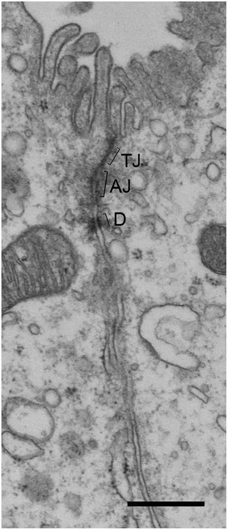
Tight junction (TJ) is the most apical member of intercellular junction systems. When viewed with transmission electron microscopy, TJ appears as a zone in which adjacent plasma membranes are fused (Fig. 1.1.1) (Farquhar & Palade, 1963). Freeze-fracture replica electron microscopy reveals that TJ is made of a continuous network of “fibrils” or “strands” on the protoplasmic (P) fracture face and complementary empty grooves on the exoplasmic (E) fracture face (Fig. 1.1.2) (Goodenough & Revel, 1970). The proteins making the TJ fibrils or strands are claudins. Claudins are tetraspan membrane proteins consisting of a family with 28 members ranging in molecular mass from 20 to 28 kDa. Claudins have four transmembrane domains, two extracellular loops (ECLs: ECL1 and ECL2), amino- and carboxyl-terminal cytoplasmic domains, and a short cytoplasmic turn. The charges in ECL1 regulate paracellular ion selectivity by electrostatic interactions. The carboxyl-terminal domain of claudin contains a PDZ (postsynaptic density 95/discs large/ZO-1) binding motif (YV) that is critical for interaction with the TJ plaque protein ZO-1 (Hou, Rajagopal, & Yu, 2013).

1.1.3 Adherens junction
Adherens junction appears under transmission electron microscopy as plasma membrane apposition separated by an intercellular cleft of 10–20 nm in width (Fig. 1.1.1). The cleft is occupied by rod-shaped molecules known as cadherins (Takeichi, 1990). The classic cadherin family comprises approximately 20 members that share a common domain organization. The cadherin protein features an amino-terminal extracellular domain or ectodomain that is followed by a transmembrane domain and a carboxyl-terminal intracellular domain. The ectodomain contains five secondary structural repeats, termed extracellular cadherin (EC) domains and numbered from EC1 to EC5. Each EC domain is made of approximately 110 amino acids and resembles the immunoglobulin domain found in antibodies (Shapiro & Weis, 2009). The intracellular domain binds to a class of cytoplasmic proteins, known as catenins. The catenins, in turn, interact with F-actin and other cytoskeletal proteins (Rimm, Koslov, Kebriaei, Cianci, & Morrow, 1995).
1.1.4 Desmosome
Desmosome is in many ways similar to adherens junction (Fig. 1.1.1). Desmosome is composed of the desmosome-intermediate filament complex, which is a network of desmosomal cadherin proteins, plaque proteins, and intermediate filaments (Garrod, 1993). Desmosomal cadherins include desmogleins and desmocollins. The extracellular domains of desmogleins and desmocollins mediate cell adhesion, whereas the cytoplasmic tails of these cadherins interact with the desmosomal plaque proteins such as plakoglobin and desmoplakin (Kowalczyk et al., 1994). The interaction between desmoplakin and intermediate filaments tether desmosomes to the cytoskeletal network. Desmogleins and desmocollins both contain four extracellular cadherin (EC1 to EC4) domains and a fifth domain termed the extracellular anchor (EA). The EA domain is not present in classic cadherin proteins.
1.1.5 Gap junction
Gap junction (GJ) is seen by transmission electron microscopy as an area of plasma membrane apposition separated by a 2–3 nm “gap” of intercellular space (Fig. 1.1.3). Freeze-fractured gap junction shows a lattice of densely packed particles on the P face and complementary pits on the E face. These particles are made of the connexin proteins (Fig. 1.1.3) (Goodenough & Paul, 2009). Connexins are tetraspan membrane proteins consisting of a family of 21 members ranging in molecular mass from 23 to 62 kDa. Connexins contain four transmembrane domains, two extracellular loops (ECLs: ECL1 a...
Table of contents
- Cover image
- Title page
- Table of Contents
- Copyright
- Dedication
- Author biography
- Preface
- Acknowledgments
- Chapter 1. Introduction
- Chapter 2. Biochemical approaches for tight junction
- Chapter 3. Biophysical approaches for tight junction
- Chapter 4. Histological approaches for tight junction
- Chapter 5. Light microscopy for tight junction
- Chapter 6. Electron microscopy for tight junction
- Chapter 7. Cell models of tight junction biology
- Chapter 8. Mouse models of tight junction physiology
- Chapter 9. Perspective
- Index
Frequently asked questions
Yes, you can cancel anytime from the Subscription tab in your account settings on the Perlego website. Your subscription will stay active until the end of your current billing period. Learn how to cancel your subscription
No, books cannot be downloaded as external files, such as PDFs, for use outside of Perlego. However, you can download books within the Perlego app for offline reading on mobile or tablet. Learn how to download books offline
Perlego offers two plans: Essential and Complete
- Essential is ideal for learners and professionals who enjoy exploring a wide range of subjects. Access the Essential Library with 800,000+ trusted titles and best-sellers across business, personal growth, and the humanities. Includes unlimited reading time and Standard Read Aloud voice.
- Complete: Perfect for advanced learners and researchers needing full, unrestricted access. Unlock 1.5M+ books across hundreds of subjects, including academic and specialized titles. The Complete Plan also includes advanced features like Premium Read Aloud and Research Assistant.
We are an online textbook subscription service, where you can get access to an entire online library for less than the price of a single book per month. With over 1.5 million books across 990+ topics, we’ve got you covered! Learn about our mission
Look out for the read-aloud symbol on your next book to see if you can listen to it. The read-aloud tool reads text aloud for you, highlighting the text as it is being read. You can pause it, speed it up and slow it down. Learn more about Read Aloud
Yes! You can use the Perlego app on both iOS and Android devices to read anytime, anywhere — even offline. Perfect for commutes or when you’re on the go.
Please note we cannot support devices running on iOS 13 and Android 7 or earlier. Learn more about using the app
Please note we cannot support devices running on iOS 13 and Android 7 or earlier. Learn more about using the app
Yes, you can access A Laboratory Guide to the Tight Junction by Jianghui Hou in PDF and/or ePUB format, as well as other popular books in Biological Sciences & Pharmaceutical, Biotechnology & Healthcare Industry. We have over 1.5 million books available in our catalogue for you to explore.