eBook - ePub
Developments and Applications for ECG Signal Processing
Modeling, Segmentation, and Pattern Recognition
- 210 pages
- English
- ePUB (mobile friendly)
- Available on iOS & Android
eBook - ePub
Developments and Applications for ECG Signal Processing
Modeling, Segmentation, and Pattern Recognition
About this book
Developments and Applications for ECG Signal Processing: Modeling, Segmentation, and Pattern Recognition covers reliable techniques for ECG signal processing and their potential to significantly increase the applicability of ECG use in diagnosis. This book details a wide range of challenges in the processes of acquisition, preprocessing, segmentation, mathematical modelling and pattern recognition in ECG signals, presenting practical and robust solutions based on digital signal processing techniques. Users will find this to be a comprehensive resource that contributes to research on the automatic analysis of ECG signals and extends resources relating to rapid and accurate diagnoses, particularly for long-term signals.
Chapters cover classical and modern features surrounding f ECG signals, ECG signal acquisition systems, techniques for noise suppression for ECG signal processing, a delineation of the QRS complex, mathematical modelling of T- and P-waves, and the automatic classification of heartbeats.
- Gives comprehensive coverage of ECG signal processing
- Presents development and parametrization techniques for ECG signal acquisition systems
- Analyzes and compares distortions caused by different digital filtering techniques for noise suppression applied over the ECG signal
- Describes how to identify if a digitized ECG signal presents irreversible distortion through analysis of its frequency components prior to, and after, filtering
- Considers how to enhance QRS complexes and differentiate these from artefacts, noise, and other characteristic waves under different scenarios
Trusted by 375,005 students
Access to over 1.5 million titles for a fair monthly price.
Study more efficiently using our study tools.
Information
Chapter 1
Classical and Modern Features for Interpretation of ECG Signal
João Paulo do Vale Madeiro⁎; Paulo César Cortez†; João Loures Salinet Jr.‡; Roberto Coury Pedrosa§; José Maria da Silva Monteiro Filho¶; Angelo Roncalli Alencar Brayner¶ ⁎Institute for Engineering and Sustainable Development (IEDS), University for the International Integration of the Afro-Brazilian Lusophony – UNILAB, Redenção, Ceará, Brazil
†Department of Teleinformatics Engineering, Federal University of Ceara, Fortaleza, Ceará, Brazil
‡Center of Engineering, Modeling, and Applied Social Sciences, Federal University of ABC, São Paulo, Brazil
§University Hospital Clementino Fraga Filho, Federal University of Rio de Janeiro, Rio de Janeiro, Rio de Janeiro, Brazil
¶Department of Computing Science, Federal University of Ceara, Fortaleza, Ceará, Brazil
Abstract
The analysis and interpretation of the ECG signal, mainly considering long monitoring examinations, is fundamental for diagnosing arrhythmias and conduction disturbances. Hence, a reliable assessment for the exam is extremely dependent on the quality of signal recording and the accuracy of extracting all the available features. It is well known that the automatic delineation of ECG characteristic waves and the quantitative analysis of timeseries, waveforms, and amplitudes derived from ECG segmentation behave as challenging tasks, considering both the broad range of observed morphologies and also the inherent noise sensibility. But what kind of metrics are desirable for a beat-to-beat analysis aiming the evaluation of myocardial contraction and heart electrical system? So in this chapter, we will detail the state-of-the-art features, which can be automatically extracted from this biomedical signal for providing useful diagnostic information or even help to predict adverse events.
Keywords
Heart electrical system; Electrophysiology; Cardiac impulse; Electrocardiogram signal; Heart rhythms; Conduction disorders
1.1 Electrical Activity of the Heart
The heart, or the cardiac muscle, has the function of pumping blood to the organs and specifically to the lungs, facilitating the exchange of gases, absorbing oxygen and eliminating carbon dioxide. It is divided into upper (atria) and lower (ventricles) chambers, and it may also be considered to be composed of two separate pumping systems: a right heart that pumps blood through the lungs, and a left heart that pumps blood through the peripheral organs. Each atrium helps to move blood into the ventricle, and the ventricles supply the main pump force needed to push blood through the pulmonary and peripheral circulation systems (Hall, 2011), see Fig. 1.1 (this figure was published in Textbook of Medical Physiology, Arthur C. Guyton and John E. Hall, Chapter 9: Heart Muscle; The Heart as a Pump and Function of the Heart Valves, Page 104, Copyright Elsevier Inc. (2006)).
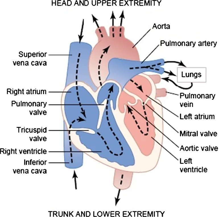
Figure 1.1 Structure of the blood pumping system of a human heart concerning heart chambers and heart valves.
In addition to the atrial and ventricle muscles, myocardial tissue consists of excitable and contractile fibers capable of developing self-regenerative electrical activity, presenting specific functions of generation, conduction, and contraction, depending on its anatomical location. There are two basic groups of cells in the myocardium that are important for cardiac function.
1.1.1 Cells Responsible for Myocardial Contraction
The contractile apparatus of cardiac fibers consists of a complex of contractile proteins, composed of actin, myosin, tropomyosin, and troponin, which—in the presence of calcium and adenosine triphosphate—interact with each other, causing contraction. Such cells possess the property of contractility, that is, the ability to shorten and then return to their original length. For a myocardial cell to contract, the cell membrane must be discharged electrically (a process called depolarization), causing a change in electrical charge across the membrane, resulting in the flow or movement of certain ions (especially sodium). The depolarization process also allows the entry of calcium into the cell, where it is responsible for the binding part between actin and myosin of the sarcomere (basic contractile unit of myocardial fibers), resulting in contraction.
1.1.2 The Electrical System
The cells that make up the electrical system of the heart are responsible for the formation of the electric current and the conduction of this impulse to the contractile cells of the myocardium, where the depolarization activates the contraction. Certain cells in the electrical system have the ability to generate an electrical impulse (a property referred to as spontaneous automaticity or depolarization). Cells possessing this property are known as “pacemaker cells”. These cells are found in the sinus node, in the cells responsible for atrial conduction, in the area immediately above the atrioventricular (AV) node, in the low portion of the AV node, in the His bundle, and in the Purkinje ventricular system.
As illustrated in Fig. 1.2 (this figure was published in Textbook of Medical Physiology, Arthur C. Guyton and John E. Hall, Chapter 10: Rhythmical Excitation of the Heart, Page 116, Copyright Elsevier Inc. (2006)) the sinus node or sinoatrial (S-A) node, in which the normal rhythmical impulse is generated, has the highest rate of spontaneous depolarization and acts as the primary pacemaker. Internodal pathways conduct the impulse from the sinus node to the AV node. The AV bundle conducts the impulse from the atria into the ventricles, and the left and right bundle branches of Purkinje fiber conduct the cardiac impulse to all parts of the ventricles (Wasilewski and Poloński, 2012).
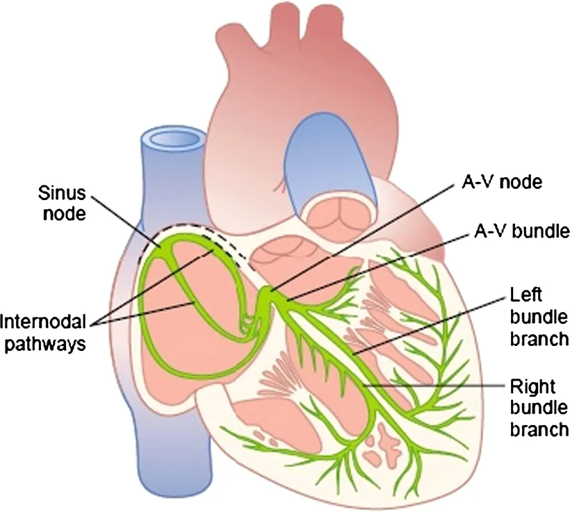
Figure 1.2 Schema of the specialized excitatory and conductive system of the heart that controls cardiac contractions.
1.1.3 Basic Electrophysiology
To better comprehend the disturbances in the electrical activity of the heart, it is important to have a basic knowledge of the electrical properties of the contractile myocardial cells and of the pacemaker cells (Fig. 1.3). With the use of a single microelectrode, the electrical event (or action potential) of a single cell can be recorded.

Figure 1.3 Schematic representation of the action potential of a ventricular myocardial cell, where the arrows indicate the movement of ions through the membrane.
The electrical activity of the cardiac fiber depends initially on the intracellular and extracellular concentration of alkaline, alkaline earth, and halogen ions. In the cellular protoplasm, low concentrations of sodium ions and high concentrations of potassium and chloride ions are maintained. The calcium ion undergoes variations in its intracellular concentration, depending on the muscular concentration. These same ions are present in the liquid that surrounds the cells, and the sodium and calcium concentrations are higher, the potassium lower, and the chlorine isotonic with the plasma. In the absence of contraction, the distribution in different concentrations intracellular and extracellular of these ions, separated by the cellular membrane, induces the formation of transmembrane ionic electrical potential known as Nernst potential, characteristic of each ionic species. The quantitative sum of the potentials of the involved ionic species generates a transmembrane potential difference that, at rest, is negative on the internal side in relation to the external face.
The transfer of sodium, potassium, and calcium ions from the medium into ...
Table of contents
- Cover image
- Title page
- Table of Contents
- Copyright
- Contributors
- Preface
- Chapter 1: Classical and Modern Features for Interpretation of ECG Signal
- Chapter 2: ECG Signal Acquisition Systems
- Chapter 3: Techniques for Noise Suppression for ECG Signal Processing
- Chapter 4: Techniques for QRS Complex Detection
- Chapter 5: Delineation of QRS Complex: Challenges for the Development of Widely Applicable Algorithms
- Chapter 6: Mathematical Modeling of T-Wave and P-Wave: A Robust Alternative for Detecting and Delineating Those Waveforms
- Chapter 7: The Issue of Automatic Classification of Heartbeats
- Index
Frequently asked questions
Yes, you can cancel anytime from the Subscription tab in your account settings on the Perlego website. Your subscription will stay active until the end of your current billing period. Learn how to cancel your subscription
No, books cannot be downloaded as external files, such as PDFs, for use outside of Perlego. However, you can download books within the Perlego app for offline reading on mobile or tablet. Learn how to download books offline
Perlego offers two plans: Essential and Complete
- Essential is ideal for learners and professionals who enjoy exploring a wide range of subjects. Access the Essential Library with 800,000+ trusted titles and best-sellers across business, personal growth, and the humanities. Includes unlimited reading time and Standard Read Aloud voice.
- Complete: Perfect for advanced learners and researchers needing full, unrestricted access. Unlock 1.5M+ books across hundreds of subjects, including academic and specialized titles. The Complete Plan also includes advanced features like Premium Read Aloud and Research Assistant.
We are an online textbook subscription service, where you can get access to an entire online library for less than the price of a single book per month. With over 1.5 million books across 990+ topics, we’ve got you covered! Learn about our mission
Look out for the read-aloud symbol on your next book to see if you can listen to it. The read-aloud tool reads text aloud for you, highlighting the text as it is being read. You can pause it, speed it up and slow it down. Learn more about Read Aloud
Yes! You can use the Perlego app on both iOS and Android devices to read anytime, anywhere — even offline. Perfect for commutes or when you’re on the go.
Please note we cannot support devices running on iOS 13 and Android 7 or earlier. Learn more about using the app
Please note we cannot support devices running on iOS 13 and Android 7 or earlier. Learn more about using the app
Yes, you can access Developments and Applications for ECG Signal Processing by Joao Paulo do Vale Madeiro,Paulo Cesar Cortez,José Maria Da Silva Monteiro Filho,Angelo Roncalli Alencar Brayner in PDF and/or ePUB format, as well as other popular books in Technology & Engineering & Electromagnetism. We have over 1.5 million books available in our catalogue for you to explore.