eBook - ePub
Characterization of Nanomaterials
Advances and Key Technologies
- 390 pages
- English
- ePUB (mobile friendly)
- Available on iOS & Android
eBook - ePub
Characterization of Nanomaterials
Advances and Key Technologies
About this book
Characterization of Nanomaterials: Advances and Key Technologies discusses the latest advancements in the synthesis of various types of nanomaterials. The book's main objective is to provide a comprehensive review regarding the latest advances in synthesis protocols that includes up-to-date data records on the synthesis of all kinds of inorganic nanostructures using various physical and chemical methods. The synthesis of all important nanomaterials, such as carbon nanostructures, Core-shell Quantum dots, Metal and metal oxide nanostructures, Nanoferrites, polymer nanostructures, nanofibers, and smart nanomaterials are discussed, making this a one-stop reference resource on research accomplishments in this area.
Leading researchers from industry, academia, government and private research institutions across the globe have contributed to the book. Academics, researchers, scientists, engineers and students working in the field of polymer nanocomposites will benefit from its solutions for material problems.
- Provides an up-to-date data record on the synthesis of all kinds of organic and inorganic nanostructures using various physical and chemical methods
- Presents the latest advances in synthesis protocols
- Presents latest techniques used in the physical and chemical characterization of nanomaterials
- Covers characterization of all the important materials groups such as: carbon nanostructures, core-shell quantumdots, metal and metal oxide nanostructures, nanoferrites, polymer nanostructures and nanofibers
- A broad range of applications is covered including the performance of batteries, solar cells, water filtration, catalysts, electronics, drug delivery, tissue engineering, food packaging, sensors and fuel cells
- Leading researchers from industry, academia, government and private research institutes have contributed to the books
Trusted by 375,005 students
Access to over 1.5 million titles for a fair monthly price.
Study more efficiently using our study tools.
Information
Chapter 1
Characterization of Carbon Nanomaterials by Raman Spectroscopy
Mirosław Szybowicz; Ariadna B. Nowicka; Anna Dychalska Poznan University of Technology, Poznan, Poland
Abstract
This chapter describes the characterization of carbon nanomaterials by Raman spectroscopy. It emphasizes the capabilities of Raman microscopy for precise and noninvasive study of carbon nanostructures and its usefulness as a diagnostic tool for the identification and characterization of different types of carbon structures. The chapter is accompanied by information on the study of nano carbon structures such as graphene, carbon nanotubes, and micro and nano diamond structures. The chapter contains the most important information related to interpretation of typical Raman spectra of carbon nanomaterials, which is correlated with describing of structure, structural defects, quality of carbon structures, and mechanical properties, etc. Additionally shown application of Raman mapping techniques for surface characterization of carbon nanomaterials.
Keywords
Carbon nanomaterials; Raman spectroscopy; Raman mapping; Graphene; Carbon nanotubes; Diamond structure; Defects in carbon nanostructure; Characterization of the surface carbon nanomaterials
1.1 Introduction
1.1.1 Raman Scattering
Raman spectroscopy is the light-scattering technique based on the interaction of light with matter, used for investigation of solids, liquids, and gases. The primary idea behind this method is observation of light scattered on crystalline lattice vibrations or oscillations of molecules. Raman spectroscopy is primarily used for investigation of oscillations. This method provides information on the chemical structure of a given material, characterization of its physical properties, and identification of substances. On the basis of Raman spectra, it is also possible to get quantitative or semiquantitative information of the content of a given compound in a studied sample. The Raman inelastic scattering means that the scattered photon can be of either lower (Stokes Raman scattering) or higher (anti-Stokes Raman scattering) energy than the incoming photon when compared with the elastic Rayleigh scattering in which the scattered photon has the same energy as the incoming photon. These bands occur symmetrically on both sides of the Rayleigh, and they are usually about 1000 times weaker than the Rayleigh band. The positions of these bands, expressed in wavenumbers or energy, depend on type of atoms, type of chemical bonds, structure, and symmetry of the molecules studied [1].
Typically, in the Raman spectroscopy, a sample is exposed to laser radiation in the range from ultraviolet (UV) to near-infrared (NIR). The incident wavelength interacts with the sample molecules, exciting them from the vibrational ground state to a virtual energy state. Oscillating molecules can couple with other molecules and perform additional vibrations, forming electron excitations [2].
1.1.2 Raman Microscopy
The Raman microscope is composed of an optical microscope; the excitation source, for example, different types of lasers; laser notch or edge filters whose function is to reject Rayleigh radiation; and a spectrometer or monochromator offering the diffraction grating with the highest spectral resolution. Additionally, the Raman microscopic system is connected to an optical detector such as a charge-coupled device (CCD) or photomultiplier tube (PMT). The modern Raman techniques allow not only measurements of a single spectrum of a sample but also Raman spatial and depth maps that consist of multiple spectra from different points (areas) on the sample. The scheme of a Raman microscope is given in Fig. 1.1A.

The advantages of Raman microscopy include high sensitivity, high spatial resolution of the order of < 1 μm, high confocal resolution of the order of < 2 μm, the possibility of Raman mapping (using motorized stage x, y, z for the study samples), and the possibility of use of different types of objectives (with different magnification and numerical aperture). Raman mapping is a powerful technique for generating detailed images based on selected spectral parameters of the Raman spectra such as position of the bands, intensities, and full width at half maximum (FWHM). The Raman mapping method is schematically shown in Fig. 1.1B.
The typical Raman system works in the backscattering geometry of the scattered light. The direction of the incident light is antiparallel to the scattered light (180 degree). Notch or edge filters minimize (cut off) Rayleigh radiation. Raman microscopy is characterized by high sensitivity, high lateral spatial and depth resolution, and fast and nondestructive measurements.
It is very important that the laser minimum focused spot size is not the same as that of the coupled Raman scattered spot. Confocal and spatial resolution is achieved by the use of a pinhole aperture (horizontal and vertical) as shown in Fig. 1.1C. Focused laser spot size is determined by the parameters such as the wavelength of the excitation light (incident light) and the numerical aperture of the microscope objective. This relationship is described as:
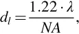
where dl is the spatial resolution of the microscope, λ is the wavelength of the incident light, and NA is the numerical aperture of the microscope objective. In the classical objective, the space between the lens and the front cover slip (glass or another material) is filled with air. The maximum numerical aperture NA of such an objective is 1, but in practice, it does not exceed 0.95. The increase in the spatial resolution is typically achieved by the use of an immersion objective whose numerical aperture varies from 1.2 to 1.6. In these objectives, the space between the lens and the front cover slip is filled with liquid. By using the liquid, which has a higher refractive index than air, we obtain a numerical aperture of NA > 1. Fig. 1.1D presents a scheme of the laser beam focus, where n is the refractive index of light and D is the diameter of the lens.
1.2 Graphene
Graphene is an allotropic form of carbon. It is a two-dimensional (2D) hexagonal lattice of carbon atoms. At room temperature, this material has a number of interesting physical properties, which makes it attractive relative to other materials. The Raman spectroscopy is an excellent, noninvasive research technique that enables identification of graphene, the number of graphene layers, stress and defects, and some other important mechanical, electrical, and optical parameters. In this section, we discuss Raman spectra of recorded graphene to determine the number of layers and types of defects, to make a Raman map of the grap...
Table of contents
- Cover image
- Title page
- Table of Contents
- Copyright
- Contributors
- Chapter 1: Characterization of Carbon Nanomaterials by Raman Spectroscopy
- Chapter 2: Raman Spectroscopy of Nanomaterials: Applications to Heterogeneous Catalysis
- Chapter 3: Characterization of Nanomaterials Using Nuclear Magnetic Resonance Spectroscopy
- Chapter 4: Electrochemical Characterization of Nanomaterials
- Chapter 5: Mammalian Cell-Based Assays for Studying Bio-Nano Interactions
- Chapter 6: Rheological Characterization of Nanocomposites
- Chapter 7: Mechanical Property Analysis of Nanomaterials
- Chapter 8: Thermoanalytical Techniques of Nanomaterials
- Chapter 9: Quantum-Dot-Filled Epoxy Nanocomposites With Enhanced Thermal, Mechanical, and Optical Properties
- Chapter 10: Optical Characterization of Nanomaterials
- Chapter 11: Nonlinear Optical Properties of Nanomaterials
- Chapter 12: Morphological Characterization of Nanomaterials
- Index
Frequently asked questions
Yes, you can cancel anytime from the Subscription tab in your account settings on the Perlego website. Your subscription will stay active until the end of your current billing period. Learn how to cancel your subscription
No, books cannot be downloaded as external files, such as PDFs, for use outside of Perlego. However, you can download books within the Perlego app for offline reading on mobile or tablet. Learn how to download books offline
Perlego offers two plans: Essential and Complete
- Essential is ideal for learners and professionals who enjoy exploring a wide range of subjects. Access the Essential Library with 800,000+ trusted titles and best-sellers across business, personal growth, and the humanities. Includes unlimited reading time and Standard Read Aloud voice.
- Complete: Perfect for advanced learners and researchers needing full, unrestricted access. Unlock 1.5M+ books across hundreds of subjects, including academic and specialized titles. The Complete Plan also includes advanced features like Premium Read Aloud and Research Assistant.
We are an online textbook subscription service, where you can get access to an entire online library for less than the price of a single book per month. With over 1.5 million books across 990+ topics, we’ve got you covered! Learn about our mission
Look out for the read-aloud symbol on your next book to see if you can listen to it. The read-aloud tool reads text aloud for you, highlighting the text as it is being read. You can pause it, speed it up and slow it down. Learn more about Read Aloud
Yes! You can use the Perlego app on both iOS and Android devices to read anytime, anywhere — even offline. Perfect for commutes or when you’re on the go.
Please note we cannot support devices running on iOS 13 and Android 7 or earlier. Learn more about using the app
Please note we cannot support devices running on iOS 13 and Android 7 or earlier. Learn more about using the app
Yes, you can access Characterization of Nanomaterials by Sneha Bhagyaraj,Oluwatobi Samuel Oluwafemi,Nandakumar Kalarikkal,Sabu Thomas,Sneha Mohan Bhagyaraj in PDF and/or ePUB format, as well as other popular books in Technology & Engineering & Materials Science. We have over 1.5 million books available in our catalogue for you to explore.