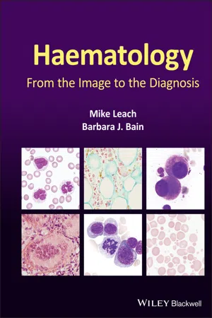
Haematology
eBook - ePub

Haematology

From the Image to the Diagnosis

  1. English
  2. ePUB (mobile friendly)
  3. Available on iOS & Android
eBook - ePub

Haematology

From the Image to the Diagnosis

About this book

Haematology

Diagnostic haematology requires the assessment of clinical and laboratory data together with a careful morphological assessment of cells in blood, bone marrow and tissue ­fluids. Subsequent investigations including flow cytometry, immunohistochemistry, cytogenetics and molecular studies are guided by the original morphological findings. These targeted investigations help generate a prompt unifying diagnosis. Haematology: From the Image to the Diagnosis presents a series of cases illustrating how skills in morphology can guide the investigative process. In this book, the authors capture a series of images to illustrate key features to recognize when undertaking a morphological review and show how they can be integrated with supplementary information to reach a final diagnosis.

Using a novel format of visual case studies, this text mimics 'real life' for the practising diagnostic haematologist – using brief clinical details and initial microscopic morphological triage to formulate a differential diagnosis and a plan for efficient and economical confirmatory investigation to deduce the correct final diagnosis. The carefully selected, high-quality photomicrographs and the clear, succinctdescriptions of key features, investigations and results will help haematologists, clinical scientists, haematology trainees and haematopathologists to make accurate diagnoses in their day-to-day work.

Covering a wide range of topics, and including paediatric as well as adult cases, Haematology: From the Image to the Diagnosis is a succinct visual guide which will be welcomed by consultants, trainees and scientists alike.

Frequently asked questions

Yes, you can cancel anytime from the Subscription tab in your account settings on the Perlego website. Your subscription will stay active until the end of your current billing period. Learn how to cancel your subscription.
At the moment all of our mobile-responsive ePub books are available to download via the app. Most of our PDFs are also available to download and we're working on making the final remaining ones downloadable now. Learn more here.
Perlego offers two plans: Essential and Complete
  • Essential is ideal for learners and professionals who enjoy exploring a wide range of subjects. Access the Essential Library with 800,000+ trusted titles and best-sellers across business, personal growth, and the humanities. Includes unlimited reading time and Standard Read Aloud voice.
  • Complete: Perfect for advanced learners and researchers needing full, unrestricted access. Unlock 1.4M+ books across hundreds of subjects, including academic and specialized titles. The Complete Plan also includes advanced features like Premium Read Aloud and Research Assistant.
Both plans are available with monthly, semester, or annual billing cycles.
We are an online textbook subscription service, where you can get access to an entire online library for less than the price of a single book per month. With over 1 million books across 1000+ topics, we’ve got you covered! Learn more here.
Look out for the read-aloud symbol on your next book to see if you can listen to it. The read-aloud tool reads text aloud for you, highlighting the text as it is being read. You can pause it, speed it up and slow it down. Learn more here.
Yes! You can use the Perlego app on both iOS or Android devices to read anytime, anywhere — even offline. Perfect for commutes or when you’re on the go.
Please note we cannot support devices running on iOS 13 and Android 7 or earlier. Learn more about using the app.
Yes, you can access Haematology by Mike Leach,Barbara J. Bain in PDF and/or ePUB format, as well as other popular books in Medicine & Hematology. We have over one million books available in our catalogue for you to explore.

Information

Year
2021
eBook ISBN
9781119777526
Edition
1
Subtopic
Hematology

1
Anaplastic large cell lymphoma with haemophagocytic syndrome

image
A 50‐year‐old man from another health board was transferred to a local hospital for a surgical biopsy of a mediastinal lymph node. He had presented 6 weeks previously with fever, sweats and weight loss. No infective or neoplastic aetiology had been identified. He had progressive pancytopenia and hyperferritinaemia and a diagnosis of idiopathic haemophagocytic syndrome had been considered. He had already been treated at the base hospital with corticosteroids and etoposide. CT imaging, however, had shown abnormal mediastinal lymph nodes, which would not have been accessible by percutaneous needle biopsy.
On arrival at the local cardiothoracic unit he was clearly unwell. The full blood count showed Hb 90 g/l, WBC 2.5 × 10⁹/l, neutrophils 1.5 × 10⁹/l and platelets 25 × 109/l. The coagulation screen showed PT 18 s, APTT 45 s, TT 18 s, fibrinogen 1.4 g/l and D dimer 5500 ng/ml (NR <230). Serum ferritin was >10 000 ÎŒg/l. The anaesthetic team phoned to ask for advice regarding management of the coagulopathy prior to surgery. The blood film showed rouleaux and was leucoerythroblastic with a few toxic granulated neutrophils but no neoplastic cell population was evident. We decided to cancel the surgery, review the imaging and perform a bone marrow aspirate and trephine biopsy. A CT scan showed definitely pathological mediastinal lymph nodes. The bone marrow aspirate showed a population of very large lymphoid cells with round or ovoid nuclei, indistinct nucleoli and partially condensed nuclear chromatin without cytoplasmic granules (top images). The cytoplasm of these cells showed prominent vacuolation, often concentrated in apparent pseudopodia (top images). In addition, there was a substantial population of macrophages showing haemophagocytosis, particularly of red cells and their precursors (all images above ×100 objective). The clinical, laboratory and morphological findings were in keeping with a haemophagocytic syndrome. Flow cytometry on the large cell population in the marrow aspirate showed these cells to express CD45, CD2, CD3, CD4 and CD30. No myeloid or B‐lineage antigens were expressed. The bone marrow trephine biopsy sections were also abnormal, showing prominent macrophages displaying haemophagocytosis on H&E staining (below, left image) and CD68R (below, centre image, immunoperoxidase), whilst the large neoplastic cells were highlighted using CD30 (below, right, immunoperoxidase) (all images below ×50). Immunohistochemistry showed the large cells to be ALK negative. The diagnosis was now confirmed as ALK‐negative anaplastic large cell lymphoma. The patient was transferred back to his local hospital with a view to commencing CHOP chemotherapy but after one cycle of treatment he deteriorated further and sadly died.
image
Haemophagocytic syndrome is a rare constellation of clinicopathological features including fever, weight loss, sweats and organomegaly, together with laboratory abnormalities including cytopenias, hyperferritinaemia, hypertriglyceridaemia, coagulopathy and bone marrow haemophagocytosis. Interestingly, according to strict diagnostic criteria, and despite the name, demonstration of the latter is not absolutely essential. In adults the typical triggers for a confirmed haemophagocytic syndrome are infective or neoplastic. In adult patients a neoplastic cause is very likely and high‐grade T‐cell and NK‐cell neoplasms are the usual culprits. In paediatric practice there are a number of inherited immunodeficiency syndromes that strongly predispose to a haemophagocytic syndrome, but neoplastic and infective triggers similar to the above have also to be considered. It is absolutely essential to consider a neoplastic cause in adult patients; this focuses attention on looking for an underlying neoplasm with subsequent targeted treatment, in which case the haemophagocytic syndrome will gradually resolve.

MCQ

  1. ALK‐negative anaplastic large cell lymphoma:
    1. Generally occurs at an older age than ALK‐positive cases
    2. Has a better prognosis than ALK‐positive anaplastic large cell lymphoma
    3. Has similar histological and immunophenotypic features to breast implant‐associated anaplastic large cell lymphoma
    4. Is usually associated with t(2;5)(p23.2‐23.1;q35.1)
    5. Usually presents with localised disease
    For answers and discussion, see page 206.

2
Bone marrow AL amyloidosis

image
A 47‐year‐old man presented with peripheral oedema due to nephrotic syndrome and a kidney biopsy was planned. Full blood count showed Hb 154 g/l, WBC 7.8 × 10⁹/l and platelets 355 × 109/l. Serum creatinine was 136 ÎŒmol/l, estimated glomerular filtration rate (eGFR) 49 ml/min/1.73 m2, albumin 17 g/l and urine protein 5.4 g/l. A serum IgA lambda paraprotein (9 g/l) and excess serum free lambda light chains at 103.5 mg/l (NR 5.7–26.3) were identified.
A bone marrow aspirate identified deposits of amorphous, purple‐staining material suggestive of amyloid protein (top left image) (all images ×50 objective). The trephine biopsy sections showed diffuse eosinophilic amorphous material (top right, H&E) in keeping with amyloid deposited around fat cells, around erythroid islands, in the walls of small vessels and throughout the interstitium. Immunohistochemistry for CD138 revealed 7% plasma cells with non‐specific binding to the amyloid deposits (bottom left, immunoperoxidase) the latter deposits staining also with Sirius red (bottom right). These findings are indicative of extensive bone marrow amyloid deposition due to primary AL amyloidosis. In view of these findings the renal biopsy was now not deemed necessary.
This case illustrates the striking morphological abnormalities encountered in amyloidosis; accurate recognition of these features is important in bringing together a unified clinical diagnosis.
A second patient presented with back pain and vertebral collapse. MRI suggested a marrow infiltrative disease such as myeloma. Indeed, he was shown to have raised serum free lambda light chains (367 mg/l) but no paraprotein was detected. The bone marrow aspirate, however, was hypocellular and showed low level (<1%) involvement by plasma cells so the diagnosis was questioned. The trephine biopsy sections showed heavy involvement by AL amyloidosis (all images below, H&E, left ×10, centre and right ×50) occupying well over 80% of the marrow space, with notable involvement of vessel walls. In the cellular areas well over 90% of cells were neoplastic plasma cells. Elsewhere small pockets of malignant plasma cells were ident...

Table of contents

  1. Cover
  2. Table of Contents
  3. Title Page
  4. Copyright Page
  5. Preface
  6. Abbreviations
  7. Normal ranges for commonly used tests (for adults)
  8. 1 Anaplastic large cell lymphoma with haemophagocytic syndrome
  9. 2 Bone marrow AL amyloidosis
  10. 3 Cup‐like blast morphology in acute myeloid leukaemia
  11. 4 Neutrophil morphology
  12. 5 Primary myelofibrosis
  13. 6 Sarcoidosis
  14. 7 Visceral leishmaniasis
  15. 8 Gelatinous transformation of the bone marrow
  16. 9 Acanthocytic red cell disorders
  17. 10 T‐cell large granular lymphocytic leukaemia
  18. 11 Pure erythroid leukaemia
  19. 12 eactive mesothelial cells
  20. 13 Plasmablastic myeloma
  21. 14 Septicaemia
  22. 15 An unstable haemoglobin and a myeloproliferative neoplasm
  23. 16 Sickle cell anaemia in crisis
  24. 17 Acute myeloid leukaemia with t(8;21)(q22;q22.1)
  25. 18 Chronic neutrophilic leukaemia
  26. 19 Essential thrombocythaemia
  27. 20 Hairy cell leukaemia
  28. 21 Mantle cell lymphoma in leukaemic phase
  29. 22 Infantile osteopetrosis
  30. 23 Reactive eosinophilia
  31. 24 Stomatocytic red cell disorders
  32. 25 Reactive lymphocytosis due to viral infection
  33. 26 Therapy‐related acute myeloid leukaemia with eosinophilia
  34. 27 Red cell fragmentation syndromes
  35. 28 NK/T‐cell lymphoma in leukaemic phase
  36. 29 Myelodysplastic syndrome with del(5q)
  37. 30 Classical Hodgkin lymphoma
  38. 31 Cryoglobulinaemia
  39. 32 Congenital dyserythropoietic anaemia
  40. 33 Acute monoblastic leukaemia with t(9;11)(p21.3;q23.3)
  41. 34 Chronic myeloid leukaemia presenting with myeloid sarcoma
  42. 35 Glucose‐6‐phosphate dehydrogenase deficiency
  43. 36 Leukaemic presentation of hepatosplenic γή T‐cell lymphoma
  44. 37 Myelodysplastic syndromes
  45. 38 Pelger–HuĂ«t anomaly
  46. 39 Russell bodies in lymphoplasmacytic lymphoma
  47. 40 T‐cell prolymphocytic leukaemia
  48. 41 Myeloid maturation arrest
  49. 42 MDS/MPN with ring sideroblasts and thrombocytosis
  50. 43 Acute myeloid leukaemia with inv(16)(p13.1q22)
  51. 44 Babesiosis
  52. 45 Haemoglobin E disorders
  53. 46 Juvenile myelomonocytic leukaemia
  54. 47 Non‐haemopoietic tumours
  55. 48 Richter transformation of chronic lymphocytic leukaemia
  56. 49 Sickle cell–haemoglobin C disease
  57. 50 T cell/histiocyte‐rich B‐cell lymphoma
  58. 51 Miliary tuberculosis
  59. 52 Pure red cell aplasia
  60. 53 Lymphoblastic transformation of follicular lymphoma
  61. 54 Primary hyperparathyroidism
  62. 55 Gamma heavy chain disease
  63. 56 Acute promyelocytic leukaemia with t(15;17)(q24.1;q21.2)
  64. 57 AA amyloidosis
  65. 58 Acquired sideroblastic anaemia
  66. 59 Diffuse large B‐cell lymphoma
  67. 60 Hickman line infection
  68. 61 Monocytes and their precursors
  69. 62 Paroxysmal cold haemoglobinuria
  70. 63 Transient abnormal myelopoiesis
  71. 64 Systemic lupus erythematosus
  72. 65 Granular blast cells in acute lymphoblastic leukaemia
  73. 66 Chronic myelomonocytic leukaemia
  74. 67 Burkitt lymphoma/leukaemia
  75. 68 Gaucher disease
  76. 69 Myelodysplastic syndrome with haemophagocytosis
  77. 70 Primary oxalosis
  78. 71 Acute myeloid leukaemia with inv(3)(q21.3q26.2)
  79. 72 Autoimmune haemolytic anaemia
  80. 73 Chronic eosinophilic leukaemia with FIP1L1‐PDGFRA fusion
  81. 74 Leukaemic phase of follicular lymphoma
  82. 75 Megaloblastic anaemia
  83. 76 Reactive bone marrow and an abnormal PET scan
  84. 77 Acute megakaryoblastic leukaemia
  85. 78 Erythrophagocytosis and haemophagocytosis
  86. 79 Hyposplenism
  87. 80 Acquired haemoglobin H disease
  88. 81 Cystinosis
  89. 82 Familial platelet disorder with a predisposition to AML
  90. 83 Nodular lymphocyte predominant Hodgkin lymphoma
  91. 84 Acute monocytic leukaemia with NPM1 mutation
  92. 85 Adult T‐cell leukaemia/lymphoma
  93. 86 Hereditary elliptocytosis and pyropoikilocytosis
  94. 87 Sézary syndrome
  95. 88 Spherocytic red cell disorders
  96. 89 Acute myeloid leukaemia and metastatic carcinoma
  97. 90 ChĂ©diak–Higashi syndrome
  98. 91 Cortical T‐lymphoblastic leukaemia/lymphoma
  99. 92 Trypanosomiasis
  100. 93 Acute myeloid leukaemia with myelodysplasia‐related changes
  101. 94 Blastic plasmacytoid dendritic cell neoplasm
  102. 95 Inherited macrothrombocytopenias
  103. 96 Persistent polyclonal B‐cell lymphocytosis
  104. 97 Acute myeloid leukaemia with t(6;9)(p23;q34.1)
  105. 98 B‐cell prolymphocytic leukaemia
  106. 99 Various red cell enzyme disorders
  107. 100 Sea‐blue histiocytosis in multiple myeloma
  108. 101 Enteropathy‐associated T‐cell lymphoma
  109. Further discussion of the themes
  110. Index
  111. End User License Agreement