**Selected for Doody's Core Titles® 2024 in Veterinary Medicine** Get an up-close look at canine anatomy with the only complete guide to the dissection of the dog. Utilizing detailed descriptions and more than 300 high-quality color anatomic drawings, Guide to the Dissection of the Dog, 8th Edition walks you through how to perform precise canine dissections while developing your understanding of basic mammalian structure and specific canine features. Each chapter offers self-contained guidance on the dissection of a specific body part, allowing you to perform dissections in whatever sequence your choose. Other helpful features include: an emphasis on the anatomical knowledge and terminology from the Nomina Anatomica Veterinaria; a comprehensive list of all figures and tables; and an extensive list of references for further research. In all, this one-of-a-kind canine dissection manual is the resource you need to better understand and review what you are learning in your first-year dissection course.- The only hands-on canine dissection guide available reinforces the information that you learn in your mandatory first-year dissection course.- Body part organization follows the order of dissection commonly taught in veterinary schools and enables you to perform dissections in any sequence.- More than 300 high-quality color anatomic drawings guide each step-by-step dissection procedure.- Radiographs, CAT scans and MR images appear throughout the text where relevant to help you visualize internal anatomic features that can only be revealed through these diagnostic methods.- Comprehensive list of tables and figures makes it easy to find key images and information at a glance.- Detailed descriptions of anatomical structures ensure the most thorough, precise canine dissections.- Clear and easy-to-follow instructions guide you in properly performing dissection techniques.- Option of a digital book on Pageburst offers high-resolution illustrations that are directly linked to the text — letting you search for any text work or anatomic clue and discover any instance of what you want to read more about.- NEW! High-resolution digital images have been added throughout the book to provide a clinical context for the drawings and to highlight internal anatomic structures with excellent contrast resolution.- NEW! Additional transverse sections of the brain give you the anatomic knowledge you need to accurately interpret MR images.- NEW! Updated figure labels and text adhere to the latest Nomina Anatomica Veterinaria.
eBook - ePub
Guide to the Dissection of the Dog - E-Book
Guide to the Dissection of the Dog - E-Book
- 344 pages
- English
- ePUB (mobile friendly)
- Available on iOS & Android
eBook - ePub
Guide to the Dissection of the Dog - E-Book
Guide to the Dissection of the Dog - E-Book
About this book
Trusted by 375,005 students
Access to over 1.5 million titles for a fair monthly price.
Study more efficiently using our study tools.
Information
Topic
MedicineSubtopic
Veterinary MedicineChapter 1
Anatomical terminology
Anatomy is the study of structure. Physiology is the study of function. Structure and function are inseparable as the foundation of the science and art of medicine. One must know the parts before one can appreciate how they work. Gross anatomy, the study of structures that can be dissected and observed with the unaided eye or with a hand lens, forms the subject matter of this guide.
The anatomy of one part in relation to other parts of the body is topographical anatomy. The practical application of such knowledge in the diagnosis and treatment of pathological conditions is applied anatomy. The study of structures too small to be seen without a light microscope is microscopic anatomy. Examination of structure in even greater detail is possible with an electron microscope and constitutes ultrastructural anatomy. When an animal becomes diseased or its organs function improperly, its deviation from the normal is studied as pathological anatomy. The study of the development of the individual from the fertilized oocyte to birth is embryology, and from the zygote to the adult it is known as developmental anatomy. The study of abnormal development is teratology.
Medical etymology and anatomical nomenclature
The student of anatomy is confronted with an array of unfamiliar terms and names of anatomical structures. A better understanding of the language of anatomy helps make its study more intelligible and interesting. For the publication of scientific papers and communication with colleagues, the mastery of anatomical terminology is a necessity. To ensure knowledge of basic anatomical terms, a medical dictionary should be kept readily accessible and consulted frequently. It is very important to learn the spelling, pronunciation, and meaning of all new terms encountered. Vertebrate structures are numerous, and in many instances common names are not available or are so vague as to be meaningless. One soon realizes why it is desirable to have an international glossary of terms that can be understood by scientists in all countries. Acquisition of a medical vocabulary can be aided by the mastery of Greek and Latin roots and affixes.
Our present medical vocabulary has a history dating back more than 2000 years and reflects the influences of the world’s languages. The early writings in anatomy and medicine were in Greek and later almost entirely in Latin. As a consequence, most anatomical terms stem from these classical languages. Latin terms are commonly translated into the vernacular of the person using them. Thus the Latin hepar becomes the English liver, the French foie, the Spanish higado, and the German Leber.
Although anatomical terminology has been rather uniform, differences in terms have arisen between the different fields and different countries. In 1895 a group of anatomists proposed a standard list of terms derived from those in use throughout the world. This list, known as the Basle Nomina Anatomica (BNA), formed the basis for the present sixth edition of Nomina Anatomica (NA) 1989, which was prepared by the International Anatomical Nomenclature Committee (IANC) and adopted by the International Congress of Anatomists in Paris in 1955. Of the 5640 standard terms, more than 80% are continued from the BNA. In response to dissatisfaction with the work of the last committee (IANC), the International Federation of Associations of Anatomists created a new committee in 1989, the Federative Committee on Anatomical Terminology (FCAT), to write Terminologia Anatomica (TA), which was published in 1998. This new list of terms gives each term in Latin accompanied by the current usage in English-speaking countries. There is an index to Latin and English terms as well as to eponyms (Thieme Publishers, Stuttgart and New York). Both the NA and TA lists are for human anatomy.
The International Committee on Veterinary Anatomical Nomenclature (ICVAN), appointed by the World Association of Veterinary Anatomists in 1957, published Nomina Anatomica Veterinaria (NAV) for domestic mammals in 1968. These terms, as revised in the fifth edition in 2005 (published on the World Wide Web), serve as the basis for the nomenclature used in this guide.
Directional terms
An understanding of the following planes, positions, and directions relative to the animal body or its parts is necessary to follow the procedures for dissection (Fig. 1-1).
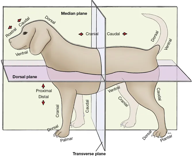
PLANE: A surface, real or imaginary, along which any two points can be connected by a straight line.
Median Plane: Divides the head, body, or limb longitudinally into equal right and left halves. Sagittal Plane: Passes through the head, body, or limb parallel to the median plane. Transverse Plane: Cuts across the head, body, or limb at a right angle to its long axis or across the long axis of an organ or a part. Dorsal Plane: Runs at right angles to the median and transverse planes and thus divides the body or head into dorsal and ventral portions.
DORSAL: Toward or relatively near the upper surface (as opposed to the supporting surface) of the head, body, and tail. This surface is opposite to the supporting surface in the standing animal. On the limbs it applies to the upper or front surface of the carpus, tarsus, metapodium, and digits (opposite to the side with the pads).
VENTRAL: Toward or relatively near the supporting surface and the corresponding surface of the head, neck, thorax, and tail. This term is never used for the limbs.
MEDIAL: Toward or relatively near the median plane.
LATERAL: Away from or relatively farther from the median plane.
CRANIAL: Toward or relatively near the head; on the limbs it applies proximal to the carpus and tarsus. In reference to the head, it is replaced by the term rostral.
ROSTRAL: Toward or relatively near the nose; applies to the head only.
CAUDAL: Toward or relatively near the tail; on the limbs it applies proximal to the carpus and tarsus. Also used in reference to the head.
The adjectives for directional terms may be modified to serve as adverbs by replacing the ending al with the ending ally, as in dorsally.
Certain terms whose meanings are more restricted are used in the description of organs and appendages.
INTERNAL or INNER: Close to, or in the direction of, the center of an organ, body cavity, or structure.
EXTERNAL or OUTER: Away from the center of an organ or structure.
SUPERFICIAL: Relatively near the surface of the body or the surface of a solid organ.
DEEP: Relatively near the center of the body or the center of a solid organ.
PROXIMAL: Relatively near the main mass or origin; in the limbs and tail, the attached end of that structure.
DISTAL: Away from the main mass or origin; in the limbs and tail, the free end of that structure.
RADIAL: On that side of the forearm (antebrachium) in which the radius is located.
ULNAR: On that side of the forearm in which the ulna is located.
TIBIAL and FIBULAR: On the corresponding sides of the leg (crus), the tibial side being medial and the fibular side being lateral.
In the dog and similar species, the paw is that part of the thoracic or pelvic limb distal to the radius and ulna or to the tibia and fibula. The human hand (manus) and foot (pes) are homologous with the forepaw and hindpaw, respectively.
PALMAR: The aspect of the forepaw on which the pads are located—the surface that contacts the ground in the standing animal—and the corresponding surface of the metacarpus and carpus.
PLANTAR: The aspect of the hindpaw on which the pads are located—the surface that contacts the ground in the standing animal—and the corresponding surface of the metatarsus and tarsus. The opposite surface of both forepaw and hindpaw is known as the dorsal surface.
AXIS: The central line of the body or any of its parts.
AXIAL, ABAXIAL: Of, pertaining to, or relative to the axis. In reference to the digits, the functional axis of the limb passes between the third and fourth digits. The axial surface of the digit faces the axis, and the abaxial surface faces away from the axis.
The following terms apply to the various basic movements of the parts of the body.
FLEXION: The movement of one bone in relation to another in such a manner that the angle formed at their joint is reduced. The limb is retracted or folded; the digit is bent; the back is arched dorsally.
EXTENSION: The movement of one bone upon another such that the angle formed at their joint increases. The limb reaches out or is extended; the digit is straightened; the back is straightened. Extension beyond 180 degrees is overextension.
ABDUCTION: The movement of a part away from the median plane.
ADDUCTION: The movement of a part toward the median plane.
CIRCUMDUCTION: The movement of a part when outlining the surface of a cone (e.g., the thoracic limb extended drawing a circle).
ROTATION: The movement of a part around its long axis (e.g., the action of the radius when using a screwdriver). The direction of rotation of a limb or segment of a limb on its long axis is designated by the direction of movement of its cranial or dorsal surface (e.g., in medial rotation of the arm, the crest of the greater tubercle is turned medially).
SUPINATION: Lateral rotation of the appendage so that the palmar or plantar surface of the paw faces medially.
PRONATION: Medial rotation of the appendage from the supine position so that the palmar or plantar surface will face the substrate.
Common regional synonyms include brachium for the arm (between shoulder and elbow), antebrachium for the forearm (between elbow and carpus), thigh for the pelvic limb (between the hip and stifle), and crus for the leg (between stifle and tarsus). The pelvic limb is not the leg. Only the crus is the leg.
On radiographs the view is described in relation to the direction of penetration by the x-ray: from point of entrance to point of exit of the body part before striking the film. Oblique views are described with combined terms. A view of the carpus with the x-ray tube perpendicular to the dorsal surface and the film on the palmar surface would be a dorsopalmar view. If the x-ray tube is turned so that it points toward the dorsomedial surface of the carpus and the film is on the palmarolateral surface, the view would be dorsomedial-palmarolateral oblique. If the animal is lying on its right side, adjacent to the radiographic film, the radiograph is a left-to-right lateral view.
Dissect...
Table of contents
- Cover image
- Title page
- Table of Contents
- Copyright
- Preface
- Acknowledgments
- Illustrations
- Tables
- 1. Anatomical terminology
- 2. The skeletal and muscular systems
- 3. The neck, thorax, and thoracic limb
- 4. The abdomen, pelvis, and pelvic limb
- 5. The head
- 6. The nervous system
- Bibliography
- Index
Frequently asked questions
Yes, you can cancel anytime from the Subscription tab in your account settings on the Perlego website. Your subscription will stay active until the end of your current billing period. Learn how to cancel your subscription
No, books cannot be downloaded as external files, such as PDFs, for use outside of Perlego. However, you can download books within the Perlego app for offline reading on mobile or tablet. Learn how to download books offline
Perlego offers two plans: Essential and Complete
- Essential is ideal for learners and professionals who enjoy exploring a wide range of subjects. Access the Essential Library with 800,000+ trusted titles and best-sellers across business, personal growth, and the humanities. Includes unlimited reading time and Standard Read Aloud voice.
- Complete: Perfect for advanced learners and researchers needing full, unrestricted access. Unlock 1.5M+ books across hundreds of subjects, including academic and specialized titles. The Complete Plan also includes advanced features like Premium Read Aloud and Research Assistant.
We are an online textbook subscription service, where you can get access to an entire online library for less than the price of a single book per month. With over 1.5 million books across 990+ topics, we’ve got you covered! Learn about our mission
Look out for the read-aloud symbol on your next book to see if you can listen to it. The read-aloud tool reads text aloud for you, highlighting the text as it is being read. You can pause it, speed it up and slow it down. Learn more about Read Aloud
Yes! You can use the Perlego app on both iOS and Android devices to read anytime, anywhere — even offline. Perfect for commutes or when you’re on the go.
Please note we cannot support devices running on iOS 13 and Android 7 or earlier. Learn more about using the app
Please note we cannot support devices running on iOS 13 and Android 7 or earlier. Learn more about using the app
Yes, you can access Guide to the Dissection of the Dog - E-Book by Howard E. Evans,Alexander de Lahunta in PDF and/or ePUB format, as well as other popular books in Medicine & Veterinary Medicine. We have over 1.5 million books available in our catalogue for you to explore.