Cartilage Surgery: An Operative Manual by Mats Brittberg, MD and Wayne Gersoff, MD is your guide to applying the most recent advances in cartilage repair, and performing cutting-edge surgical procedures. An internationally diverse collection of authors offers a global perspective on timely topics such as cartilage biologics. Clinical pearls, operative video clips, and detailed, full-color intraoperative photographs offer step-by-step guidance on essential techniques. You can access the full content and videos online at expertconsult.com.- Stay current with the recent advances in cartilage repair including surgical and non-surgical treatments as well as biologic management of cartilage lesions.- Get unmatched visual guidance from an unparalleled video collection online that demonstrates how to perform a variety of key techniques.- Quickly reference essential topics with a templated, focused format that includes clinical pearls to help you make a confident diagnosis and select the best treatment.- Benefit from the knowledge, experience, and global perspective of a diverse collection of leading international authors. Access the book from any computer at ExpertConsult.com, complete with the full text, entire image bank, and video library.
eBook - ePub
Cartilage Surgery E-Book
An Operative Manual, with Expert Consult
- 320 pages
- English
- ePUB (mobile friendly)
- Available on iOS & Android
eBook - ePub
About this book
Trusted by 375,005 students
Access to over 1.5 million titles for a fair monthly price.
Study more efficiently using our study tools.
Information
Topic
MedicineSubtopic
OrthopedicsChapter 1 Cartilage Morphology
Hyaline cartilage provides the diarthrodial joint with a low-friction surface, resilience, and compressive stiffness, and this unique tissue is, under normal conditions, wear resistant.
Loss of cartilage function may lead to a painful joint with a decreased mobility. Many factors (epidemiological, biochemical, and morphological) are associated with cartilage destruction. However, only trauma is known directly to cause osteoarthritis.1 It is well known that once the cartilaginous tissue has been destroyed, the intrinsic reparative ability is poor. Therefore, it is of uttermost importance to increase knowledge about the cartilage, the tissue reaction to trauma, and the intrinsic attempts to repair the defects as well as extrinsic methods.
Cartilage Biochemistry and Morphology
The hyaline cartilage could be regarded as a composite gel with relatively low percentage chondrocytes (5%) embedded in a rich extracellular matrix consisting in negatively charged hydrophilic proteoglycans constrained by a three-dimensional collagen network.
The negatively charged proteoglycans have the ability to form large aggregates, which can bind water molecules within the positively charged collagen fibrils, thus generating a high osmotic pressure within the gel.
The collagen fibers are responsible for the structure of cartilage and consist mainly of collagen type II. They are highly cross-linked via collagen type IX fibers.2
Chondrocytes are the producers of the surrounding ground substance: matrix.
The cells have different appearances depending on where in the cartilage they are situated. The cells in the top layer appear flattened, whereas the cells in the deeper layer are more rounded and aligned along vertically orientated type II collagen.3
Collagen is the most important scaffolding material in the body, existing in several types. The major type in hyaline cartilage is named type II. It is built by three identical polypeptide alpha-chains. These chains are coiled to form a triple-helix and are produced by the chondrocyte in the form of procollagen. Outside the cell, this procollagen is transformed to tropocollagen, and these molecules aggregate to form the much larger molecule: collagen.
In the hyaline cartilage there also exist minor collagens like types IX, XI, V, and VI. Type IX contributes with covalent cross-linking of the type II fibrils, whereas type XI is thought to control the diameter of type II fibrils. The collagen gives the cartilage its strength and tensile stiffness.
Proteoglycans are large protein-polysaccharide molecules making up 5% to 10% of the wet weight of the cartilage.4 They are composed by chains of the glycosaminoglycans keratan sulfate and chondroitin sulfate covalently bound to a central protein core molecule. Large aggregates are formed with several proteoglycan monomers via a link protein connecting the central protein cores to a chain of hyaluronic acid.
All the components of the proteoglycan aggregates are synthesized by the chondrocytes.
The proteoglycans are unevenly distributed throughout the cartilage layers with the highest concentration in the middle part and the lowest concentration in the superficial layers.5 The proteoglycans give the cartilage its elasticity and resilience.
There is a difference in cartilage composition between the cartilage surface and the subchondral bone plate. These structural differences give rise to four separate layers or zones (see Fig. 1-1).
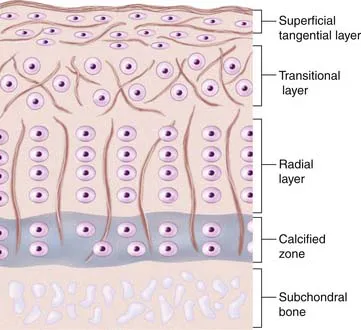
FIGURE 1-1 Schematic drawing of the different layers in a full-thickness osteocartilaginous biopsy.
In the top zone, the superficial zone, there is first a cell-free fibril-layer, called the lamina splendens.6 Beneath this thin layer, chondrocytes are dispersed in an elongated manner parallel to the surface, reflecting as well the tangential orientation of the collagen fibers. This is the tangential layer.
In the second zone, often called the transitional layer, the cells are larger, rounded, and randomly distributed between the oblique-oriented collagen fiber. In the third zone, the chondrocytes are even larger and arranged in typical columns because of the radial collagen fiber courses, the radial zone.
The fourth layer, finally, which is mineralized, is called the calcified zone. There exists a visible border between the third and fourth zone, the tidemark with a special affinity for basic dyes (e.g., toluidine blue).
The calcified zone provides an important transition to the less resilient subchondral bone. For a long time this was regarded more or less as an inactive zone, until Hunziker (1992)7 noted that also the chondrocytes here could take up and incorporate (35S) sulfate into the pericellular and territorial matrix. Hunziker speculated that, following trauma, the metabolic activity here becomes temporarily impaired.7
Regarding experimental animals, it is important to know that it is only in adult animals that the division into zone I to zone III is possible.8 In the immature animal, the cells are more randomly distributed with a gradient in cell size from the top to the calcified zone, with the cells in the deeper parts being largest. Thus, the articular cartilage organization during prepubertal growth imitates the structure of the growth plate, and during that time the biomechanical properties of the cartilage change with an increase in stiffness and in shearing and compressive resistance.7,9
Metabolic Events in the Cartilage
Under normal conditions, the components of the matrix have a slow turnover. The collagen has the slowest turnover rate compared to the much faster turnover of the proteoglycans.
The majority of the proteoglycans have a life span of about 600 days, but a small proportion of the proteoglycans in adult cartilage act as a fast fraction with a half-life of about 8 days. The proteoglycans are thus also more vulnerable to enzymatic degradation.10,11
The chondrocytes secrete different enzymes called metalloproteinases (collagenases, gelatinases, and stromelysin), which all control the degree of degradation. The degradation of proteoglycans is followed by an increased synthesis...
Table of contents
- Cover
- Title Page
- Front Matter
- Copyright
- Dedication
- Contributors
- Preface
- Table of Contents
- Chapter 1: Cartilage Morphology
- Chapter 2: Patient Evaluation and Treatment Algorithms
- Chapter 3: Debridement of the Injured Cartilage
- Chapter 4A: Bone Marrow Stimulating Techniques: Drilling, Abrasion Arthroplasty, and Microfracture
- Chapter 4B: Bone Marrow Stimulating Techniques: Carbon Fiber Resurfacing
- Chapter 4C: Bone Marrow Stimulating Techniques: Autologous Matrix-Induced Chondrogenesis (AMIC)
- Chapter 4D: Bone Marrow Stimulating Techniques: TRUFIT Plugs
- Chapter 5: Osteochondral Mosaicplasty
- Chapter 6: Mega-OATS
- Chapter 7: Osteochondral Allografts
- Chapter 8: Fixation of Osteochondral Fragments
- Chapter 9A: Autologous Chondrocyte Implantation: Cartilage Biopsy Handling
- Chapter 9B: Autologous Chondrocyte Implantation: Quality Assurance of Cells for Chondrogenic Implantation
- Chapter 9C: Autologous Chondrocyte Implantation: ACI First and Second Generation
- Chapter 9D: Autologous Chondrocyte Implantation: Transarthroscopic Implantation of Hyalograft (Hyaff 11) with Autologous Chondrocytes
- Chapter 9E: Autologous Chondrocyte Implantation: Matrix-Induced Autologous Chondrocyte Implantation (MACI)
- Chapter 10: Allograft Particulate Cartilage Transplantation: DeNovo Natural Tissue (NT) Graft
- Chapter 11: Cartilage Fragment Implantation
- Chapter 12: Unloading Osteotomies: Effect on Cartilage and Cartilage Repair
- Chapter 13: Unloading the Patellofemoral Joint for Cartilage Lesions
- Chapter 14: Meniscal Allografts, Cartilage Repair, and Concomitant Procedures
- Chapter 15: Bone Grafting around an Articular Joint
- Chapter 16: Postoperative Cartilage Repair Rehabilitation
- Index
- Instructions for online access
Frequently asked questions
Yes, you can cancel anytime from the Subscription tab in your account settings on the Perlego website. Your subscription will stay active until the end of your current billing period. Learn how to cancel your subscription
No, books cannot be downloaded as external files, such as PDFs, for use outside of Perlego. However, you can download books within the Perlego app for offline reading on mobile or tablet. Learn how to download books offline
Perlego offers two plans: Essential and Complete
- Essential is ideal for learners and professionals who enjoy exploring a wide range of subjects. Access the Essential Library with 800,000+ trusted titles and best-sellers across business, personal growth, and the humanities. Includes unlimited reading time and Standard Read Aloud voice.
- Complete: Perfect for advanced learners and researchers needing full, unrestricted access. Unlock 1.5M+ books across hundreds of subjects, including academic and specialized titles. The Complete Plan also includes advanced features like Premium Read Aloud and Research Assistant.
We are an online textbook subscription service, where you can get access to an entire online library for less than the price of a single book per month. With over 1.5 million books across 990+ topics, we’ve got you covered! Learn about our mission
Look out for the read-aloud symbol on your next book to see if you can listen to it. The read-aloud tool reads text aloud for you, highlighting the text as it is being read. You can pause it, speed it up and slow it down. Learn more about Read Aloud
Yes! You can use the Perlego app on both iOS and Android devices to read anytime, anywhere — even offline. Perfect for commutes or when you’re on the go.
Please note we cannot support devices running on iOS 13 and Android 7 or earlier. Learn more about using the app
Please note we cannot support devices running on iOS 13 and Android 7 or earlier. Learn more about using the app
Yes, you can access Cartilage Surgery E-Book by Mats Brittberg,Wayne Gersoff in PDF and/or ePUB format, as well as other popular books in Medicine & Orthopedics. We have over 1.5 million books available in our catalogue for you to explore.