The new edition of this introductory-level textbook continues to offer a concise and approachable bridge between student lecture notes and more detailed clinical reference works. All aspects of equine medicine, surgery and reproduction are covered in a single, convenient volume. The second edition has been subject to an extensive revision, with each chapter updated and new chapters added to cover wound management, critical care, anaesthesia and sedation, and diagnostic imaging. While offering key information in an easily and quickly digested format for clinical veterinary students and practising veterinary surgeons, this second edition of Equine Medicine, Surgery and Reproduction will also be relevant to students undertaking equine science degrees, and to professional horse owners and trainers.The wide range of international contributors, highly experienced and all experts in their fields, ensures that the new edition of this popular all-in-one resource remains as indispensable as ever.- Comprehensive coverage of all areas of equine medicine, surgery and reproduction- Easy-to-use format- Completely revised since the first edition with new chapters added- Now with over 100 new illustrations including colour photographs- Includes diagnostic and therapeutic information as well as descriptions of commonly employed clinical techniques- Includes lists of important differential diagnoses for common clinical signs
eBook - ePub
Equine Medicine, Surgery and Reproduction - E-Book
Equine Medicine, Surgery and Reproduction - E-Book
- 624 pages
- English
- ePUB (mobile friendly)
- Available on iOS & Android
eBook - ePub
Equine Medicine, Surgery and Reproduction - E-Book
Equine Medicine, Surgery and Reproduction - E-Book
About this book
Trusted by 375,005 students
Access to over 1.5 million titles for a fair monthly price.
Study more efficiently using our study tools.
Information
Chapter 1
Upper alimentary system
J. Geoffrey Lane and Robert Pascoe
Contents
1.1 Normal upper alimentary tract function: deglutition
Oral, pharyngeal and oesophageal phases of deglutition
Prehension
Mastication
Lingual function
Elevation of palate
Pharyngeal constriction
Laryngeal protection
Crico-pharyngeal relaxation
Primary and secondary oesophageal peristalsis
1.2 Diagnostic approach to cases of dysphagia
History – signs of dysphagia
Physical examination, external and oral inspection
Endoscopy per nasum
Radiography and fluoroscopy
Oral examination under general anaesthesia
1.3 Aetiology of dysphagia: oral phase abnormalities
Facial palsy and lip lesions
Temporo-mandibular joint and hyoid disorders
Lingual abnormalities
Dental disorders
Congenital and acquired palatal defects
Other oral conditions: foreign bodies, neoplasia
1.4 Aetiology of dysphagia: pharyngeal phase abnormalities
Pharyngeal paralysis
Pharyngeal compression: strangles abscessation
Pharyngeal cysts, palatal cysts
Epiglottal lesions, including sub-epiglottic cysts
Laryngeal abnormalities
Fourth branchial arch defects (4-BAD)
1.5 Aetiology of dysphagia: oesophageal phase abnormalities
Megaoesophagus
Oesophageal obstruction (‘choke’)
Oesophageal strictures/stenosis
Intra-mural oesophageal cysts
Oesophageal rupture
Neoplasia
‘Wind-sucking’
Grass sickness
1.6 Oral trauma, mandibular fractures etc.
1.7 Oesophageal obstruction
1.8 Anatomy of the oral cavity
Oral cavity
Normal dental anatomy
Triadan system
Eruption of teeth
1.9 Abnormalities of wear – abrasion and attrition
1.10 Periodontal disease
1.11 Dental caries
1.12 Endodontic disease including dental abscessation
1.13 Tumours of the upper alimentary tract
Odontogenic tumours
Other tumours of the jaw
1.14 Diagnostic approach to dental disorders
Ageing of horses by dentition
Clinical signs of dental disease
Oral examination
Radiography of teeth
Other ancillary diagnostic techniques
Indications for dental extraction
Options for the extraction of incisors, canines and wolf teeth
Options for the extraction of cheek teeth
Further reading
1.1 Normal upper alimentary tract function: deglutition
Normal deglutition comprises the prehension and mastication of ingesta followed by its transfer from the oro-pharynx to the stomach.
Oral, pharyngeal and oesophageal phases of deglutition
Deglutition is divided into three stages:
1. The oral phase – which includes the gathering of food, movements within the oral cavity, mastication and the formation of boluses of ingesta at the base of the tongue – is under voluntary control.
2. The presence of a bolus gathered at the tongue base triggers the sequence of reflexes, collectively known as swallowing, which propels the ingesta from the pharynx – the pharyngeal phase – into the oesophagus. The glosso-pharyngeal nerve (IX) and the pharyngeal branches of the vagus (X) innervate the pharynx and larynx, and their afferent and efferent pathways are co-ordinated in the swallowing centre in the brainstem.
3. Waves of peristalsis convey the ingesta along the oesophagus to the stomach – the oesophageal phase of deglutition.
Prehension
Prehension in the horse relies on the incisor teeth to grasp and section herbage and on the lips to pick up smaller pieces of ingesta as well as to contain it within the mouth and to manipulate food towards the cheek teeth.
Mastication
The molar and premolar teeth are responsible for the mechanical crushing of the fibrous diet.
The tongue and buccal musculature assist in manipulating the ingesta between the maxillary and mandibular dental arcades.
Mastication requires free opening and closure of the temporo-mandibular joints (TMJs) through the action of the masticatory muscles – the masseter, pterygoid and temporal muscles close the jaws, and gravity, assisted by the digastric muscles, opens them. The masticatory muscles receive their innervation through the mandibular branch of the trigeminal nerve (V).
The shape of the articular surfaces of the TMJs together with the presence of menisci permit lateral movements by the mandibular teeth across the wearing surfaces of the upper cheek teeth.
Lingual function
The tip of the tongue assists in prehension and moves the ingesta between the cheek teeth.
Contraction of the tongue base helps in the formation of boluses and, once collected, each bolus is driven caudally; this triggers the involuntary phases of deglutition by driving food and fluid caudally from the oro-pharynx.
The tongue is attached to the hyoid apparatus, and free movement at the tympano-hyoid articulation is required for the craniocaudal tongue motion which facilitates bolus formation in the oro-pharynx.
The glossal musculature receives its motor supply via the hypoglossal nerve (XII).
Elevation of palate
The action of the levator palatini muscles draws the soft palate dorsally to close off the naso-pharynx and prevents the nasal reflux of ingesta; this marks the onset of the involuntary stages of deglutition.
The horse has an intra-narial larynx at all times other than during the momentary disengagement for deglutition. (See 5.18 and 5.21.)
The levator palatini muscles lie parallel with the drainage ostia of the auditory tube diverticula (ATDs) so that when they contract the ostia open to allow exchanges of air for pressure equilibration across the ear drum.
Pharyngeal constriction
The constrictor action of the circular muscles of the pharyngeal walls embraces both oro-pharynx and naso-pharynx – the latter can be appreciated during endoscopic examinations of the naso-pharynx. A wave of constriction follows the contraction of the tongue base and passes from rostral to caudal efficiently to empty the oro-pharynx – the pharyngeal ‘stripping’ wave – leaving minimal quantities of ingesta at the base of the tongue.
Laryngeal protection
Aspiration of food and fluid through the rima glottidis is prevented primarily by the tight adduction of the vocal folds and arytenoid cartilages and to a lesser extent by the retroversion of the apex of the epiglottis.
Crico-pharyngeal relaxation
The upper oesophageal sphincter is formed by the thyro- and crico-pharyngeus muscles, and these are maintained in a state of contraction to prevent involuntary aerophagia, especially during forced exercise. Relaxation of the crico-pharynx simultaneous with the pharyngeal stripping wave permits the food and fluid boluses to pass caudally into the proximal oesophagus.
Primary and secondary oesophageal peristalsis
After each bolus has passed through into the proximal oesophagus, primary peristaltic waves are initiated by active closure of the cricopharynx.
Primary oesophageal peristalsis carries individual boluses to the cardia, but the process is not completely efficient and small quantities of ingesta are left at variable levels in both the cervical and thoracic oesophagus, even in normal horses. These ingesta are either picked up in the bolus of a subsequent primary wave or by locally generated secondary peristalsis.
1.2 Diagnostic approach to cases of dysphagia
History – signs of dysphagia
The signs of dysphagia include:
• an unwillingness to eat.
• slow, messy feeding.
• halitosis.
• rejection of semi-masticated food onto the ground (quidding).
• productive coughing.
• nasal reflux of saliva, ingesta and fluids.
Obviously, horses that are unable to eat and swallow food are likely to lose weight rapidly, but this process is accelerated if the horse develops secondary inhalation pneumonia, which is a common sequel to dysphagia. A moist cough is typical of animals aspirating food or saliva into the rima glottidis. In addition to a clear case history, careful observation of the patient's attempts to eat and drink should be made.
If the horse shows return of ingesta from its mouth, the site of the lesion causing the dysfunction must lie in the oral cavity or oropharynx.
Nasal reflux of ingesta points to an abnormality of the pharyngeal or oesophageal phase of deglutition (Figure 1.1).
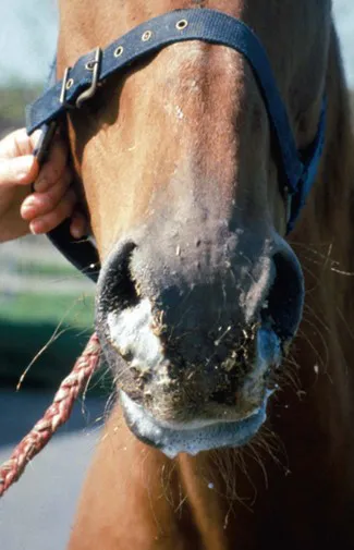
Figure 1.1 Nasal reflux of saliva and ingesta in a case of acute oesophageal obstruction.
Physical examination, external and oral inspection
Evidence of systemic and/or toxic disease, including Streptococcus equi infection, botulism, grass sickness, rabies, upper motor neur...
Table of contents
- Cover image
- Title page
- Table of Contents
- Copyright
- Contributors
- Preface
- Chapter 1: Upper alimentary system
- Chapter 2: Gastroenterology 1. Colic
- Chapter 3: Gastroenterology 2. Hepatic and intestinal disorders
- Chapter 4: Abdominal cavity
- Chapter 5: Disorders of the ear, nose and throat
- Chapter 6: Lower respiratory tract
- Chapter 7: Cardiovascular system
- Chapter 8: Diseases of the equine urinary tract
- Chapter 9: Endocrinology
- Chapter 10: Haematopoietic and immune systems
- Chapter 11: Neurology
- Chapter 12: Ophthalmology
- Chapter 13: Dermatology
- Chapter 14: Reproduction
- Chapter 15: Orthopaedics 1. Diagnosis of lameness/diseases of joints and bones
- Chapter 16: Orthopaedics 2. Diseases of the foot and distal limbs
- Chapter 17: Orthopaedics 3. The proximal limbs
- Chapter 18: Orthopaedics 4. The back and pelvis
- Chapter 19: Infectious diseases and parasitology
- Chapter 20: Diseases of the foal
- Chapter 21: Muscle disorders and performance problems
- Chapter 22: Metabolic diseases and toxicology
- Chapter 23: Principles of wound management
- Chapter 24: Sedation and anaesthesia
- Chapter 25: Equine diagnostic imaging
- Chapter 26: Common problems and techniques in equine critical care
- Index
Frequently asked questions
Yes, you can cancel anytime from the Subscription tab in your account settings on the Perlego website. Your subscription will stay active until the end of your current billing period. Learn how to cancel your subscription
No, books cannot be downloaded as external files, such as PDFs, for use outside of Perlego. However, you can download books within the Perlego app for offline reading on mobile or tablet. Learn how to download books offline
Perlego offers two plans: Essential and Complete
- Essential is ideal for learners and professionals who enjoy exploring a wide range of subjects. Access the Essential Library with 800,000+ trusted titles and best-sellers across business, personal growth, and the humanities. Includes unlimited reading time and Standard Read Aloud voice.
- Complete: Perfect for advanced learners and researchers needing full, unrestricted access. Unlock 1.5M+ books across hundreds of subjects, including academic and specialized titles. The Complete Plan also includes advanced features like Premium Read Aloud and Research Assistant.
We are an online textbook subscription service, where you can get access to an entire online library for less than the price of a single book per month. With over 1.5 million books across 990+ topics, we’ve got you covered! Learn about our mission
Look out for the read-aloud symbol on your next book to see if you can listen to it. The read-aloud tool reads text aloud for you, highlighting the text as it is being read. You can pause it, speed it up and slow it down. Learn more about Read Aloud
Yes! You can use the Perlego app on both iOS and Android devices to read anytime, anywhere — even offline. Perfect for commutes or when you’re on the go.
Please note we cannot support devices running on iOS 13 and Android 7 or earlier. Learn more about using the app
Please note we cannot support devices running on iOS 13 and Android 7 or earlier. Learn more about using the app
Yes, you can access Equine Medicine, Surgery and Reproduction - E-Book by Tim Mair,Sandy Love,James Schumacher,Roger K. W. Smith,Grant Frazer in PDF and/or ePUB format, as well as other popular books in Medicine & Equine Veterinary Science. We have over 1.5 million books available in our catalogue for you to explore.