Patients with cardiac conditions routinely present for noncardiac surgeries, requiring special protocols for perioperative assessment and management by the anesthesiologist. Essentials of Cardiac Anesthesia for Noncardiac Surgery: A Companion to Kaplan's Cardiac Anesthesia provides current, easily accessible information in this complex area, ideal for general anesthesiologists and non-cardiac subspecialists. From preoperative assessment through postoperative care, this practical reference covers all perioperative approaches to today's patients with cardiac conditions.- Provides guidance on the anesthetic diagnosis and management of the full range of cardiac lesions, helping minimize adverse outcomes and reduce complications for patients with common, complex, or uncommon cardiac conditions.- Includes complete coverage of echocardiography and current monitoring techniques needed for thorough perioperative assessment – all from the anesthesiologist's perspective.- Discusses safe and effective perioperative anesthetic management of patients presenting with advanced levels of cardiac care such as drug-eluting stents, multiple antiplatelet drugs, ventricular assist devices, multiple drugs for end-stage heart failure, and implanted electrical devices that produce cardiac resynchronization therapy, as well as patients with complicated obstetric problems or other significant cardiovascular issues.- Features a concise, easy-to-navigate format and Key Points boxes in each chapter that help you find answers quickly.
eBook - ePub
Essentials of Cardiac Anesthesia for Noncardiac Surgery E-Book
A Companion to Kaplan's Cardiac Anesthesia
- 796 pages
- English
- ePUB (mobile friendly)
- Available on iOS & Android
eBook - ePub
Essentials of Cardiac Anesthesia for Noncardiac Surgery E-Book
A Companion to Kaplan's Cardiac Anesthesia
About this book
Trusted by 375,005 students
Access to over 1.5 million titles for a fair monthly price.
Study more efficiently using our study tools.
Information
Topic
MedicineSubtopic
Anesthesiology & Pain ManagementSection II
Anesthesia for Noncardiac Surgery
Chapter 9
Cardiovascular Monitoring in Noncardiac Surgery
Gerard R. Manecke Jr, MD, Timothy M. Maus MD
Keywords
hemodynamic monitoring; electrocardiogram; central venous pressure; cardiac output; goal-directed therapy
Key Points
- 1. Excellent cardiac and hemodynamic management is essential to achieving good outcomes in patients with cardiovascular disease, particularly those undergoing high-risk noncardiac surgery.
- 2. Much cardiovascular information can be obtained from the standard American Society of Anesthesiologists monitors, including those usually associated with evaluation of respiratory function (pulse oximetry, capnography). The pulse oximeter plethysmograph can be used to assess adequacy of the peripheral circulation; expired capnography reflects pulmonary blood flow and cardiac output.
- 3. The five-electrode electrocardiographic system commonly used perioperatively allows rapid diagnosis of a wide variety of cardiac abnormalities, including rhythm disturbances, conduction abnormalities, myocardial ischemia, myocardial infarction, and electrolyte abnormalities.
- 4. Although often unreliable as an intravascular volume monitor, invasive monitoring of the central venous pressure (CVP) can be useful in the management of cardiac patients. CVP provides information about the systolic and diastolic performance of the heart in response to fluid administration, as well as waveform information that can aid in the diagnosis of abnormalities such as tricuspid regurgitation and junctional rhythms.
- 5. The pulmonary artery catheter is a very powerful monitor, providing a wide array of data that include right-sided pressures, cardiac performance, and a surrogate for left atrial pressure (pulmonary capillary wedge pressure). Although its use has declined in noncardiac surgery, it is still very useful in select patients such as those with pulmonary hypertension or right ventricular failure. It is also useful for monitoring left ventricular function and solving hemodynamic problems when transesophageal echocardiography is unavailable.
- 6. Minimally invasive and noninvasive means of continuously monitoring arterial blood pressure, as well as cardiac output and dynamic parameters such as stroke volume variation, are now widely used. They are particularly useful in cardiac patients undergoing high-risk surgery. They facilitate perioperative goal-directed therapy (PGDT), enhanced recovery from surgery, and rapid diagnosis of hemodynamic problems.
- 7. Noninvasive monitors that assess tissue oxygenation, pH, and perfusion are likely to be further developed and used. Because the purpose of circulation is tissue perfusion, it is logical to quantify tissue perfusion and oxygenation. Somatic near-infrared spectroscopy is currently used for this purpose in PGDT algorithms.
Perioperative care includes effective cardiac, hemodynamic, and fluid management. Excellent cardiovascular management is particularly important in patients undergoing major noncardiac surgery and those with preexisting cardiovascular disease. It is only with meaningful, accurate monitoring that appropriate cardiac, hemodynamic, and fluid therapy can be provided. This chapter focuses on the various means by which the cardiac and hemodynamic status can be monitored, ranging from noninvasive to highly invasive techniques. Other indicators of cardiovascular function, such as urine output, are discussed as well. Echocardiography is not discussed here; it is presented in Chapter 10.
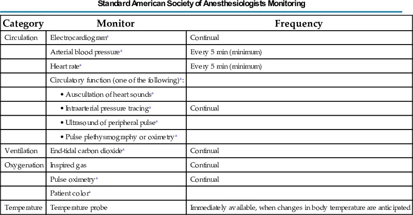
Standard American Society of Anesthesiologists Monitors
Most of the standard American Society of Anesthesiologists (ASA) monitors provide information about the cardiovascular system (Box 9.1). Electrocardiogram (ECG), arterial blood pressure, heart rate, and intraarterial pressure tracings are obviously useful, but those used to monitor respiratory function, such as end-tidal carbon dioxide (ETCO2) and pulse oximetry with its plethysmograph tracing, can also provide valuable cardiovascular information. The standard ASA monitors are listed in Table 9.1.

Basic Perioperative Monitors of Cardiovascular Function
- • Electrocardiogram
- • Heart rate
- • Noninvasive blood pressure
- • Pulse oximetry with plethysmograph analysis
- • Perfusion index
- • Pleth variability index
- • End-tidal carbon dioxide
- • Pulmonary blood flow
- • Auscultation of heart sounds
- • Amplitude and frequency of S1 for inotropic state
- • Amplitude and frequency of S2 for systemic blood pressure
Table 9.1
| Category | Monitor | Frequency |
|---|---|---|
| Circulation | Electrocardiograma | Continual |
| Arterial blood pressurea | Every 5 min (minimum) | |
| Heart ratea | Every 5 min (minimum) | |
| Circulatory function (one of the following)a: | ||
• Auscultation of heart soundsa | ||
• Intraarterial pressure tracinga | Continual | |
• Ultrasound of peripheral pulsea | ||
• Pulse plethysmography or oximetrya | ||
| Ventilation | End-tidal carbon dioxidea | Continual |
| Oxygenation | Inspired gas | Continual |
| Pulse oximetrya | Continual | |
| Patient colora | ||
| Temperature | Temperature probe | Immediately available, when changes in body temperature are anticipated |
aParameters that are useful in cardiovascular monitoring.
From American Society of Anesthesiologists Standards for Basic Monitoring, http://www.asahq.org.
Electrocardiogram
The ECG is a mainstay for monitoring cardiac status. Continuously monitoring cardiac electrical activity, it provides heart rate and rhythm data, as well as assessment of cardiac conduction (PR interval, QRS duration) and repolarization (ST segment, T-wave morphology, and QT interval). The normal morphologies of the ECG signal and the ECG intervals are shown in Fig. 9.1.
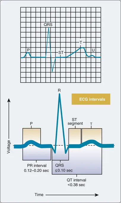
Fig. 9.1 Electrocardiographic morphology of one cardiac cycle and intervals.
A three-lead system, using three or four electrodes (right arm, left arm, left leg, ground), allows monitoring of limb leads I, II, or III, providing primarily rhythm and conduction data. This can suffice for healthy patients, but a five-electrode system (right arm, left arm, left leg, precordial, ground) is usually used perioperatively and in intensive care units. This system allows simultaneous monitoring of a limb lead (usually lead II) and a precordial “V” lead that enhances the detection of myocardial ischemia. The sensitivity for detecting myocardial ischemia when using a combination of leads II and V5 has been reported to be 80%. The V lead can be placed according to the particular area of interest, ranging from anterior (V1) to lateral (V6) (Fig. 9.2), with V3 to V5 generally being the most sensitive for anterior-lateral myocardial ischemia (lead II is used for inferior wall ischemia).
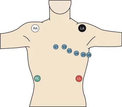
Fig. 9.2 Placement of the five-electrode system commonly used in operating rooms and intensive care units. The precordial lead (V) can be placed according to the area of interest, with the V3 to V5 positions generally being the most sensitive for myocardial ischemia.
Myocardial ischemia most often manifests as ST-segment depression, although elevated ST segments, change in T-wave morphology, new conduction defects, or frequent premature ventricular contractions may also be signs of myocardial ischemia. ECG monitoring systems have automated digital signal processing to continuously display heart rate, QT interval, and ST-segment depression or elevation, as well as alarm systems for these parameters.
Abnormal rhythms, such as sinus bradycardia and tachycardia, junctional rhythms, atrial fibrillation, right and left bundle branch blocks, and heart block, are not uncommon in cardiac patients. All these abnormalities can be detected using a five-electrode system (Tables 9.2 and 9.3). Whereas a limb lead such as lead II is preferred for conduction and rhythm detection, the precordial leads are preferred for diagnosis of myocardial ischem...
Table of contents
- Cover image
- Title Page
- Table of Contents
- Copyright
- Dedication
- Contributors
- Preface
- Section I Perioperative Medicine
- Section II Anesthesia for Noncardiac Surgery
- Section III Critical Care Medicine
- Index
Frequently asked questions
Yes, you can cancel anytime from the Subscription tab in your account settings on the Perlego website. Your subscription will stay active until the end of your current billing period. Learn how to cancel your subscription
No, books cannot be downloaded as external files, such as PDFs, for use outside of Perlego. However, you can download books within the Perlego app for offline reading on mobile or tablet. Learn how to download books offline
Perlego offers two plans: Essential and Complete
- Essential is ideal for learners and professionals who enjoy exploring a wide range of subjects. Access the Essential Library with 800,000+ trusted titles and best-sellers across business, personal growth, and the humanities. Includes unlimited reading time and Standard Read Aloud voice.
- Complete: Perfect for advanced learners and researchers needing full, unrestricted access. Unlock 1.5M+ books across hundreds of subjects, including academic and specialized titles. The Complete Plan also includes advanced features like Premium Read Aloud and Research Assistant.
We are an online textbook subscription service, where you can get access to an entire online library for less than the price of a single book per month. With over 1.5 million books across 990+ topics, we’ve got you covered! Learn about our mission
Look out for the read-aloud symbol on your next book to see if you can listen to it. The read-aloud tool reads text aloud for you, highlighting the text as it is being read. You can pause it, speed it up and slow it down. Learn more about Read Aloud
Yes! You can use the Perlego app on both iOS and Android devices to read anytime, anywhere — even offline. Perfect for commutes or when you’re on the go.
Please note we cannot support devices running on iOS 13 and Android 7 or earlier. Learn more about using the app
Please note we cannot support devices running on iOS 13 and Android 7 or earlier. Learn more about using the app
Yes, you can access Essentials of Cardiac Anesthesia for Noncardiac Surgery E-Book by Joel A. Kaplan in PDF and/or ePUB format, as well as other popular books in Medicine & Anesthesiology & Pain Management. We have over 1.5 million books available in our catalogue for you to explore.