Quickly reference the answers you need to the most important clinical questions in orthopedics with Orthopedic Secrets. Fully updated throughout, this classic medical reference book covers the entire range of essential topics in orthopedics, organized by subspecialty, for rapid access to the knowledge you need for success both in practice and on board and recertification exams.- Consult this title on your favorite e-reader, conduct rapid searches, and adjust font sizes for optimal readability.- Zero in on key orthopedic information with a question-and-answer format, bulleted lists, mnemonics, and practical tips from the authors.- Enhance your reference power with "Key Points" boxes and lists of useful websites.- Review essential material efficiently with a "Top 100 Secrets" chapter, perfect for last-minute study or self-assessment.- Take a fresh, updated approach to orthopedics with new editors and authors from the world-class orthopedic program at the University of Pennsylvania.- Focus on the details most relevant to your needs through a new case-based approach that's perfect for student or resident reference/review, or for any practitioner looking for a broad overview of the field.
eBook - ePub
Orthopedic Secrets E-Book
Orthopedic Secrets E-Book
- 472 pages
- English
- ePUB (mobile friendly)
- Available on iOS & Android
eBook - ePub
Orthopedic Secrets E-Book
Orthopedic Secrets E-Book
About this book
Trusted by 375,005 students
Access to over 1.5 million titles for a fair monthly price.
Study more efficiently using our study tools.
Information
Topic
MedicineSubtopic
Clinical MedicineChapter 1
Adult Reconstruction
Pramod B. Voleti, Atul F. Kamath
Knee
- 1. What is the differential diagnosis?
The differential diagnosis for this patient includes osteoarthritis, rheumatoid arthritis, crystalline arthropathies such as gout or pseudogout (calcium pyrophosphate deposition disease), spondyloarthropathies such as psoriatic arthritis and ankylosing spondylitis, and septic arthritis. Given the patient's age and clinical presentation, osteoarthritis is the most likely diagnosis.Case 1-1ContinuedThe patient is moderately obese with a Body Mass Index (BMI) of 32. Her left knee is not warm or swollen, but there is crepitus and medial joint line tenderness. Range of motion of the left knee is from 5° to 90°. Plain films of the left knee demonstrate narrowing of the medial joint space, subchondral sclerosis, and osteophyte formation (Fig. 1.1).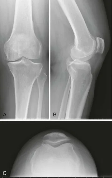 Figure 1.1 Anteroposterior (A), lateral (B), and merchant view (C) knee radiographs demonstrating medial joint space narrowing, subchondral sclerosis, and osteophyte formation, consistent with osteoarthritis.
 - 2. What is the likely diagnosis?
Osteoarthritis (also known as OA, osteoarthrosis, degenerative joint disease) is the most common form of joint disease. Osteoarthritis is characterized by loss of articular cartilage, which results in damage to the underlying bone. The process results in pain, stiffness, and loss of joint mobility. The pain is typically worse with use of the joint and improves with rest. Loss of the smooth articulating surface accounts for the finding of crepitus when the joint is moved. The most common joints affected are the hips, knees, and proximal and distal interphalangeal joints (Bouchard's and Heberden's nodes, respectively), with the knee being the most commonly involved joint. Radiographs of the affected joint typically show joint space narrowing, subchondral sclerosis, osteophyte formation, and subchondral cysts. The patient's symptoms, physical exam findings, and radiographs are most consistent with osteoarthritis. - 3. What is the pathogenesis of osteoarthritis?
Osteoarthritis is characterized by degeneration of articular cartilage and often is associated with overuse or trauma to the joint. Chondrocytes produce and maintain type II collagen, which is the primary component of articular cartilage. Osteoarthritis is thought to be a result of a failed attempt of chondrocytes to repair damaged articular cartilage. When the articular cartilage is not properly maintained, the joint space narrows, and the bones in the diarthrodial knee joint may come into direct contact with one another. The resulting wear and tear leads to bony proliferation, with formation of subchondral sclerosis and osteophytes. Subchondral cysts arise secondary to microfractures and may contain amorphous gelatinous material. - 4. What changes occur in the cartilage of osteoarthritic joints?
Osteoarthritic cartilage is characterized by increased water content (in contrast with the decreased water content seen with aging), alterations in proteoglycans (decrease in overall content, shorter chain structure, an increase in the chondroitin/keratin sulfate ratio), and collagen abnormalities. - 5. What are the anatomic sources of the joint pain in osteoarthritis?
Although articular cartilage is the primary site of injury in this disease, cartilage is aneural, and, therefore, no pain is transmitted from this tissue. The pain of osteoarthritis primarily originates from the periosteum surrounding the bone. As the articular cartilage wears away and the bones of the joint begin to rub against one another, the highly innervated periosteum becomes damaged and results in the joint pain seen in osteoarthritis. Other potential anatomic sources of osteoarthritic pain include subchondral bone, capsule, synovium, and periarticular tendons and bursae. - 6. What are the risk factors associated with developing osteoarthritis?
Obesity, joint trauma, and muscle weakness are some of the risk factors for osteoarthritis. These factors all increase the mechanical forces to which the articular cartilage is subjected. Gender, hormones, metabolic disorders, and genetics also play a role. Elderly populations are affected by this disease more frequently and more severely than younger populations. Obesity is the strongest modifiable risk factor for osteoarthritis.
Note: Osteoarthritis can be classified as primary (idiopathic disease caused by intrinsic defect, the most common form), or secondary, with an underlying cause (e.g., trauma, infection, congenital deformity). - 7. What are the initial treatment options for osteoarthritis of the knee?
Treatment begins with supportive measures, including weight loss and activity modification. Bracing, including compartmental unloader bracing, and/or ambulatory assistive devices may also be prescribed. Oral pain medications (such as NSAIDs), corticosteroid injections, viscosupplementation, and topical analgesics have been shown to alleviate the pain associated with osteoarthritis. While not demonstrating a clear benefit in the literature, supplements such as glucosamine and chondroitin sulfate may be tried. Moderate physical therapy may provide some symptomatic benefit, but it may only aggravate more advanced disease. Low-impact or aquatic therapy, in conjunction with stretching and isometric strengthening, may prove helpful. Other “joint protection” programs, those that cause low loads across the joint, include swimming, bicycling, walking, or tai chi; these activities increase muscle mass while protecting joints from undue stresses. Alternative therapies such as acupuncture may provide benefit in some patients, but there are no well-controlled data regarding efficacy in advanced osteoarthritis of the knee. Below is Table 1.1 summarizing the strong and moderate recommendations of the American Academy of Orthopaedic Surgeons (AAOS) with regard to treatment of knee osteoarthritis.
Table of contents
- Cover image
- Title Page
- Table of Contents
- Copyright
- Preface to the 4th Edition
- Contributors
- 1 Adult Reconstruction
- 2 Basic Science
- 3 Foot and Ankle
- 4 Hand
- 5 Orthopedic Oncology
- 6 Pediatric Orthopedics
- 7 Rehabilitation and Neuro-Orthopedic Surgery
- 8 Shoulder and Elbow
- 9 Spine
- 10 Sports
- 11 Orthopedic Trauma
- Index
Frequently asked questions
Yes, you can cancel anytime from the Subscription tab in your account settings on the Perlego website. Your subscription will stay active until the end of your current billing period. Learn how to cancel your subscription
No, books cannot be downloaded as external files, such as PDFs, for use outside of Perlego. However, you can download books within the Perlego app for offline reading on mobile or tablet. Learn how to download books offline
Perlego offers two plans: Essential and Complete
- Essential is ideal for learners and professionals who enjoy exploring a wide range of subjects. Access the Essential Library with 800,000+ trusted titles and best-sellers across business, personal growth, and the humanities. Includes unlimited reading time and Standard Read Aloud voice.
- Complete: Perfect for advanced learners and researchers needing full, unrestricted access. Unlock 1.5M+ books across hundreds of subjects, including academic and specialized titles. The Complete Plan also includes advanced features like Premium Read Aloud and Research Assistant.
We are an online textbook subscription service, where you can get access to an entire online library for less than the price of a single book per month. With over 1.5 million books across 990+ topics, we’ve got you covered! Learn about our mission
Look out for the read-aloud symbol on your next book to see if you can listen to it. The read-aloud tool reads text aloud for you, highlighting the text as it is being read. You can pause it, speed it up and slow it down. Learn more about Read Aloud
Yes! You can use the Perlego app on both iOS and Android devices to read anytime, anywhere — even offline. Perfect for commutes or when you’re on the go.
Please note we cannot support devices running on iOS 13 and Android 7 or earlier. Learn more about using the app
Please note we cannot support devices running on iOS 13 and Android 7 or earlier. Learn more about using the app
Yes, you can access Orthopedic Secrets E-Book by Surena Namdari,Stephan G. Pill,Samir Mehta,Stephan Pill in PDF and/or ePUB format, as well as other popular books in Medicine & Clinical Medicine. We have over 1.5 million books available in our catalogue for you to explore.