The central themes of Cell Boundaries concern the structural and organizational principles underlying cell membranes, and how these principles enable function. By building a biological and biophysical foundation for understanding the organization of lipids in bilayers and the folding, assembly, stability, and function of membrane proteins, the book aims to broaden the knowledge of bioscience students to include the basic physics and physical chemistry that inform us about membranes. In doing so, it is hoped that physics students will find familiar territory that will lead them to an interest in biology. Our progress toward understanding membranes and membrane proteins depends strongly upon the concerted use of both biology and physics. It is important for students to know not only what we know, but how we have come to know it, so Cell Boundaries endeavours to bring out the history behind the central discoveries, especially in the early chapters, where the foundation is laid for later chapters. Science is far more interesting if, as students, we can appreciate and share in the adventures—and misadventures—of discovering new scientific knowledge.
Cell Boundaries was written with advanced undergraduates and beginning graduate students in the biological and physical sciences in mind, though this textbook will likely have appeal to researchers and other academics as well.
Highlights the history of important central discoveries
Early chapters lay the foundation for later chapters to build on, so knowledge is amassed
High-quality line diagrams illustrate key concepts and illuminate molecular mechanisms
Box features and spreads expand on topics in main text, including histories of discoveries, special techniques, and applications
Trusted by 375,005 students
Access to over 1.5 million titles for a fair monthly price.
Why are cell membranes and their lipids and proteins worth knowing about? Simply put, membranes enable life; they organize cells into protected compartments, control the flow of nutrients and information between compartments, generate and store energy, and define cells structurally and phylogenetically. These functions make understanding membranes a key to understanding cell biology. From the point of view of physics and physical chemistry, the extreme thinness and chemical heterogeneity of cell membranes open new vistas—and challenges—at the nano scale.
The biology and physics of membranes are not easily separated; together, they have laid the foundations for understanding the structural principles of membrane function. What were the key discoveries that built the foundation? This chapter is devoted to answering this question. We will see that many of the early insights into membranes and cells came from biophysical studies based solidly on thermodynamics. Indeed, thermodynamics helps us appreciate in a deeper way the elegance of life, whose very existence must be compatible with the laws of thermodynamics. We have therefore provided a primer on thermodynamics at the beginning of this book (Chapter 0). As an aid to our discussion of foundations, we begin with a brief overview of cell structure.
1.1 Membranes Define Cell Anatomy
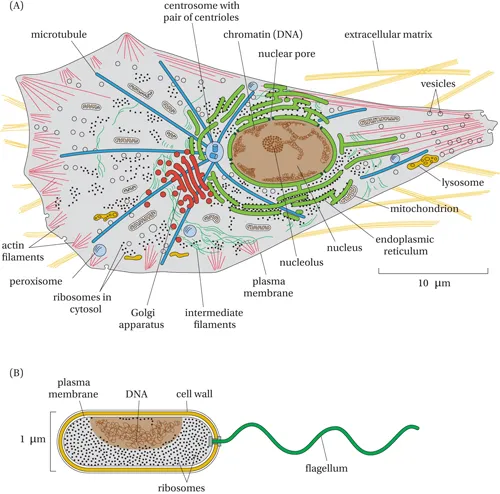
All living creatures are divided into two broad groups: prokaryotes and eukaryotes (Greek: pro = before, eu = true, and karyon = kernel, meaning nucleus). For both groups, the cellular interior (cytoplasm) is isolated from the external environment by the plasma membrane, which protects the cell biochemically by controlling the movement of all chemical substances between an often fickle external environment and the cytoplasm. This control, exercised by membrane proteins, allows biochemical processes essential for life to proceed in an organized fashion. Eukaryotes (Figure 1.1A), which include all multi-cellular organisms, are distinguished from prokaryotes (e.g., bacteria, Figure 1.1B) by having a double membrane (nuclear membrane) that isolates the genetic material (DNA) and its regulatory machinery from the cytoplasm (except during cell division).
Figure 1.1 Anatomies of two types of biological cells. (A) Eukaryotic cell. (B) Prokaryotic cell. All cells have two features in common: a surface (plasma) membrane, which separates the cell interior (cytoplasm) from the external world, and DNA (deoxyribonucleic acid), which carries the inheritable genetic information. In eukaryotes, the DNA is separated from the cytoplasm by a double membrane (nuclear envelope) except during cell division. Prokaryotes do not have a nuclear membrane; the DNA resides directly in contact with the cytoplasm. A particularly important difference between prokaryotes and eukaryotes is the presence of numerous membrane-enclosed compartments in eukaryotes, such as mitochondria and the Golgi apparatus. (From Alberts B, Johnson A, Lewis JH et al. [2014] Molecular Biology of the Cell, 6th Edition. Garland Science, New York. With permission from W. W. Norton.)
1.1.1 Prokaryotes Have a Minimum Complement of Membranes
Prokaryotes, further subdivided into eubacteria and archaea, generally have no internal membrane-delimited compartments but only a plasma membrane that surrounds the cytoplasm (Figure 1.1B). In many prokaryotes, the plasma membrane is protected by a semi-rigid cell wall of variable composition and architecture. There are five different types of walls, ranging from a single layer of protein or glycoprotein (the S layer) to more complex structures comprised of an S layer plus an additional layer of chondroitin-like molecules or polysaccharides. Eubacteria are protected by a tough peptidoglycan layer, which is a composite mesh-like material built from linear polysaccharide chains that are crosslinked by short peptides. For a long time, microbiologists classified eubacteria as either Gram-positive or Gram-negative based on whether their surfaces stain blue or pink in a staining procedure invented by H.C. Gram in the late 19th century. This classification is still with us, although the staining procedure has now largely been superseded by phylogenetic analysis based on the sequence of the ribosomal 16S ribonucleic acid (RNA) molecule. In most Gram-positive bacteria (such as Streptococcus aureus), the cell wall is composed of a thick layer of peptidoglycan. Some simple Gram-positive bacteria (such as Mycoplasma genitalium) lack the peptidoglycan and only have a single lipid membrane to protect their cytoplasmic compartment.
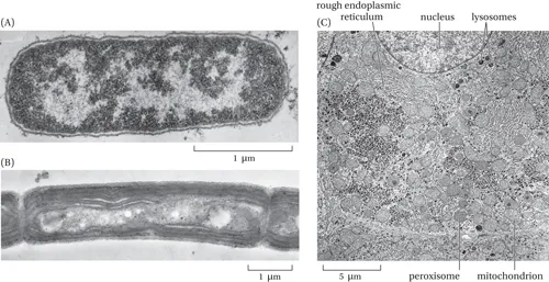
Gram-negative bacteria (such as Escherichia coli) have two membranes, the plasma (or inner) membrane and an outer membrane that contains polysaccharides as well as lipids and proteins. This second membrane allows some control over the immediate environment, thus providing protection to the inner membrane and cytoplasm. The space between the two membranes defines the periplasm, where important biochemical processes necessary for survival occur. Some bacteria also have specialized membrane-bounded intracytoplasmic compartments for special activities such as photosynthesis (thylakoids) or nitrogen fixation (respiratory membranes) (Figure 1.2B). Like Gram-negative bacteria, plant cells (which are eukaryotes) are protected by a tough cell wall. Unlike in bacteria, however, this cell wall is formed from cellulose and polysaccharides. It is not generally considered to be a cell membrane, because it lacks the characteristic thin lipid matrix of the cell wall of Gram-negative bacteria and archaea.
Figure 1.2 Electron micrographs of cells and their membranes. (A) The periplasmic space and the protective cell wall of Escherichia coli are visible in this electron micrograph. The lighter area of the cytoplasm is the cell’s DNA. (B) Intracellular membranes of the photosynthetic bacterium Phormidium laminosum that are evolutionary harbingers of eukaryotic intracellular membranes. In this case, the intracellular membranes are the sites of photosynthesis. (C) The interiors of eukaryotic cells, in this case from human liver, are filled with membranous structures such as mitochondria that produce ATP, peroxisomes that break down fatty acids, and the endoplasmic reticulum from which membrane proteins originate. (A, From Alberts B, Johnson A, Lewis JH et al. [2014] Molecular Biology of the Cell, 6th Edition. Garland Science, New York. With permission from W. W. Norton. B, From Alberts B, Gray D, Hopkin K et al. [2013] Essential Cell Biology, 4th Edition. Garland Science, New York. With permission from W. W. Norton. C, From Alberts B, Johnson A, Lewis JH et al. [2014] Molecular Biology of the Cell, 6th Edition. Garland Science, New York. With permission from W. W. Norton.)
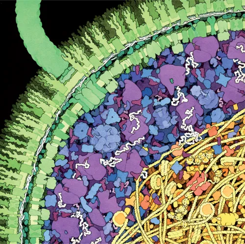
We often draw cells as containing a dilute, water-rich cytoplasm surrounded by a lipid bilayer membrane in which a few dispersed proteins float around, but that is highly inaccurate. The cell interior is actually a concentrated solution of macromolecules and small metabolites, and the cell membrane is stuffed full of proteins. Suggestive images, such as Figure 1.3, are good to keep in mind when thinking about what goes on inside a cell, and especially when comparing biochemical data obtained in dilute solutions in the test tube with data obtained from studies in vivo.
Figure 1.3 An artist’s scale rendition of a cross-section of an E. coli cell. The outer and inner membranes are shown in green, ribosomes in purple, and DNA in yellow. This illustration makes clear that cell interiors are much more crowded and regional than a simple aqueous solution. Illustration by David S. Goodsell. (With permission from the Scripps Research Institute.)
1.1.2 Eukaryotic Cells Have Many Compartments
The number of specialized intracellular compartments is greatly expanded in eukaryotic cells in order to sequester critical biochemical processes to specialized regions of the cell with distinctive chemical characteristics such as pH, ionic composition, and ATP/ADP ratio (Figure 1.4). These specialized compartments, referred to as organelles, greatly increase the membrane surface area available for organizing functional membrane-associated protein complexes. The compartments are interconnected by various transport processes that allow proteins, lipids, and other molecules to move between compartments in a highly regulated fashion. Historically, the biochemical and structural characterization of intracellular organelles was pioneered by Albert Claude, George Palade, and Christian de Duve using the ultracentrifuge to separate different subcellular fractions and the electron microscope to visualize the cell architecture. They shared the 1974 Nobel Prize for physiology or medicine.
Figure 1.4 Schematic picture of an animal epithelial cell, such as those lining the gut. The cell is polarized, i.e., the membrane on the apical side is insulated by a protein barrier—a tight junction—from the membrane covering the basolateral side that faces adjacent cells and connective tissue, permitting functional differences. The apical side faces the lumen of the gut. (From Alberts B, Johnson A, Lewis JH et al. [2014] Molecular Biology of the Cell, 6th Edition. Garland Science, New York. With permission from W. W. Norton.)
Some of the cell’s compartments are connected biosynthetically. The nucleus houses the cell’s DNA and is surrounded by a double membrane that is continuous with the endoplasmic reticulum (ER). Proteins destined for secretion from the cell and most of the cell’s integral membrane proteins are synthesized by ER-bound ribosomes. Most proteins are then transported from the ER along the secretory pathway, first through the Golgi apparatus to the trans-Golgi network, and onwards to the plasma membrane. Proteins and other cargo can also be transported backwards from the plasma membrane to intracellular endosomes and then to lysosomes, where they can be degraded by digestive enzymes.
Other intracellular organelles include lipid droplets that are composed of a hydrophobic core of stored lipids surrounded by a protein-rich phospholipid monolayer, mitochondria containing the enzymes of the respiratory chain that converts nutrient energy into ATP, and peroxisomes that protect the cell from hydrogen peroxide as well as being involved in lipid biosynthesis. Plant cells also contain chloroplasts, the organelle that houses the photosynthetic apparatus.
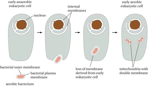
Mitochondria are surrounded by an outer and an inner membrane. Chloroplasts also have both an outer and an inner membrane, plus an internal thylakoid membrane system. Both mitochondria and chloroplasts are thought to have arisen through endosymbiosis, an evolutionary process through which a eukaryotic cell “engulfs” a prokaryotic cell or the prokaryotic cell invades a eukaryotic cell (as is seen in some modern infections), and the two organisms mutually benefit (Figure 1.5). After the initial endosymbiotic event, large parts of the genome of the prokaryotic cells moved into the nuclear genome of the eukaryotic cell, resulting in an intracellular organelle with only a minimal genome left as a witness to its prokaryotic ancestry. There is evidence that the mitochondrial ancestors may have come from a Gram-negative Rickettsiae-like bacterium. Chloroplasts likely arose from photosynthetic cyanobacteria.
Figure 1.5 Scenario for the evolution of mitochondria or chloroplasts from an invasion by a free-living ba...
Table of contents
Cover
Half-Title
Title
Copyright
Contents
List of Boxes
Preface
Acknowledgements
Chapter 0 The “E” Words: A Concise Guide to Thermodynamics
Chapter 1 Foundations of Membrane Structure
Chapter 2 Lipid Bilayers
Chapter 3 Interactions of Peptides with Lipid Bilayers
Chapter 4 Membrane Protein Folding and Stability
Chapter 5 Protein Trafficking in Cells
Chapter 6 Biosynthesis and Assembly of Membrane Proteins
Chapter 7 How Proteins Shape Membranes
Chapter 8 Membrane Protein Bioinformatics
Chapter 9 Primer on Biomolecular Structure Determination
Chapter 10 Small-Molecule Channels
Chapter 11 Ion Channels
Chapter 12 Primary Transporters: Transport against Electrical and Chemical Gradients
Chapter 13 Secondary Transport
Chapter 14 Bioenergetics
Chapter 15 Information Transfer: Signaling in Cells
Electrostatics Appendix
Index
Frequently asked questions
Yes, you can cancel anytime from the Subscription tab in your account settings on the Perlego website. Your subscription will stay active until the end of your current billing period. Learn how to cancel your subscription
No, books cannot be downloaded as external files, such as PDFs, for use outside of Perlego. However, you can download books within the Perlego app for offline reading on mobile or tablet. Learn how to download books offline
Perlego offers two plans: Essential and Complete
Essential is ideal for learners and professionals who enjoy exploring a wide range of subjects. Access the Essential Library with 800,000+ trusted titles and best-sellers across business, personal growth, and the humanities. Includes unlimited reading time and Standard Read Aloud voice.
Complete: Perfect for advanced learners and researchers needing full, unrestricted access. Unlock 1.5M+ books across hundreds of subjects, including academic and specialized titles. The Complete Plan also includes advanced features like Premium Read Aloud and Research Assistant.
Both plans are available with monthly, semester, or annual billing cycles.
We are an online textbook subscription service, where you can get access to an entire online library for less than the price of a single book per month. With over 1.5 million books across 990+ topics, we’ve got you covered! Learn about our mission
Look out for the read-aloud symbol on your next book to see if you can listen to it. The read-aloud tool reads text aloud for you, highlighting the text as it is being read. You can pause it, speed it up and slow it down. Learn more about Read Aloud
Yes! You can use the Perlego app on both iOS and Android devices to read anytime, anywhere — even offline. Perfect for commutes or when you’re on the go. Please note we cannot support devices running on iOS 13 and Android 7 or earlier. Learn more about using the app
Yes, you can access Cell Boundaries by Stephen White,Gunnar von Heijne,Donald Engelman,Stephen H White,Donald M Engelman in PDF and/or ePUB format, as well as other popular books in Médecine & Biochimie en médecine. We have over 1.5 million books available in our catalogue for you to explore.