Laminitis, a horse-centred approach describes in depth the current mainstream thinking on laminitis and suggests ways of reframing our understanding of this challenging condition. New thinking based on putting the horse at the centre of the problem is presented, allowing a better understanding of the biomechanics of laminitis. The book suggests ways in which damaged feet can recover, and also helps the reader to understand the pathological processes within the horse as a whole that lead to laminitis occurring, starting with an understanding of the horse's innate ability to heal itself and working towards interventions that create an environment that is conducive to healing. The book also explores the concept that laminitis, rather than being a disease in its own right, is merely a symptom of a range of underlying health problems that affect the whole horse.
- 208 pages
- English
- ePUB (mobile friendly)
- Available on iOS & Android
eBook - ePub
About this book
Trusted by 375,005 students
Access to over 1.5 million titles for a fair monthly price.
Study more efficiently using our study tools.
Information
Topic
MedicinaSubtopic
Ciencias veterinarias equinasPart 1
What is Laminitis?
1Essential Anatomy
It is not possible to discuss the subject of laminitis without first having a reasonable understanding of the anatomy of the equine foot. This chapter attempts, without going into excessive detail, to provide a quick guide to those aspects of basic anatomy that are necessary to understand the remainder of the book. As well as covering the names for the key structures, this chapter also looks at what functions these structures have. This helps to make the rather dry subject of anatomy a little more interesting.

Fig. 1 The bones of the lower foreleg of the horse compared to the equivalent bones in the human hand. The proximal and distal sesamoids are not present in the human hand.
BONES
Horses and humans have evolved from common ancestors and share similar bone structures. The bones of the horse’s lower leg (from the knee/hock downwards) closely map to the bones of the human hand/foot, with the horse having largely lost all but the middle finger/toe. The horse’s knee is equivalent to our wrist, and the horse’s hock is equivalent to our ankle. For that reason the lowest part of the horse’s leg is often referred to as the digit (just as our fingers and toes are digits).
The bones most relevant in the discussion of laminitis are the three phalanges (singular phalanx) – these are the finger and toe bones in a human, but in a horse they provide the underlying bone structure to the pastern and foot.
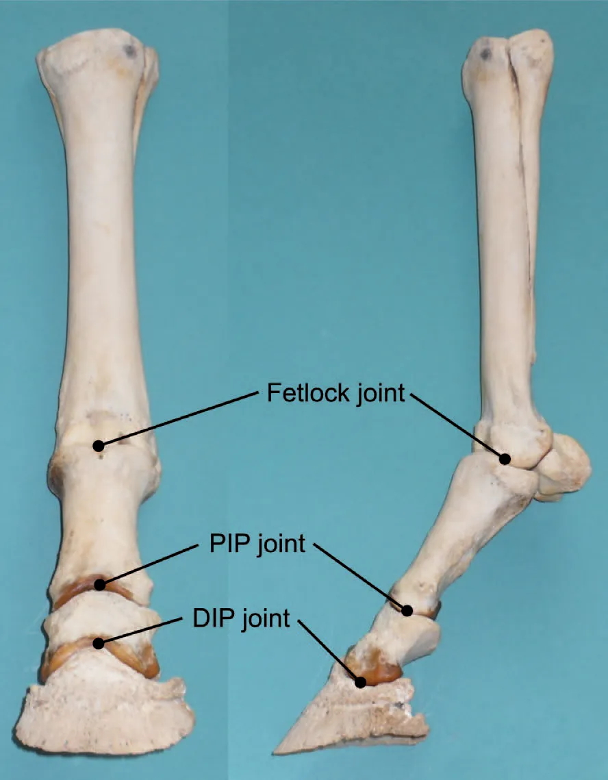
Fig. 2 A set of bones of a lower foreleg wired into their correct positions.
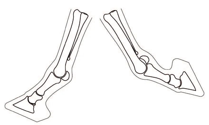
Fig. 3 The bones of the lower leg in extension (left) and flexion (right).
JOINTS
Between each adjacent pair of bones in the lower leg there is a hinge joint – a joint which can extend (curl forwards) or flex (curl backwards). Those parts of each bone that are involved in a joint are covered with articular cartilage, which is smooth and shiny. This allows the two bones to move in relation to each other. To further minimize friction, the joint is lubricated with an oily liquid called synovial fluid. The entire joint is then encased in a membranous sac called a bursa so as to keep the synovial fluid in place. Where two or more joints are very close together, they sometimes share a single bursa.
The pastern is made up of the long and short pastern bones (often abbreviated to P1 and P2 for first and second phalanges). Between these two bones sits the pastern joint. The technical term for this joint is the ‘proximal interphalangeal joint’ – often abbreviated to PIP joint. The pastern joint can extend and flex but only by very small amounts, which is why most horse owners are unaware that the pastern has a joint in the middle of it. The main role of this joint is to allow absorption of some of the impact of the foot hitting the ground by extending very slightly.
The third phalanx is the pedal bone (sometimes also called the distal phalanx or coffin bone, and often abbreviated to P3). Between P2 and P3 is the distal interphalangeal joint, or DIP joint (sometimes also referred to as the coffin joint). Unlike the PIP joint, the DIP joint can extend and flex through quite a large range of movement.
At the back of the DIP joint, between P2 and P3, sits the navicular bone. This bone is tightly attached to the back of P3 by the impar ligament and so for the most part it moves with P3 as the DIP joint flexes and extends. What little movement there is between P3 and the navicular bone allows the flexor tendon (see below) to pass into the foot at the most advantageous angle as the foot lands on uneven ground.
TENDONS AND LIGAMENTS
Bones alone are not enough to allow a horse to move. To start with, something has to keep the joints from dislocating, and this is the task of ligaments. Ligaments are connective tissues that, in most cases, join bone to bone. They can be thought of as like strong rubber bands. Each of the joints in the lower leg has collateral (on each side) ligaments that hold the joint together whilst allowing it to move. Another important ligament is the suspensory ligament, which passes down the back of the cannon bone, under the fetlock and attaches to the back of the pastern, as well as splitting to form two branches that pass either side of the pastern and attach at the front. The role of this ligament is to support the weight of the horse at rest and prevent the fetlock from dropping to the ground. The horse uses the suspensory ligament rather like a spring, and much of the efficiency of movement in a horse comes from its ability to bounce off the lower legs like a pogo stick. The arrangement of the pastern, fetlock and suspensory ligament works in rather the same way as the running blades used by human amputees.
There are many other ligaments in the lower leg that hold joints together and act as support bandages, keeping important structures where they should be. The end result of all these ligaments, as well the joints being held together, is that the bone column has a ‘neutral’ position that it will adopt when no muscles are brought into play.
For the horse to move around, it needs to be able to flex and extend the lower limb under the control of muscles. But muscles are heavy structures, and having them in the lower leg would make the leg heavier and hence reduce the horse’s ability to run away quickly from predators. So instead, the muscles are located higher up the leg and pull on the relevant bones using tendons. Tendons are similar to ligaments but act more like strings than rubber bands. Each tendon is pulled by a muscle at one end and is attached to a bone at the other, allowing the muscle to move the bone from a distance.
There are three main tendons in the lower leg. The extensor tendon (its full name is ‘common digital extensor tendon’ or ‘CDET’ on a front leg, and ‘long digital extensor tendon’ or ‘LDET’ on the hind leg) sits at the front of the leg and acts to extend the lower leg. The horse typically pulls on the extensor tendon just before the foot hits the ground to lift the toe. Its main role is to ensure that the foot hits the ground with the heel first, with no risk of the horse tripping over the toe.
At the back of the leg are two flexor tendons. The deep digital flexor tendon (DDFT) connects to the back of the pedal bone and has the effect of flexing the DIP joint. The superficial digital flexor tendon (SDFT) – so named because it is closest to the surface at the back of the cannon bone – acts largely to flex the fetlock joint. These two tendons between them have two roles. The first is to fold the foot and pastern up behind the leg while the limb is in flight, so minimizing the risk of catching the foot on uneven ground, vegetation and suchlike. The second is to assist in propelling the horse forwards during breakover (the point at which the heels have left the ground but the toe is still in contact). Much of this propulsion comes from the release of energy stored during the previous impact in the suspensory ligament, but the flexor tendons play an important role in controlling the release of this energy.

Fig. 4 The role of the tendons. LEFT: The limb extended prior to impact as a result of tension in the CDET. CENTRE: The limb at the point of breakover, with the SDFT and the DDFT contributing to propulsion. RIGHT: The limb flexed during flight as a result of tension in the SDFT and the DDFT.
MAJOR FOOT STRUCTURES
Of the bones of the lower limb, two and a half sit within the foot: the pedal bone, the navicular bone, and the lower half of the short pastern bone. The pedal bone in particular gives the front portion of the foot its shape. There is, however, no bone in the back of the foot. Given that a sound horse on hard ground lands heel first, this makes sense – the horse does not want to jar bones by landing directly on to them. Instead, the shape of the back of the foot is formed by two lateral cartilages (each of which controls the shape of one heel), between which lies the digital cushion.
The shape and position of the lateral cartilages in a healthy foot supports the pedal bone such that it is tipped slightly forwards (the optimal angle is believed to be between 3 and 5 degrees), rather than being oriented flat to the ground. The result is that, with the outer horny structures removed, the internal structures of the foot form an arch similar to that seen in a human foot. As the foot hits the ground (heel first), the lateral cartilages flex slightly, which allows the arch to flatten somewhat, so absorbing and storing some of the energy of impact. As the foot leaves the ground again, the arch springs back to its normal shape, releasing much of that stored energy in a way that helps to propel the horse forwards. You can think of the internal arch as a form of spring on the bottom of the foot, with which the horse can bounce off the ground. Although this is a much smaller effect than the bounce the horse gets from the suspensory ligament, it nevertheless contributes to saving the horse energy and making movement more efficient.

Fig. 5 The role of the lateral cartilages at impact, mid-stance and breakover. The internal arch is indicated in red.
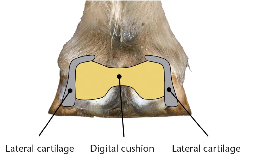
Fig. 6 The position of the digital cushion and lateral cartilages (viewed from the back of the foot).
The digital cushion sits between the lateral cartilages, and the top part of it can be felt by pressing down on the foot above the heel bulbs and between the lateral cartilages. Its main role is to absorb the vibration associated with the impact of the foot on hard ground so that this vibration doesn’t reach the bone column (where it could cause inflammation and damage, typically in the form of arthritis in places such as the navicular bone, coffin joint and so on). In a healthy foot, the digital cushion should fill the space between the lateral cartilages such that there is little or no depression in the centre of the back part of the foot. It should ideally also feel like a block of hard rubber. Where it is small and feels more like chicken fat, this suggests that it has atrophied and that it won’t work so well as a shock absorber.

Fig. 7 The major arteries of the foot.
THE BLOOD SUPPLY
The details of the blood vessels in the foot are beyond the scope of this book, but two arteries within the foot are worth mentioning. The terminal arch circles through the middle of the pedal bone approximately half way up, and is the key arterial blood supply to the pedal bone. The circumflex artery of the sole (usually just referred to as the circumflex artery) passes around the rim of the pedal bone just outside and just below the bottom of the bone. The circumflex artery supplies both the lower portions of the pedal bone and the sole. It is important to note that the main way in which blood reaches the sole is via the circumflex artery, as there are no significant channels allowing blood to pass through the bottom surface of the pedal bone to supply the sole directly. As such, any damage to the circumflex artery will have a direct impact on the growth of the sole. The relevance of these two arteries in laminitis will become clear in later chapters.
The other arteries relevant to laminitis are the two palmar digital arteries, which provide the main blood supply to the foot and sit one on each side of the leg towards the back.
THE HOOF
The foot is defined as everything (including the hoof) below the hairline, in addition to the part of the lateral cartilages that protrudes above the hairline. The hoof (sometimes called the hoof capsule because it encapsulates the foot) is defined as all the avascular (not containing blood vessels) structures of the foot – which essentially means everything that is made of horn. The pedal bone, lateral cartilages and digital cushion form the shape of the foot. The external hoof then forms a hard casing around these internal structures. The hoof has a number of important roles, including the following:
•Protection of internal structures from mechanical trauma
•Prevention of infection
•Durability (the foot must not wear away too fast)
•Traction (so the horse doesn’t slip)
•Adaptation to environment (a horse living in a desert needs a different foot from one living in a bog)

Fig. 8 A cut-away dissection showing the structures of the hoof. All hoof horn has been removed on one side of the foot so as to expose the underlying coria that produce the hoof (see below). The damage to the coronary band at the heel and the knife cut along the join between the solar and laminar coria are artefacts of ...
Table of contents
- Cover
- Halftitle
- Title
- Copyright
- Contents
- Introduction
- Part One: What is Laminitis?
- Part Two: What Causes Laminitis?
- Part Three: Management of the Laminitic Horse
- Conclusion
- Abbreviations Used in This Book
- Glossary
- References
- Further Information
- Index
Frequently asked questions
Yes, you can cancel anytime from the Subscription tab in your account settings on the Perlego website. Your subscription will stay active until the end of your current billing period. Learn how to cancel your subscription
No, books cannot be downloaded as external files, such as PDFs, for use outside of Perlego. However, you can download books within the Perlego app for offline reading on mobile or tablet. Learn how to download books offline
Perlego offers two plans: Essential and Complete
- Essential is ideal for learners and professionals who enjoy exploring a wide range of subjects. Access the Essential Library with 800,000+ trusted titles and best-sellers across business, personal growth, and the humanities. Includes unlimited reading time and Standard Read Aloud voice.
- Complete: Perfect for advanced learners and researchers needing full, unrestricted access. Unlock 1.5M+ books across hundreds of subjects, including academic and specialized titles. The Complete Plan also includes advanced features like Premium Read Aloud and Research Assistant.
We are an online textbook subscription service, where you can get access to an entire online library for less than the price of a single book per month. With over 1.5 million books across 990+ topics, we’ve got you covered! Learn about our mission
Look out for the read-aloud symbol on your next book to see if you can listen to it. The read-aloud tool reads text aloud for you, highlighting the text as it is being read. You can pause it, speed it up and slow it down. Learn more about Read Aloud
Yes! You can use the Perlego app on both iOS and Android devices to read anytime, anywhere — even offline. Perfect for commutes or when you’re on the go.
Please note we cannot support devices running on iOS 13 and Android 7 or earlier. Learn more about using the app
Please note we cannot support devices running on iOS 13 and Android 7 or earlier. Learn more about using the app
Yes, you can access Laminitis by Richard Vialls in PDF and/or ePUB format, as well as other popular books in Medicina & Ciencias veterinarias equinas. We have over 1.5 million books available in our catalogue for you to explore.