A simple case-by-case approach to the different types of orthopedic fractures. The book has been written for residents, registrars, newly qualified doctors and medical students, primarily targeted to those in the fields of diagnostic radiology, orthopedic surgery and emergency medicine. There are a variety of texts that exist but they lack simple images and explanations from which a fundamental base of skeletal trauma can be formed. Some of the standard textbooks are still too advanced and lack the basic knowledge. This book is divided into sections by anatomic location, each containing a number of cases which includes a brief clinical description, physical examination, followed by radiological images. Several questions are then asked, including the most likely diagnosis and followed by brief discussions on radiological findings and clinical management. With this format readers can test their knowledge on important orthopedic fractures as seen on a plain film. Furthermore, they can integrate this information with the standard clinical management involved with that specific disease entity.
- English
- ePUB (mobile friendly)
- Available on iOS & Android
eBook - ePub
About this book
Trusted by 375,005 students
Access to over 1.5 million titles for a fair monthly price.
Study more efficiently using our study tools.
Information
Topic
MedicineChapter 1
Spine
Case 1
Clinical presentation
A 36-year-old man presents after a high-speed motor vehicle accident. He was a restrained passenger. No air bags deployed in the vehicle. At the scene, the patient was found to have a GCS of 13 due to confusion, but was hemodynamically stable. In the trauma bay, his GCS is improving, but he is complaining of neck pain. He was placed in a hard collar at the scene.
Physical examination
The patient is alert, oriented to person and year, but not to date. He has lacerations on his forehead, and he has peri-orbital swelling and ecchymosis. When you palpate his spine after removing the collar, there is tenderness in the upper midline. You promptly replace the collar. There is no blood on your glove when you remove your hand from palpating his neck. You find no deficits on your neurologic exam.
Review the image below [Figure 1]. Describe your findings.
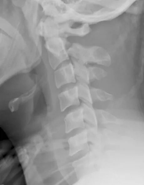
Figure 1.
Questions
| 1. | What is the diagnosis? |
| 2. | How do these injuries occur? |
| 3. | Describe the commonly used classification system for these injuries. |
| 4. | What are other associated injuries? |
Radiology findings
A lateral radiograph through the cervical spine demonstrates a mild kyphotic angulation at C2/3. There is a fracture through the pars interarticularis of C2. There is 5mm gapping and 12° of angulation of fracture fragments. There is also malalignment of the spinal laminar line, with gapping between the posterior spinous processes at C1/2.
Answers
| 1. | Hangman’s fracture (Type II). |
| 2. | In adults, this injury typically results from hyperextension and distraction, as seen in hanging or a high-energy motor vehicle accident (head striking the dashboard). Sometimes in adults and commonly in children, the mechanism is a combination of flexion and distraction as appears to be the case in this example. |
| 3. | The classification system proposed by Levine is most commonly used to organize these fractures: |
| i) | a Type I injury (labeled ‘A’ in Figure 2) consists of a fracture of the C2 pedicles (pars interarticularis), with the fracture line orientated either vertical or near vertical. There is less than 3mm translation and no angulation. The C2-3 disc space remains intact; |
| ii) | in a Type II injury (most common subtype), there is greater than 3mm translation and greater than 10° of angulation of fracture fragments. These fractures are unstable and also demonstrate anterior displacement of the C2 vertebral body. There may also be a compression of the anterosuperior corner of the C3 body (labeled ‘B’ in Figure 2). Type IIa injuries (labeled ‘C’ in Figure 2) differ from Types I and II because the fracture line is more oblique and there is minimal translation but severe angulation. These injuries also have a different mechanism of injury, as they occur due to a flexion distraction force; |
| iii) | Type III injuries are Type II injuries (angulation and translation) with additional bilateral interfacetal dislocation (labeled ‘D’ in Figure 2). There is a higher incidence of neurologic deficits. |
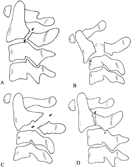
Figure 2. The Levine classification system is pictured; with Type I through III injuries depicted (images A-D, respectively). Reprinted with permission from Elsevier Ltd. Levine AM, Eismont FJ, Garfin SR, Zigler JE. Spine Trauma. Philadelphia: W. B. Saunders Co., 1998. © Elsevier.
| 4. | Associated injuries include craniofacial injuries (as seen in this patient), vertebral artery injuries, and cranial nerve injuries. |
Management
The stability of this injury depends on the integrity of the C2/3 intervertebral disc.
| ♦ | Type I: cervical collar for up to six weeks. |
| ♦ | Type II: requires halo traction, with serial X-rays to verify the reduction. The traction is worn for ~6-8 weeks. |
| ♦ | Type IIA: traction is contraindicated, as it may cause exacerbation of this condition. Patients are immobilized in a hard collar. |
| ♦ | Type III: while the patient is in the Emergency Room (ER) they should be placed in traction. Definitive treatment will require fixation by fusion of C2 and C3. |
Overall, the stability of this injury depends on the integrity of the C2-C3 disc. An MRI can be obtained to further evaluate this. If >50% of the disc is disrupted, the injury is considered to be unstable, and surgical fixation is warranted. (See also the threecolumn classification by Francis Denis, page 13.)
Key points
| ♦ | Associated injuries inc... |
Table of contents
- Cover Page
- Title Page
- Copyright Page
- Contents
- Foreword
- Acknowledgements
- Chapter 1. Spine
- Chapter 2. Shoulder girdle and proximal humerus
- Chapter 3. Elbow and distal humerus
- Chapter 4. Wrist and forearm
- Chapter 5. Hand
- Chapter 6. Pelvis, acetabulum, hip, and femur
- Chapter 7. Knee and leg
- Chapter 8. Ankle and foot
- Chapter 9. Pediatric trauma
- Appendix
- Index
Frequently asked questions
Yes, you can cancel anytime from the Subscription tab in your account settings on the Perlego website. Your subscription will stay active until the end of your current billing period. Learn how to cancel your subscription
No, books cannot be downloaded as external files, such as PDFs, for use outside of Perlego. However, you can download books within the Perlego app for offline reading on mobile or tablet. Learn how to download books offline
Perlego offers two plans: Essential and Complete
- Essential is ideal for learners and professionals who enjoy exploring a wide range of subjects. Access the Essential Library with 800,000+ trusted titles and best-sellers across business, personal growth, and the humanities. Includes unlimited reading time and Standard Read Aloud voice.
- Complete: Perfect for advanced learners and researchers needing full, unrestricted access. Unlock 1.5M+ books across hundreds of subjects, including academic and specialized titles. The Complete Plan also includes advanced features like Premium Read Aloud and Research Assistant.
We are an online textbook subscription service, where you can get access to an entire online library for less than the price of a single book per month. With over 1.5 million books across 990+ topics, we’ve got you covered! Learn about our mission
Look out for the read-aloud symbol on your next book to see if you can listen to it. The read-aloud tool reads text aloud for you, highlighting the text as it is being read. You can pause it, speed it up and slow it down. Learn more about Read Aloud
Yes! You can use the Perlego app on both iOS and Android devices to read anytime, anywhere — even offline. Perfect for commutes or when you’re on the go.
Please note we cannot support devices running on iOS 13 and Android 7 or earlier. Learn more about using the app
Please note we cannot support devices running on iOS 13 and Android 7 or earlier. Learn more about using the app
Yes, you can access Musculoskeletal Trauma Simplified by Gupta, Shivani,Diwan, Amna,Perone, Richard W,Smith, R Malcolm in PDF and/or ePUB format, as well as other popular books in Medicine & Medical Theory, Practice & Reference. We have over 1.5 million books available in our catalogue for you to explore.