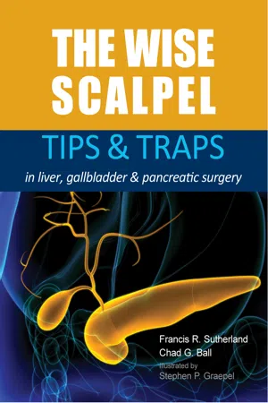
The Wise Scalpel
eBook - ePub

The Wise Scalpel

Tips & Traps in liver, gallbladder & pancreatic surgery

Sutherland, Francis, Ball, Chad G.

  1. English
  2. ePUB (mobile friendly)
  3. Available on iOS & Android
eBook - ePub

The Wise Scalpel

Tips & Traps in liver, gallbladder & pancreatic surgery

Sutherland, Francis, Ball, Chad G.

Book details
Book preview
Table of contents
Citations

About This Book

Surgery remains a challenge for young learners. Perhaps the most difficult of all surgeries are those done on the liver, biliary tree and pancreas. These organs are difficult to expose, difficult to operate and the patients are often difficult to recover. There are significant consequences for any missteps on what is a narrow pathway to a successful outcome.The skills needed to master surgery on these organs are hard to acquire. The volume of information presented to students is enormous and complex to parse. HPB textbooks contain a large amount of information from various authors but are often poorly coordinated. Surgical manuals contain significant anatomic details and descriptions but have difficulty transferring key concepts. Experienced surgeons are often oblivious to their skills and communicate poorly to their students regarding the things they do or understand that make them wise. This book tries to capture this wisdom.The topics in this book are most applicable to hepatobiliary and pancreatic surgeons but really extend to all general surgeons. Anyone who operates in the abdomen must have a basic understanding of these dominant organs, general surgery trainees and staff are an additional focus of this book. The skills needed for a modern surgeon to succeed are much broader than the scalpel tip. Section I, "HPB Surgical Craft", imparts important lessons on the laws of human interactions and cognitive aspects of surgery. Sections on the anatomy and operations on the liver, pancreas and bile ducts are presented. The expanded gallbladder section and pancreatitis chapters are of particular importance to general surgery residents. This book has been written in a readable and entertaining style, with many fun historical and contemporary quotes dotted throughout.The Wise Scalpel is an attempt to impart knowledge and understanding to students and practicing surgeons at all levels. The book focuses on tips, traps and underlying truths about the diseases of these organs and their surgical treatment. While this book will not supplant the struggle needed to move from novice to expert surgeon, hopefully it will quicken the process and help avoid some of the dangers and frustrations along the way.

Frequently asked questions

How do I cancel my subscription?
Simply head over to the account section in settings and click on ā€œCancel Subscriptionā€ - itā€™s as simple as that. After you cancel, your membership will stay active for the remainder of the time youā€™ve paid for. Learn more here.
Can/how do I download books?
At the moment all of our mobile-responsive ePub books are available to download via the app. Most of our PDFs are also available to download and we're working on making the final remaining ones downloadable now. Learn more here.
What is the difference between the pricing plans?
Both plans give you full access to the library and all of Perlegoā€™s features. The only differences are the price and subscription period: With the annual plan youā€™ll save around 30% compared to 12 months on the monthly plan.
What is Perlego?
We are an online textbook subscription service, where you can get access to an entire online library for less than the price of a single book per month. With over 1 million books across 1000+ topics, weā€™ve got you covered! Learn more here.
Do you support text-to-speech?
Look out for the read-aloud symbol on your next book to see if you can listen to it. The read-aloud tool reads text aloud for you, highlighting the text as it is being read. You can pause it, speed it up and slow it down. Learn more here.
Is The Wise Scalpel an online PDF/ePUB?
Yes, you can access The Wise Scalpel by Sutherland, Francis, Ball, Chad G. in PDF and/or ePUB format, as well as other popular books in Medizin & Chirurgie & chirurgische Medizin. We have over one million books available in our catalogue for you to explore.

Information

Year
2021
ISBN
9781913755133
image
Section II
Liver
67
image
Chapter 13
Liver anatomy
Simple can be harder than complex: you have to work hard to
get your thinking clean to make it simple. But it's worth it in the
end because once you get there, you can move mountains.
Steve Jobs
Liver cover and attachments
The liver parenchyma is encased in Glissonā€™s capsule and in most areas, peritoneum. The capsule and peritoneum are not the same thing. The capsule is a very thin fibrous layer attached directly to the liver parenchyma. It can bleed quite vigorously when stripped. The capsule extends entirely around the liver including the bare area and gallbladder fossa where no peritoneum exists. Glissonā€™s capsule fuses with hepatic vein walls on one end and the plate system under the liver on the other.
The liverā€™s outer covering consists of peritoneum which folds down from the diaphragm and abdominal/chest wall to encapsulate the liver; this effectively suspends the liver in the subdiaphragmatic space. The most immediately obvious attachment is the falciform ligament that connects the left liver to the anterior abdominal wall. It is basically a fold of peritoneum that hold the ligamentum teres on its way to become the left portal vein in the umbilical fossa. There is a variable amount of fat in this ligament. The left
69
image
THE WISE SCALPEL: TIPS & TRAPS in liver, gallbladder & pancreatic surgery
triangular/coronary ligament is also a partly fused fold of peritoneum that holds segment 2 next to the diaphragm. It is a wonder that we continue to label these peritoneal folds as ligaments, which they are not (including the coronary ligament, triangular ligament, splenorenal ligament, etc.). On the right side there is a similar fold but it is incomplete and does not cover the ā€˜bare areaā€™ of the liver posteriorly. The first milestone of any trainee is to take down these attachments without doing damage. The two most common ā€˜damagesā€™ done are making a hole in the left hepatic vein while taking down the left triangular ligament or making a tear in the liver parenchyma while dissecting the right bare area. The diaphragm (with the phrenic veins) from which the liver is suspended, is also easily damaged by the ā€˜learning handā€™.
70
image
Chapter 13: Liver anatomy
External liver orientation
Knowledge of the general layout of the liver is an important foundation on which to build anatomic understanding. The right liver is the largest and has an anterior to posterior orientation as it extends into the right subdiaphragmatic space. The left liver, with the stomach and spleen filling the left upper quadrant, tends to have a more right to left or horizonal orientation. It is thinner and more superficial and hence much more operable.
There are a few external clues to internal liver anatomy. Fissures are the major anatomic dividing planes between the different areas of the liver. The division of the portal vein is the most consistent compared to hepatic artery and bile ducts and is therefore used to label the major fissures. The gallbladder and space between the middle and right hepatic vein mark the main portal fissure. The origin of the right hepatic vein marks the right portal fissure. The falciform ligament and umbilical fissure demarcate the plane between segments 4 and 2/3. The posterior crease between segment 2/3 and the caudate lobe can be clearly seen with the ligamentum venosum deep in this groove. This is also where the lesser omentum originates.
Operating on the right liver requires that it be mobilised from its deep posterior location into the same horizontal plane as the left. The attachments to the diaphragm must be divided to allow the liver to rotate on the vena cava and porta hepatis. This rotation is only possible if the diaphragm attachments of the left liver (triangular ligaments) are first taken down. Mobilization changes the position of the right liver segments and hepatic veins relative to the preoperative imaging. With more extreme mobilization to visualize the vena cava, the posterior segments 6 and 7 become anterior. A lesion on the inferolateral side of the inferior vena cava (segment 7) is now above the vena cava!
The use of intraoperative ultrasound is important in reorienting after mobilization. Mobilization may also allow the surgeon to identify and orient the tumor. Finger tips are extremely effective tumor probes. With manual or
71
image
THE WISE SCALPEL: TIPS & TRAPS in liver, gallbladder & pancreatic surgery
bimanual palpation of a mobilized liver, surgeons can usually feel the tumor with their fingers.
Wisdom
First get oriented and then get operating.
72
image
Chapter 13: Liver anatomy
Internal liver orientation (obliquity)
A three-dimensional understanding of the internal liver is not possible without a word on ā€˜obliquityā€™. The fissural planes of the liver, in situ, are not vertical. They are actually oblique in several directions. The main portal fissure, containing the middle hepatic vein, is at an angle from left to right. In situ, it is also tipped to the patientā€™s right shoulder, off the coronal plane. The right portal fissure, containing the right hepatic vein, is almost horizonal in situ. The right and main portal fissures angle in towards the underlying caudate. This is intuitive when one realizes that none of the segments are square. In the right anterior sector, they are pie-shaped. They have a large surface area on the outside of the liver that narrows down as they close in on the vena cava. The shape of the other segments are all unique and unclassifiable in three dimensions. Even the hilum is oblique, tipped towards the right posterior subdiaphragmatic space.
When you divide the inflow tracts on either side of the liver, the demarcation line between segments and sectors is irregular. Rather than a clean plane, the fissural planes are an inexact wavy separation. The main portal fissure does not always end in the centre of the hilum (portal bifurcation). In about half of cases, it is either to the right or to the left. Surgeons should remember that the demarcation line (Cantlieā€™s line) is just that, a line on the liver surface. It is a good start, but once one moves into the parenchyma plane, demarcation is indistinct and difficult to see. This is usually not a real problem as a moderate amount of devascularized liver tissue at the resection margins is well tolerated. Even larger areas of dead tissue do not seem to create a consistent issue; however, we should still strive to preserve only the vascularized tissue.
Wisdom
The liver is separated by oblique irregular planes into odd- shaped segments.
73
image
THE WISE SCALPEL: TIPS & ...

Table of contents

Citation styles for The Wise Scalpel

APA 6 Citation

Sutherland, Francis, Ball, & Chad, G. (2021). The Wise Scalpel ([edition unavailable]). tfm Publishing Ltd. Retrieved from https://www.perlego.com/book/3277226/the-wise-scalpel-tips-traps-in-liver-gallbladder-pancreatic-surgery-pdf (Original work published 2021)

Chicago Citation

Sutherland, Francis, Ball, and G Chad. (2021) 2021. The Wise Scalpel. [Edition unavailable]. tfm Publishing Ltd. https://www.perlego.com/book/3277226/the-wise-scalpel-tips-traps-in-liver-gallbladder-pancreatic-surgery-pdf.

Harvard Citation

Sutherland et al. (2021) The Wise Scalpel. [edition unavailable]. tfm Publishing Ltd. Available at: https://www.perlego.com/book/3277226/the-wise-scalpel-tips-traps-in-liver-gallbladder-pancreatic-surgery-pdf (Accessed: 15 October 2022).

MLA 7 Citation

Sutherland et al. The Wise Scalpel. [edition unavailable]. tfm Publishing Ltd, 2021. Web. 15 Oct. 2022.