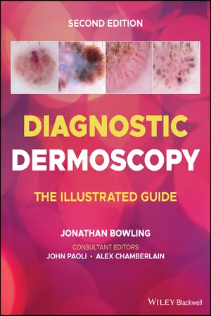
Diagnostic Dermoscopy
eBook - ePub

Diagnostic Dermoscopy

The Illustrated Guide

  1. English
  2. ePUB (mobile friendly)
  3. Available on iOS & Android
eBook - ePub

Diagnostic Dermoscopy

The Illustrated Guide

About this book

DIAGNOSTIC DERMOSCOPY

DIAGNOSTIC DERMOSCOPY

Diagnostic Dermoscopy delivers a focused, practical and user-friendly guide to diagnosing the most frequently seen presentations of skin lesions practitioners encounter in clinic. The new edition offers a comprehensive collection of strategies to find and diagnose skin cancers at an earlier point in their evolution, and to increase confidence in diagnosing benign lesions to reduce both patient morbidity and mortality.

The many full-colour illustrations included within are standardised to aid in recognition, while the adjoining dermoscopy images focus on critical diagnostic features which, when combined with the provided clinical information, are sufficient for specific diagnoses or narrow differential diagnoses. The new edition is formatted to aid teledermoscopy education, including many illustrated cases for reference. Diagnostic Dermoscopy offers:

  • A comprehensive overview of melanocytic lesions, melanoma presentations, and non-melanocytic lesions
  • Thorough explorations of basal cell carcinoma and keratinocyte dysplasia
  • Practical discussions of several special sites, including acral, nail, facial, and scalp lesions, as well as trichoscopy
  • In-depth examinations of vascular lesions, inflammoscopy for general dermatology, and entomodermoscopy
  • Detailed illustrations of genetic and iatrogenic lesions
  • Tips from international experts in melanoma diagnosis

Diagnostic Dermoscopy is the ideal resource for dermatologists, primary care physicians, plastic surgeons and allied health practitioners involved in skin lesion and melanoma diagnosis.

Dermoscopy UK

Further information on dermoscopy education, and online courses can be found at www.dermoscopy.co.uk

Trusted by 375,005 students

Access to over 1.5 million titles for a fair monthly price.

Study more efficiently using our study tools.

Information

Year
2022
Print ISBN
9781118930489
eBook ISBN
9781118931622
Edition
2
Subtopic
Dermatology

1
Introduction

  1. Introduction
  2. Dermoscopy and diagnosis
  3. Dermoscopy principle I – illumination
  4. Dermoscopy principle II – magnification
  5. Dermoscopy colours I – eumelanin
  6. Dermoscopy colours II – non‐eumelanin
  7. Imaging modes I – colours
  8. Imaging modes II – structures
  9. Dermoscopy of normal skin
  10. Dermoscopy of photodamaged skin
  11. Vascular morphologies
  12. Characteristic vascular patterns
  13. Polarised dermoscopy – shiny white structures

Introduction

Dermoscopy has been fully embraced by dermatologists and all those involved in skin cancer diagnosis as the gold standard tool used for clinical examination and diagnosis. It is simple to utilise, quick to apply and requires very little additional resource apart from the dermoscopy device/dermoscope itself.
However, without understanding the utility of dermoscopy, in wider clinical practice, the full diagnostic potential for the clinician may not be reached.

Format and imaging

This book is specifically a textbook with an emphasis on diagnosis, and diagnosis alone. In this way we are able to provide a comprehensive guide to the common presentations of skin lesions seen in clinics in addition to some of the more uncommon. From this starting point of diagnosis the rest of medical management flows. With greater confidence in diagnosis, a reduction in unnecessary biopsies can be achieved which frees resources to meet the increasing demand from rising skin cancer rates. Additionally, finding skin cancers at an earlier point in their evolution consumes less resources in surgical and medical management, and reduces patient morbidity and mortality.
For each topic covered, the clinical images are aimed to be standardised for illumination and orientation to aid recognition whilst illustrating the main clinical features. The adjoining dermoscopy image focuses upon key diagnostic features, which in combination with the clinical information should be all that is required for a specific diagnosis or at least a narrow differential diagnosis to be made.
Whenever possible, images of conditions have been taken across skin tones and with different dermoscope devices in different imaging modes to Illustrate the broad variety of presentations to the clinician.
Therefore, we anticipate that this book becomes a user‐friendly guide, to be used in clinic by not only clinicians and nurses but also by any allied paramedical staff involved in skin lesion diagnosis.

Teledermoscopy

This format also lends itself to the growing field of teledermoscopy.
With teledermoscopy becoming an integral part of clinical practice it is helpful to have a collection of illustrated cases for reference. Therefore, throughout this book, for each topic covered, a minimal dataset, is included. This dataset includes a brief clinical description of the lesion, anatomical site and age and sex of the patient, a clear clinical image and a corresponding dermoscopic image highlighting a diagnostic feature. This textbook therefore becomes a useful reference manual, of over 500 cases, for anyone involved in teledermoscopy consultations.

Dermoscopy devices

Over the last 10 years, the greatest change in dermoscopes has been the standardisation and incorporation of both polarising and non‐polarising imaging modes into the majority of dermoscopes. This leads to an increase in dermoscopic diagnostic information available for interpretation, with a simple toggle/push of a button to change between the two imaging modes. By switching between imaging modes, a composite ‘occipital cortex’ image can be created for interpretation and diagnosis.
There are multiple devices available for the clinician to choose. This book does not aim to advise on which device is best suited for the clinician. All we would advise is that the device sought is of a quality that allows the user to see a clear, bright, image for interpretation. The device must be robust enough for regular daily use and ergonomically designed so that it feels good in the clinician’s hand. High‐quality optics will ensure a crisp focused image for interpretation or image capture with a camera. The field ...

Table of contents

  1. Cover
  2. Table of Contents
  3. Dedication
  4. Title Page
  5. Copyright
  6. Preface
  7. 1 Introduction
  8. 2 Melanocytic naevi
  9. 3.1 Melanoma – clinical variants
  10. 3.2 Melanoma – pigment variants
  11. 3.3 Melanoma – dermoscopic features
  12. 3.4 Melanoma – high‐risk scenarios
  13. 4 Non‐melanocytic lesions
  14. 5 Basal cell carcinoma
  15. 6 Keratinocyte dysplasia
  16. 7.1 Acral melanocytic lesions
  17. 7.2 Onychoscopy
  18. 7.3 Facial lesions
  19. 7.4 Scalp lesions
  20. 7.5 Trichoscopy
  21. 8 Vascular lesions
  22. 9 Inflammoscopy
  23. 10 Genodermatoses
  24. 11 Entomodermoscopy
  25. 12 Miscellaneous
  26. Index
  27. End User License Agreement

Frequently asked questions

Yes, you can cancel anytime from the Subscription tab in your account settings on the Perlego website. Your subscription will stay active until the end of your current billing period. Learn how to cancel your subscription
No, books cannot be downloaded as external files, such as PDFs, for use outside of Perlego. However, you can download books within the Perlego app for offline reading on mobile or tablet. Learn how to download books offline
Perlego offers two plans: Essential and Complete
  • Essential is ideal for learners and professionals who enjoy exploring a wide range of subjects. Access the Essential Library with 800,000+ trusted titles and best-sellers across business, personal growth, and the humanities. Includes unlimited reading time and Standard Read Aloud voice.
  • Complete: Perfect for advanced learners and researchers needing full, unrestricted access. Unlock 1.5M+ books across hundreds of subjects, including academic and specialized titles. The Complete Plan also includes advanced features like Premium Read Aloud and Research Assistant.
Both plans are available with monthly, semester, or annual billing cycles.
We are an online textbook subscription service, where you can get access to an entire online library for less than the price of a single book per month. With over 1.5 million books across 990+ topics, we’ve got you covered! Learn about our mission
Look out for the read-aloud symbol on your next book to see if you can listen to it. The read-aloud tool reads text aloud for you, highlighting the text as it is being read. You can pause it, speed it up and slow it down. Learn more about Read Aloud
Yes! You can use the Perlego app on both iOS and Android devices to read anytime, anywhere — even offline. Perfect for commutes or when you’re on the go.
Please note we cannot support devices running on iOS 13 and Android 7 or earlier. Learn more about using the app
Yes, you can access Diagnostic Dermoscopy by Jonathan Bowling in PDF and/or ePUB format, as well as other popular books in Medicine & Dermatology. We have over 1.5 million books available in our catalogue for you to explore.