- 132 pages
- English
- ePUB (mobile friendly)
- Available on iOS & Android
eBook - ePub
About this book
Indocyanine green (ICG) fluorescence has been used for imaging purposes for more than half a century; First employed by ophthalmologists for visualizing the retinal artery in the late 1960s, the application of ICG fluorescence imaging has since been continuously expanded. Recently, advances in imaging technologies have led to renewed attention regarding the use of ICG in the field of hepatobiliary surgery, as a new tool for visualizing the biliary tree and liver tumors. This book introduces cutting-edge knowledge about fluorescence imaging techniques using both ICG and other new promising chemicals. After an introductory chapter on the history and basic technique of fluorescence imaging for hepatobiliary-pancreatic surgery, various clinical applications of ICG fluorescence imaging are discussed. These range from the identification of various malignancies to the use of imaging in surgery. The last part of this publication is dedicated to an outlook on near-future technology.
Tools to learn more effectively

Saving Books

Keyword Search

Annotating Text

Listen to it instead
Information
Clinical Applications of Indocyanine Green Fluorescence Imaging
Kokudo N, Ishizawa T (eds): Fluorescent Imaging: Treatment of Hepatobiliary and Pancreatic Diseases.
Front Gastrointest Res. Basel, Karger, 2013, vol 31, pp 10-17 (DOI: 10.1159/000348601)
Front Gastrointest Res. Basel, Karger, 2013, vol 31, pp 10-17 (DOI: 10.1159/000348601)
______________________
Identification of Hepatocellular Carcinoma
Takeaki Ishizawa · Norihiro Kokudo
Hepato-Biliary-Pancreatic Surgery Division, Department of Surgery, Graduate School of Medicine, The University of Tokyo, Tokyo, Japan
______________________
Abstract
Fluorescence imaging using indocyanine green (ICG) enables highly sensitive identification of hepatocellular carcinoma (HCC) by allowing visualization of impaired biliary excretion of ICG in differentiated HCC tissues and/or in noncancerous liver parenchyma around the tumor. In this technique, ICG is administered intravenously at the dose of 0.5 mg/kg for routine liver function testing within 2 weeks prior to surgery. Intraoperatively, liver cancer can be easily identified by fluorescence imaging of the liver surface prior to resection and on the resected specimen. Intraoperative ICG fluorescence imaging is useful for detecting superficially located small HCCs and confirming that these lesions have been removed with sufficient surgical margins. The present technique also enables identification of new lesions of HCC that have not been diagnosed preoperatively; however, additional resection should be considered only after re-evaluation by visual inspection and palpation or intraoperative ultrasonography because the positive predictive values of such newly detected lesions are 50% or lower, especially when the ICG is administered on the day before the surgery in patients with liver cirrhosis.
Copyright © 2013 S. Karger AG, Basel
In 2007, we developed a fluorescence imaging technique for intraoperative cholangiography using intrabiliary injection of indocyanine green (ICG) [1]. While developing this technique, we noticed that cancerous tissues on the liver surface emitted their own fluorescence even before the intraoperative administration of ICG for cholangiography. Actually, in all the patients at our department, ICG is administered intravenously before surgery in order to measure the ICG retention rate at 15 min as a routine liver function test. Thus, it was assumed that the intraoperative visualization of liver cancer by ICG fluorescence imaging was caused by accumulation of the ICG injected intravenously prior to the surgery in cancerous tissues and/or surrounding noncancerous liver tissues at the time of surgery. Then, a prospective clinical study was initiated to evaluate the efficacy of fluorescence imaging utilizing preoperatively injected ICG to detect liver cancer during surgery [2]. Here, we focus on the mechanistic background and clinical applications of intraoperative ICG fluorescence imaging of hepatocellular carcinoma (HCC). Other chapters in this volume detail the use of the ICG fluorescence imaging technique for the identification of metastatic liver cancer during surgery.
Principle of Indocyanine Green Fluorescence Imaging of Hepatocellular Cancer
Fluorescence imaging of liver cancer using preoperative intravenous administration of ICG is based on the fact that ICG is exclusively excreted into the bile and emits fluorescence that peaks at about 840 nm when protein-bound ICG is exposed to an excitation light in the range of 750-810 nm [3]. Because visualization at this wavelength is scarcely affected due to absorption by hemoglobin or water, biological structures that contain ICG can be visualized through tissue thicknesses of 5-10 mm with the use of an appropriate filter and a camera that is sensitive in the infrared region. On the other hand, a certain pathological type of HCC, termed ‘green hepatoma’, is known to retain the ability to produce bile. Furthermore, previous studies of delayed magnetic resonance imaging obtained 10-24 h after the administration of a contrast material excreted via bile suggested the presence of impaired bile excretion in HCC tissues as well as in noncancerous liver parenchyma surrounding the tumor, resulting in hyperenhancement of well-differentiated HCCs and rim enhancement of metastatic liver cancer [4-7]. Based on the above findings, we developed an intraoperative ICG fluorescence imaging technique aimed at visualizing liver cancer on the liver surface during surgery or on resected specimens based on the impaired biliary excretion in HCC tissues and in the noncancerous liver parenchyma around the tumor [2].
In our previous series consisting of 37 patients with HCC and 12 patients with colorectal liver metastasis, fluorescence imaging following preoperative intravenous administration of ICG at the dose of 0.5 mg/kg identified all of the microscopically confirmed HCCs (n = 63) and colorectal liver metastases (n = 28) on the cut surfaces of the resected specimens. The fluorescence patterns of these tumors were classifiable into the total fluorescence type (all of the cancer tissues showed uniform fluorescence), partial fluorescence type (a part of the cancer tissues showed fluorescence) and rim fluorescence type (the cancer tissues were negative for fluorescence, but the surrounding liver parenchyma showed fluorescence; fig. 1) [2, 8]. The fluorescence patterns were closely associated with the characteristics of the liver cancers: the total fluores-cence-type tumors included all of the well-differentiated HCCs, while the rim fluorescence-type tumors consisted of only poorly differentiated HCCs and colorectal liver metastases. Furthermore, fluorescence microscopy confirmed the presence of fluorescence in the cytoplasm and pseudoglands of the HCC cells and in the noncancerous liver parenchyma surrounding the poorly differentiated HCCs and metastases (fig. 2).
These results are consistent with the previously proposed mechanism of ICG accumulation in cancerous tissues and/or noncancerous liver parenchyma around the tumor. Such pharmacokinetics of ICG in the liver involving HCC may be proven by gene expression analysis and immunohistochemical staining, as used in the previous study conducted to reveal the background of magnetic resonance imaging of HCC with gadolinium ethoxybenzyl diethylenetriamine pentaacetic acid [9]. Our preliminary results suggested that the expression levels of portal uptake transporters of ICG (organic anion-transporting polypeptides and Na+/taurocholate cotransporting poly-peptide [10]) are well-preserved in HCCs showing fluorescence of ICG in the cancerous tissues as compared with the impaired gene expression levels in the cancerous tissues of rim fluorescence-type HCCs (unpubl. data).
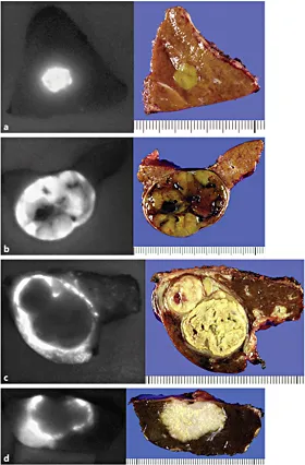
Fig. 1. Fluorescence patterns of liver cancers on cut surfaces of the liver (left) and their gross appearances (right) [8]. a Total fluorescence type (well-differentiated HCC, 7 mm in diameter). b Partial fluorescence type (moderately differentiated HCC, 35 mm in diameter). c Rim fluorescence type (poorly differentiated HCC, 30 mm in diameter). d Rim fluorescence type (metastasis of colorectal cancer, 25 mm in diameter).
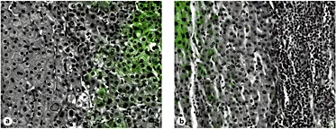
Fig. 2. Fluorescence microscopy. Fluorescence microscopy reveals that fluorescence of ICG (indicated in green) exists in cancerous tissues of well-differentiated HCC (a) and in noncancerous liver parenchyma around the tumor in poorly differentiated HCC (b).
Actually, the fluorescence imaging technique using ICG to identify liver cancer was included in a patent obtained by a group at the University of Rochester (WO 2008/043101 A2). Although there have been no detailed articles concerning liver cancer imaging, except for a recent article on ICG fluorescence imaging of renal cancer [11], their method is probably not based on the disordered biliary excretion of ICG, but on the difference in hemodynamics between cancerous tissues and noncancerous liver parenchyma that may occur in the earlier phase after intravenous administration of ICG.
Advantages and Limitations of the Use of Indocyanine Green
The major advantage of ICG fluorescence imaging is its sensitivity and feasibility: once the ICG retention test has been performed within 2 weeks prior to the surgery, surgeons can obtain fluorescence images of the liver cancer with a commercially available small imaging system at any time during the surgical procedures in order to detect cancerous tissues on the liver surface before resection or on the resected liver specimens. Although the ICG retention rate at 15 min has not been widely used as a preoperative liver function test in Western countries, this test is practically the only way to estimate the acceptable limit of liver volume to be removed in each patient [12]. Especially in liver resection for patients with background liver disease, it is strongly recommended that the ICG retention rate at 15 min be evaluated not only for intraoperative ICG fluorescence imaging of liver cancer, but also to ensure the safety of liver resection [13].
In contrast, it should be noted that when 0.5 mg/kg of ICG is administered intravenously for a liver function test on the day before surgery, washout from the noncancerous liver parenchyma is inadequate and there may be many false-positive nodules; the poorer the liver function, the more marked this tendency [2]. Further studies are needed to determine the optimal interval between ICG injection and surgery on the basis of the patient's liver function. Moreover, this technique does not use a cancer-specific antigen-antibody reaction. Instead, it just allows visualization of the impaired bile excretion in HCC tissues and/or noncancerous liver tissues around the tumor. Thus, benign lesions, such as regenerating nodules, bile duct proliferation and expanding liver cysts, may also exhibit fluorescence if there is delayed bile excretion. In fact, in previous reports, 40-50% of the lesions newly identified by fluorescence imaging during surgery were pathologically proven to be noncancerous lesions [2, 14, 15]. Even when new lesions are detected by ICG fluorescence imaging during surgery, additional resection should be considered only after the lesions have also been confirmed to be cancerous by inspection and palpation, and/or by an intraoperative ultrasonography.
Clinical Application of Indocyanine Green Fluorescence Imaging
Considering the advantages and limitations of ICG fluorescence imaging for liver cancer, its major expected roles in liver resection for HCC are to identify: (1) peripherally located, but invisible HCC diagnosed preoperatively, (2) new lesions to be considered for additional resection, (3) HCC tissues left on the raw surface of the liver after resection, and (4) small HCCs on the resected specimens.
In liver resection for HCC, especially during repeated resection for rec...
Table of contents
- Cover Page
- Front Matter
- History and Basic Technique of Fluorescence Imaging for Hepatobiliary-Pancreatic Surgery
- Clinical Applications of Indocyanine Green Fluorescence Imaging
- Near-Future Technology
- Author Index
- Subject Index
Frequently asked questions
Yes, you can cancel anytime from the Subscription tab in your account settings on the Perlego website. Your subscription will stay active until the end of your current billing period. Learn how to cancel your subscription
No, books cannot be downloaded as external files, such as PDFs, for use outside of Perlego. However, you can download books within the Perlego app for offline reading on mobile or tablet. Learn how to download books offline
Perlego offers two plans: Essential and Complete
- Essential is ideal for learners and professionals who enjoy exploring a wide range of subjects. Access the Essential Library with 800,000+ trusted titles and best-sellers across business, personal growth, and the humanities. Includes unlimited reading time and Standard Read Aloud voice.
- Complete: Perfect for advanced learners and researchers needing full, unrestricted access. Unlock 1.4M+ books across hundreds of subjects, including academic and specialized titles. The Complete Plan also includes advanced features like Premium Read Aloud and Research Assistant.
We are an online textbook subscription service, where you can get access to an entire online library for less than the price of a single book per month. With over 1 million books across 990+ topics, we’ve got you covered! Learn about our mission
Look out for the read-aloud symbol on your next book to see if you can listen to it. The read-aloud tool reads text aloud for you, highlighting the text as it is being read. You can pause it, speed it up and slow it down. Learn more about Read Aloud
Yes! You can use the Perlego app on both iOS and Android devices to read anytime, anywhere — even offline. Perfect for commutes or when you’re on the go.
Please note we cannot support devices running on iOS 13 and Android 7 or earlier. Learn more about using the app
Please note we cannot support devices running on iOS 13 and Android 7 or earlier. Learn more about using the app
Yes, you can access Fluorescent Imaging by N. Kokudo,T. Ishizawa,N., Kokudo,T., Ishizawa, G. Rogler,G., Rogler in PDF and/or ePUB format, as well as other popular books in Medicine & Oncology. We have over one million books available in our catalogue for you to explore.