eBook - ePub
Locking Plates in Veterinary Orthopedics
- English
- ePUB (mobile friendly)
- Available on iOS & Android
eBook - ePub
Locking Plates in Veterinary Orthopedics
About this book
Locking Plates in Veterinary Orthopedics is a comprehensive and state-of-the-art guide to all aspects of using locking plates to treat orthopedic conditions in dogs, cats, and large animals.
• Offers a proven approach to using locking plates in veterinary practice
• Highlights practical clinical applications with illustrative clinical cases
• Includes information on the history, principles, and materials as well as specific techniques
• Presents data on both traumatic and non-traumatic applications
• Provides instructive color photographs to demonstrate the procedures
Tools to learn more effectively

Saving Books

Keyword Search

Annotating Text

Listen to it instead
Information
1
A Brief History of Veterinary Locking Plates Applications
Karl C. Maritato
As with all of medicine, orthopedics is an ever‐evolving science. While locking implants are a relatively recent addition to veterinary orthopedics, they have been used for humans for some time. To better understand where we are, and where we are going, with fracture repair and locking implants, we first need to look back on the history of fracture fixation − a fascinating journey through the brilliant minds of our predecessors.
In the mid‐1700s, John Hunter was the first surgeon to define the four stages of callus formation during fracture repair. Around the same time, Albrecht von Haller noted that bone healing was dependent on the vascularity around the fractured region of the bone, emphasizing the role of blood supply in fracture healing. Henri Duhamel disagreed, thinking that all bone arose from the periosteum, and coined the term cambium layer[1].
In 1736, John Belchier was the first to identify the important role of osteoblasts in fracture healing, and in the 1840s, John Goodsir confirmed that osteoblasts were the true bone‐forming cells [1]. This led some, including Sir William Macewen, to focus strongly on the osteoblast and ignore the role of the periosteum [1]. In the late 1800s, Louis Ollier, like Duhamel, felt more than osteoblasts were in play in fracture repair. He believed that in addition to osteoblasts, the periosteum and the bone marrow all contributed to bone repair; he recommended the periosteum be protected during surgery [1].
In 1886, Carl Hansmann invented the first bone plate and screws. Ironically, it was a locking plate that protruded through the skin [1]. By contrast, Halsted in 1893 and Lane in 1894, utilized the first completely implanted plates [1].
In 1912, William Sherman, who was a surgeon for the Pittsburgh Steel Company, designed plates with better metallurgy and engineering due to this connection. Because of his improved production knowledge, his plates did not corrode or break and were the most widely used plates until the Association for Osteosynthesis (AO) plates were introduced 50 years later [1].
Up until this point, the plates in use were not designed with compression in mind; rather, they served only to stabilize and align the bone as a replacement for external splinting, similar to current locking plates. In 1946, Eggers performed experiments on animals with induced fractures to show the effects of fracture site compression on the rate of healing and, in 1949, Robert Danis was the first to apply compression plating to human patients [1].
A decade later, George Bagby was the first to use a plate similar in design to the dynamic compression plates (DCP) used today. His plates had oval‐shaped holes with beveled edges that allowed the plate to slide into compression as the screw was tightened [1].
On November 6, 1958, a critical moment in orthopedics history occurred: Arbeitsgemeinschaft fur Osteosynthesenfragen (Association for Osteosynthesis) was formed by 13 surgeons in Switzerland [1, 2]. This group’s unprecedented collective focus on the study of biomechanics, osteogenesis, implants, and instruments, as well as orthopedic techniques and postoperative care, ushered in a new era. Additionally, they focused on orthopedic continuing education through instructional courses and labs and published the first AO manual in 1963 [2, 3] followed by Techniques of Internal Fixation of Fractures, published in 1965 [2]. The critical principles of primary focus in these early editions were that of anatomic reduction and rigid fixation [2]. It was thought that fracture healing with no callus formation was most desired, and that the presence of callus formation was considered a sign of instability and inappropriate repair. Willeneger and Schenk’s research on direct bone healing reinforced this theory [2, 3].
On 31 August 1969, AOVET was founded in Waldenburg, Switzerland (Figure 1.1a–d) [3]. In the decade prior to its formation, a beautiful collaboration between veterinary and human surgeons and engineers had blossomed. Transfer of knowledge between disciplines was initiated as never before in veterinary surgery, most notably by Dr. Guggenbuhl, one of the original 13 AO founders, and Dr. von Salis, a large animal veterinarian and first president of AOVET. This relationship and collaboration with von Salis’s colleagues led to the AOVET formation [3]. One of the first documented fracture cases in a dog repaired using AO principles and plates was a femur fracture in a Spitz, performed on 3 February 1969 by Dr. Geri Kasa with a four‐hole 4.5 mm round hole plate (Figure 1.2) [3].

Figure 1.1 (a–d) Four of the primary founders of AOVEET. Geri Kasa, Feri Kasa, Ortun Pohler, Bjorn von Salis.
(Source: Courtesy of AO Foundation.)
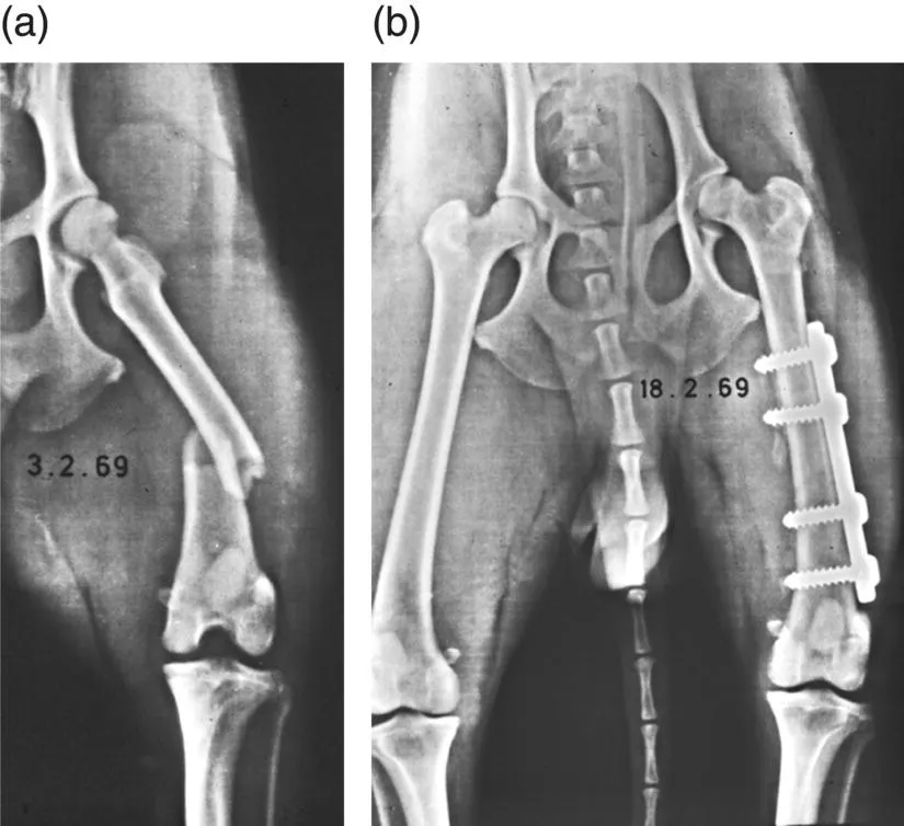
Figure 1.2 (a, b) One of the first documented fracture cases in a dog repaired using AO principles and plates; a femur fracture in a Spitz, performed on February 3, 1969, by Dr. Geri Kasa with a four‐hole 4.5 mm round hole plate.
(Source: Courtesy of AO Foundation.)
In 1970, Allgowar and Perren continued research on the Bagby design and developed a sophisticated plate called the dynamic compression unit. Compression could be achieved in any equally spaced hole on either side of the fracture. Consistent fracture healing and early return to function were noted in patients treated with these plates, and the previously common “fracture disease” problem was disappearing [2]. It was also noted that complications such as sepsis, sequestrum formation, union difficulties, and re‐fracture were occurring using these rigid techniques. Focus was redirected toward the effect of the plate on the bone surface and its blood supply [2].
At this same time, Dr. Hohn began the first, and soon to become annual, AOVET course held in the United States at The Ohio State University (Figure 1.3). Dr. Hohn had been the first to perform a canine fracture repair using AO principles in the United States at the Animal Medical Center in New York, as a part of a fantastic collaboration with Dr. Rosen, the first AO principled human orthopedic surgeon in the U...
Table of contents
- Cover
- Table of Contents
- Foreword
- Preface
- 1 A Brief History of Veterinary Locking Plates Applications
- Section I: Principles of Locking Plate Application
- Section II: Principles of Locking Plate Applications in Large Animals
- Section III: Current Veterinary Locking Plate Instrumentation and Implants
- Section IV: Trauma Applications: Clinical Case Examples
- Section V: Nontrauma Applications: Clinical Case Examples
- Index
- End User License Agreement
Frequently asked questions
Yes, you can cancel anytime from the Subscription tab in your account settings on the Perlego website. Your subscription will stay active until the end of your current billing period. Learn how to cancel your subscription
No, books cannot be downloaded as external files, such as PDFs, for use outside of Perlego. However, you can download books within the Perlego app for offline reading on mobile or tablet. Learn how to download books offline
Perlego offers two plans: Essential and Complete
- Essential is ideal for learners and professionals who enjoy exploring a wide range of subjects. Access the Essential Library with 800,000+ trusted titles and best-sellers across business, personal growth, and the humanities. Includes unlimited reading time and Standard Read Aloud voice.
- Complete: Perfect for advanced learners and researchers needing full, unrestricted access. Unlock 1.4M+ books across hundreds of subjects, including academic and specialized titles. The Complete Plan also includes advanced features like Premium Read Aloud and Research Assistant.
We are an online textbook subscription service, where you can get access to an entire online library for less than the price of a single book per month. With over 1 million books across 990+ topics, we’ve got you covered! Learn about our mission
Look out for the read-aloud symbol on your next book to see if you can listen to it. The read-aloud tool reads text aloud for you, highlighting the text as it is being read. You can pause it, speed it up and slow it down. Learn more about Read Aloud
Yes! You can use the Perlego app on both iOS and Android devices to read anytime, anywhere — even offline. Perfect for commutes or when you’re on the go.
Please note we cannot support devices running on iOS 13 and Android 7 or earlier. Learn more about using the app
Please note we cannot support devices running on iOS 13 and Android 7 or earlier. Learn more about using the app
Yes, you can access Locking Plates in Veterinary Orthopedics by Matthew D. Barnhart, Karl C. Maritato, Matthew D. Barnhart,Karl C. Maritato in PDF and/or ePUB format, as well as other popular books in Medicine & Veterinary Medicine. We have over one million books available in our catalogue for you to explore.