- Fully updated, with new chapters on the assessment of the post-Fontan procedure patient and on pregnancy and heart disease
- Each lesion chapter includes new section highlighting the key elements of the echocardiogram(s)
- Written by experts from the leading centers around the world, with numerous new authors
- Revision emphasizes new technologies and quality of images
- Comprehensive content contains overview of ultrasound physics, discussion of laboratory set-up, protocol for a standard pediatric echocardiogram and quantitative methods of echocardiographic evaluation, including assessment of diastolic function
- Also includes special techniques and topics including 3D echocardiography, intraoperative echocardiography, and fetal echocardiography
eBook - ePub
Echocardiography in Pediatric and Congenital Heart Disease
From Fetus to Adult
- English
- ePUB (mobile friendly)
- Available on iOS & Android
eBook - ePub
Echocardiography in Pediatric and Congenital Heart Disease
From Fetus to Adult
About this book
This comprehensive textbook on the echocardiographic assessment of pediatric and congenital heart disease has been updated for a second edition with an emphasis on new technologies. This highly-illustrated full-color reference contains over 1200 figures, and offers over 600 video clips on a companion website.
Trusted by 375,005 students
Access to over 1.5 million titles for a fair monthly price.
Study more efficiently using our study tools.
Information
PART I
Introduction to Cardiac Ultrasound Imaging
CHAPTER 1
Ultrasound Physics
Jan D'hooge1 and Luc L. Mertens2
1Department of Cardiovascular Sciences, Catholic University of Leuven; Medical Imaging Research Center, University Hospital Gasthuisberg, Leuven, Belgium
2Cardiology, The Hospital for Sick Children; Department of Pediatrics, University of Toronto, Toronto, ON, Canada
Physics and technology of echocardiography
Echocardiography is the discipline of medicine in which images of the heart are created by using ultrasound waves. Knowledge of the physics of ultrasound helps us to understand how the different ultrasound imaging modalities operate and also is important when operating an ultrasound machine to optimize the image acquisition.
This section describes the essential concepts of how ultrasound waves can be used to generate an image of the heart. Certain technological developments will also be discussed as well as how systems settings influence image characteristics. For more detailed treatment of ultrasound physics and imaging there is dedicated literature, to which readers should refer, for example [1, 2].
How the ultrasound image is created
The pulse–echo experiment
To illustrate how ultrasound imaging works, the acoustic “pulse–echo” experiment can be used:
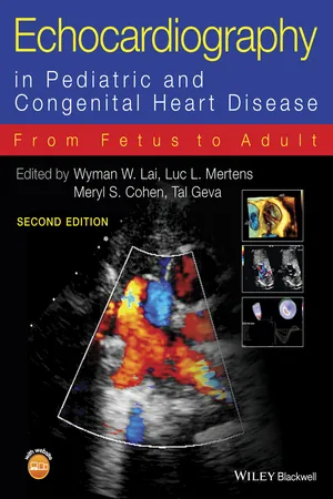
- A short electric pulse is applied to a piezoelectric crystal. This electric field will induce a shape change of the crystal through reorientation of its polar molecules. In other words, due to application of an electric field the crystal will momentarily deform.
- The deformation of the piezoelectric crystal induces a local compression of the tissue with which the crystal is in contact: that is, the superficial tissue layer is briefly compressed resulting in an increase in local pressure; this is the so-called acoustic pressure (Figure 1.1).
- Due to an interplay between tissue elasticity and inertia, this local tissue compression (with subsequent decompression, i.e., rarefaction) will propagate away from the piezoelectric crystal at a speed of approximately 1530 m/s in soft tissue (Figure 1.2). This is called the acoustic wave. The rate of compression/decompression determines the frequency of the wave and is typically 2.5–10 MHz (i.e., 2.5–10 million cycles per second) for diagnostic ultrasonic imaging. As these frequencies cannot be perceived by the human ear, these waves are said to be “ultrasonic.” The spatial distance between subsequent compressions is called the wavelength (λ) and relates to the frequency (f) and sound velocity (c) as: λf = c. During propagation, acoustic energy is lost mostly as a result of viscosity (i.e., friction) resulting in a reduction in amplitude of the wave with propagation distance. The shorter the wavelength (i.e., the higher the frequency), the faster the particle motion and the larger the viscous effects. Higher frequency waves will thus attenuate more and penetrate less deep into the tissue.
- Spatial changes in tissue density or tissue elasticity will result in a disturbance of the propagating compression (i.e., acoustic) wave and will cause part of the energy in the wave to be reflected. These so-called “specular reflections” occur, for example, at the interface between different types of tissue (e.g., blood and myocardium) and behave in a similar way to optic waves in that the direction of the reflected wave is determined by the angle between the reflecting surface and the incident wave (cf. reflection of optic waves on a water surface). When the spatial dimensions of the changes in density or compressibility become small relative to the wavelength (i.e., below ∼100 μm), these inhomogeneities will cause part of the energy in the wave to be scattered, that is, retransmitted in all possible directions. The part of the scattered energy that is retransmitted back into the direction of origin of the wave is called backscatter. Both the specular and backscattered reflections propagate back towards the piezoelectric crystal.
- When the reflected (compression) waves impinge upon the piezoelectric crystal, the crystal deforms. This results in relative motion of its (polar) molecules and generation of an electric field, which can be detected and measured. The amplitude of this electric signal is directly proportional to the amount of compression of the crystal, that is, the amplitude of the reflected/backscattered wave. This electric signal is called the radio-frequency (RF) signal and represents the amplitude of the reflected ultrasound wave as a function of time (Figure 1.3). Because reflections occurring further away from the transducer need to propagate further, they will be received later. As such, the time axis in Figure 1.3 can be replaced by the propagation distance of the wave (i.e., depth). The signal detected by the transducer is typically electronically amplified. The amount of amplification has a preset value but can be modified on an ultrasound system by using the “gain” button (typically the largest button on the operating panel). Importantly, the overall gain will amplify both the signal and potential measurement noise and will thus not affect the signal-to-noise ratio.
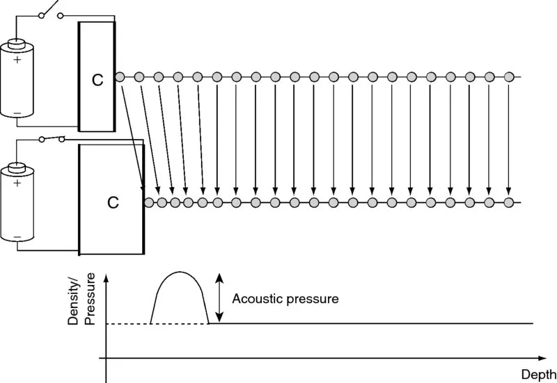
Figure 1.1 Local tissue compression due to deformation of the piezoelectric crystal when applying an electric field.
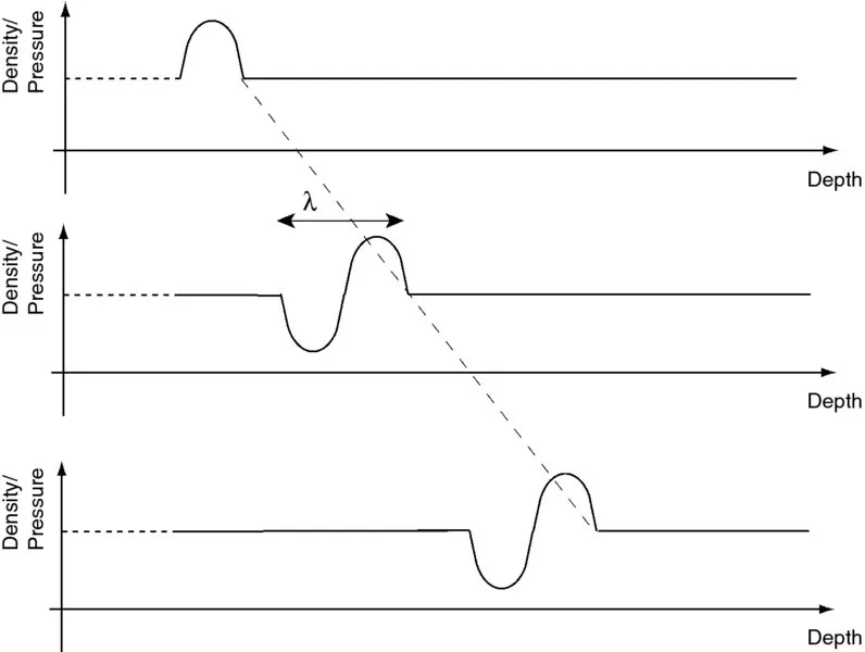
Figure 1.2 The local tissue compression/ decompression propagates away from its source at a speed of approximately 1530 m/s in soft tissue.
Figure 1.3 The reflected amplitude of the reflected ultrasound waves as a function of time after transmission of the ultrasound pulse is called the radio-frequency (RF) signal.
In the example shown in Figure 1.3, taken from a water tank experiment, two strong specular reflections can be observed (around 50 and 82 μs, respectively) while the lower amplitude reflections in between are scatter reflections. In clinical echocardiography, the most obvious specular reflection is the strong reflection coming from the pericardium observed in the parasternal views as a consequence of its increased stiffness with respect to the surrounding tissues. The direction of propagation of the specular reflection is determined by the angle between the incident wave and the reflecting surface. Thus, the strength of the observed reflection will depend strongly on the exac...
Table of contents
- Cover
- Dedication
- Titlepage
- Copyright
- Contributors
- Preface
- About the Companion Website
- Part I: Introduction to Cardiac Ultrasound Imaging
- Part II: Quantitative Methods
- Part III: Anomalies of the Systemic and Pulmonary Veins, Septa, and Atrioventricular Junction
- Part IV: Anomalies of the Ventriculo-arterial Junction and Great Arteries
- Part V: Miscellaneous Cardiovascular Lesions
- Part VI: Anomalies of Ventricular Myocardium
- Part VII: Acquired Pediatric Heart Disease
- Part VIII: Special Techniques and Topics
- Appendix: Normal Echocardiographic Values for Cardiovascular Structures
- Index
- EULA
Frequently asked questions
Yes, you can cancel anytime from the Subscription tab in your account settings on the Perlego website. Your subscription will stay active until the end of your current billing period. Learn how to cancel your subscription
No, books cannot be downloaded as external files, such as PDFs, for use outside of Perlego. However, you can download books within the Perlego app for offline reading on mobile or tablet. Learn how to download books offline
Perlego offers two plans: Essential and Complete
- Essential is ideal for learners and professionals who enjoy exploring a wide range of subjects. Access the Essential Library with 800,000+ trusted titles and best-sellers across business, personal growth, and the humanities. Includes unlimited reading time and Standard Read Aloud voice.
- Complete: Perfect for advanced learners and researchers needing full, unrestricted access. Unlock 1.5M+ books across hundreds of subjects, including academic and specialized titles. The Complete Plan also includes advanced features like Premium Read Aloud and Research Assistant.
We are an online textbook subscription service, where you can get access to an entire online library for less than the price of a single book per month. With over 1.5 million books across 990+ topics, we’ve got you covered! Learn about our mission
Look out for the read-aloud symbol on your next book to see if you can listen to it. The read-aloud tool reads text aloud for you, highlighting the text as it is being read. You can pause it, speed it up and slow it down. Learn more about Read Aloud
Yes! You can use the Perlego app on both iOS and Android devices to read anytime, anywhere — even offline. Perfect for commutes or when you’re on the go.
Please note we cannot support devices running on iOS 13 and Android 7 or earlier. Learn more about using the app
Please note we cannot support devices running on iOS 13 and Android 7 or earlier. Learn more about using the app
Yes, you can access Echocardiography in Pediatric and Congenital Heart Disease by Wyman W. Lai,Luc L. Mertens,Meryl S. Cohen,Tal Geva in PDF and/or ePUB format, as well as other popular books in Medicine & Cardiology. We have over 1.5 million books available in our catalogue for you to explore.