Comparative Cardiac Imaging
eBook - ePub

Comparative Cardiac Imaging

A Case-based Guide

  1. English
  2. ePUB (mobile friendly)
  3. Available on iOS & Android
eBook - ePub

About this book

Provides the information necessary to guide clinicians to more efficient and appropriate use of the cardiac imaging modalities at their disposal

Most books currently available on cardiac imaging focus on just modality. As the number of imaging options has increased, the choice of procedure has become more complicated. This comprehensive book will guide the practitioner in choosing the most appropriate test when confronted by various cardiac symptoms and diseases and to understand the benefits and limitations of each imaging modality. It demonstrates the advantages and disadvantages of various imaging modalities such as echocardiography, computer tomography, MRI, and nuclear cardiology in the evaluation of various disease states both commonly and infrequently seen in a standard practice.

Comparative Cardiac Imaging—A Case-based Guide utilizes actual case examples to demonstrate the state of the art in comparative cardiac imaging. It offers in-depth chapter coverage of Aortic Diseases; Aortic Valvular Diseases; Mitral Valvular Diseases; Prosthetic Valves; Coronary Artery Disease; Pulmonary Artery Diseases; Congenital Heart Disease; Cardiac Tumor; Infective Disease; Cardiomypathy; and Cardiac Trauma.

  • Uniquely focuses on and compares the many different modalities for cardiac imaging
  • Breaks the topic down by anatomy and pathophysiology in order to cover all aspects of non-invasive cardiac imaging
  • Covers newer and lesser known modalities like speckle tracking and velocity vector imaging
  • Offers coverage of more controversial topics, such as CT angiography

Comparative Cardiac Imaging—A Case-based Guide presents a level of data that is appropriate for the practicing cardiologist and cardiology trainee, as well as residents, internists, and other primary care clinicians.

Frequently asked questions

Yes, you can cancel anytime from the Subscription tab in your account settings on the Perlego website. Your subscription will stay active until the end of your current billing period. Learn how to cancel your subscription.
No, books cannot be downloaded as external files, such as PDFs, for use outside of Perlego. However, you can download books within the Perlego app for offline reading on mobile or tablet. Learn more here.
Perlego offers two plans: Essential and Complete
  • Essential is ideal for learners and professionals who enjoy exploring a wide range of subjects. Access the Essential Library with 800,000+ trusted titles and best-sellers across business, personal growth, and the humanities. Includes unlimited reading time and Standard Read Aloud voice.
  • Complete: Perfect for advanced learners and researchers needing full, unrestricted access. Unlock 1.4M+ books across hundreds of subjects, including academic and specialized titles. The Complete Plan also includes advanced features like Premium Read Aloud and Research Assistant.
Both plans are available with monthly, semester, or annual billing cycles.
We are an online textbook subscription service, where you can get access to an entire online library for less than the price of a single book per month. With over 1 million books across 1000+ topics, we’ve got you covered! Learn more here.
Look out for the read-aloud symbol on your next book to see if you can listen to it. The read-aloud tool reads text aloud for you, highlighting the text as it is being read. You can pause it, speed it up and slow it down. Learn more here.
Yes! You can use the Perlego app on both iOS or Android devices to read anytime, anywhere — even offline. Perfect for commutes or when you’re on the go.
Please note we cannot support devices running on iOS 13 and Android 7 or earlier. Learn more about using the app.
Yes, you can access Comparative Cardiac Imaging by Jing Ping Sun, Xing Sheng Yang, Bryan P. Yan, Jing Ping Sun,Xing Sheng Yang,Bryan P. Yan in PDF and/or ePUB format, as well as other popular books in Medicine & Cardiology. We have over one million books available in our catalogue for you to explore.

Information

Year
2018
Print ISBN
9780470656372
eBook ISBN
9781119453208
Edition
1
Subtopic
Cardiology

Part I
Congenital Heart Disease in the Adult

1
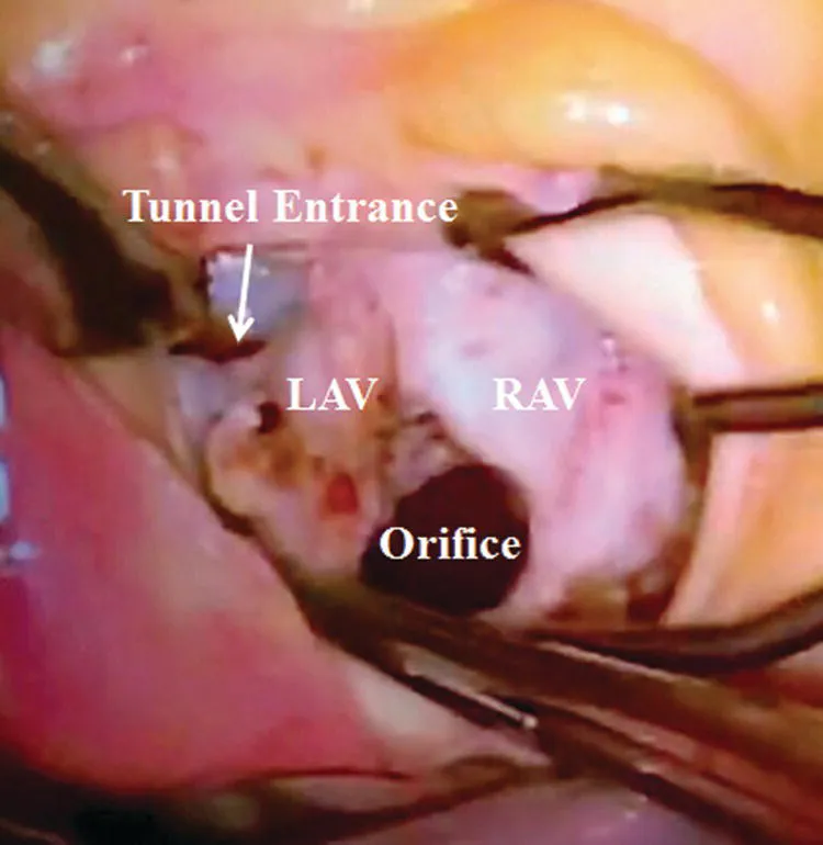
Aneurysmal Aorto–Left Ventricular Tunnel and Bicuspid Aortic Valve with Severe Stenosis

Fanxia Meng1, Bo Zhang1, Nan Xu1, and Jing Ping Sun2
1 Tongji University, Shanghai, China
2 The Chinese University of Hong Kong, Hong Kong

History

A 60‐year‐old male presented with increasing shortness of breath over 2 years.

Physical Examination

The heart rate was 80 bpm, with frequent premature beats. On auscultation, there were 3/6 systolic and diastolic murmurs at the left parasternal edge in the third intercostal space.

Transthoracic Echocardiogram

Image
A transthoracic echocardiogram showed that the aortic valves were severely calcified (Figure 1‐1*) with stenosis (peak velocity was 6 m/s) and mild regurgitation. An aorto–left‐ventricular tunnel (ALVT) (T) with entry from the aorta (thick arrow) and exit (thin arrow) into the left ventricle (LV) was seen (Figure 1). Blood flows from the aorta into the ALVT (T), and then into the LV cavity which can be seen by the color Doppler (Figure 1‐1B). Left ventricle hypertrophy with normal systolic function was noted (Video 1‐1). Nonvalvular regurgitation (thick arrow) and a shunt from aorta into ALVT (thin arrow) was seen by color Doppler (Figure 1‐1D, Video 1.2).
Image described by caption.
Figure 1‐1 Transthoracic echocardiogram showed: A. Aortic valves displayed severe calcification (Figure 1‐1.*). An aorto‐left ventricular tunnel (ALVT) (T) was seen with entry from the aorta (thin arrow) and exit (thick arrow) into the left ventricle (LV). B. The blood flow from the aorta into the ALVT (T) and out of the tunnel into the LV were seen with color Doppler. C. The outline of the tunnel (T) with the entrance from the aorta was clearly seen. D. A nonvalvular regurgitation (thick arrow) and a shunt from the aorta into the ALVT (thin arrow) were seen with color Doppler.
Image
A computer angiogram showed that the ascending aorta was dilated, with an aneurysmal ALVT (T) arising from the aortic root and connected with the LV (*) (Figure 1‐2A). The aortic annulus, aortic valves, and tunnel (T) were severely calcified (arrow), (Figure 1‐2B). The connection (*) between the aortic root and ALVT (T) could be seen (Figure 1‐2C). Three‐dimensional reconstruction computed tomography imaging showed aneurysmal ALVT (arrow), left coronary artery (LCA) arising from aortic root (arrow) behind the aneurysmal ALVT, and significantly dilated ascending AO (Figure 1‐2D).
Image described by caption and surrounding text.
Figure 1‐2 Computer angiogram showed: A. Computed tomography scan, long‐axis view, showed the dilated ascending aorta, an aneurysmal ALVT (T) arising from aortic root and connected with the LV (*). B. The short‐axis view showed the aortic annulus with calcification, and calcificatiive connection (arrow) between the LV and tunnel (T). C. The short‐axis view showed the connection (*) between the aortic root and ALVT (T). D. Computed tomography three‐dimensional reconstruction imaging showed aneurysmal ALVT (arrow), left coronary artery (LCA) arising from the aortic root (arrow) behind the aneurysmal ALVT, and significantly dilated ascending AO.

Management

The patient underwent open heart surgery. Surgical inspection revealed a 3 cm aneurysmal tunnel with smooth surface, it was (white arrow) arising from the left Valsalva sinus, located at below the LCA orifice (Figure 1‐3). The aortic valve was bicuspid and severelycalcified. The aortic and LV orifices of the ALVT were repaired, and the aortic valve was replaced. The patient recovered well after the operation. A follow‐up echocardiogram showed a well‐seated and functioning prosthetic aortic valve.
Image described by caption.
Figure 1‐3 Intraoperative picture showed the entrance of tunnel (white arrow) arising from the left Valsalva sinus, located at below and in front of the LCA orifice in the ascending aorta.

Discussion

An aorto–left‐ventricular tunnel (ALVT) is a congenital extracardiac channel connecting the ascending aorta to either left or right ventricular cavity. It is extremely rare with incidence as low as 0.001% of all congenital heart diseases [1]. Hovaguimian and colleagues [2] classified the ALVT into four types (I, II, III, and IV) that have a bearing on the appropriate surgical techniques of repair: A slitlike opening at the aortic end with no valve distortion in type I (prevalence, 24%), a large extracardiac aneurysm in type II (44%), an intracardiac aneurysm of the septal portion of the tunnel with or without rightventricular outflow tract obstruction in type III (24%), and a combination of types II and III in type IV (8%). In most cases, the aortic orifice of the tunnel arises from the right coronary sinus and is on the anterolateral side of the ascending aorta [2].The ALVT rarely arises from the left coronary sinus, and there are only a few reports of this presentation [3, 4].
In our patient the aortic orifice of the aneurysmal tunnel arises from the left coronary sinus below the orifice of the left coronary artery.The exit of the tunnel into the left ventricle appears narrow and has severe calcification. This patient had congenital ALVT associated with a bicuspid aortic valve with severe calcification and stenosis. To the best of our knowledge, this is very rare in the literature.

Associated Anomalies

Aortic valve abnormalities like dysplastic or bicuspid valve with stenosis are frequent [5], but aortic atresia has rarely been reported [6]. Stenosis of the pulmonary valve [7] and subvalvular pulmonary obstruction due to a tunnel have been reported [8]. Proximal coronary anomalies like coronary ostium lying within the tunnel or atresia of coronary ostium have been documented [9].

Clinical Presentation

The clinical presentation of ALVT varies depending upon the compression of the coronary arteries, the presence of right or left ventricular outflow obstruction, and the diameter of the tunnel [10–12]. Congestive heart failure freq...

Table of contents

  1. Cover
  2. Title Page
  3. Table of Contents
  4. Notes on Contributors
  5. Foreword
  6. Preface
  7. Abbreviations
  8. About the Companion Website
  9. Part I: Congenital Heart Disease in the Adult
  10. Part II: Artery Disease
  11. Part III: Cardiac Mass
  12. Part IV: Cardiomyopathy and Myocarditis
  13. Part V: Diversification
  14. Index
  15. End User License Agreement