Implant Site Development
eBook - ePub

Implant Site Development

Michael Sonick, Debby Hwang, Michael Sonick, Debby Hwang

Compartir libro
  1. English
  2. ePUB (apto para móviles)
  3. Disponible en iOS y Android
eBook - ePub

Implant Site Development

Michael Sonick, Debby Hwang, Michael Sonick, Debby Hwang

Detalles del libro
Vista previa del libro
Índice
Citas

Información del libro

With the desire for dental implant therapy ever escalating, clinicians are faced with the challenge of augmenting deficient natural physiology to provide effective sites for implantation. Implant Site Development helps the clinician decide if, when, and how to create a ridge site amenable to implantation. This practical book offers solutions to many implant site preservation scenarios, discussing different treatment options, timing, a variety of materials and techniques, and their application to the clinical practice. With a unique integrated clinical approach, Implant Site Development covers a range of site development techniques.

Highly illustrated, Implant Site Development presents diagrams and clinical photographs to aid with clinical judgment and will prove useful for any dental professional involved in implant therapy, from general practitioners to prosthodontists, but especially surgeons. This literature-based, yet user-friendly, reference will be indispensable to the novice or veteran clinician.

Preguntas frecuentes

¿Cómo cancelo mi suscripción?
Simplemente, dirígete a la sección ajustes de la cuenta y haz clic en «Cancelar suscripción». Así de sencillo. Después de cancelar tu suscripción, esta permanecerá activa el tiempo restante que hayas pagado. Obtén más información aquí.
¿Cómo descargo los libros?
Por el momento, todos nuestros libros ePub adaptables a dispositivos móviles se pueden descargar a través de la aplicación. La mayor parte de nuestros PDF también se puede descargar y ya estamos trabajando para que el resto también sea descargable. Obtén más información aquí.
¿En qué se diferencian los planes de precios?
Ambos planes te permiten acceder por completo a la biblioteca y a todas las funciones de Perlego. Las únicas diferencias son el precio y el período de suscripción: con el plan anual ahorrarás en torno a un 30 % en comparación con 12 meses de un plan mensual.
¿Qué es Perlego?
Somos un servicio de suscripción de libros de texto en línea que te permite acceder a toda una biblioteca en línea por menos de lo que cuesta un libro al mes. Con más de un millón de libros sobre más de 1000 categorías, ¡tenemos todo lo que necesitas! Obtén más información aquí.
¿Perlego ofrece la función de texto a voz?
Busca el símbolo de lectura en voz alta en tu próximo libro para ver si puedes escucharlo. La herramienta de lectura en voz alta lee el texto en voz alta por ti, resaltando el texto a medida que se lee. Puedes pausarla, acelerarla y ralentizarla. Obtén más información aquí.
¿Es Implant Site Development un PDF/ePUB en línea?
Sí, puedes acceder a Implant Site Development de Michael Sonick, Debby Hwang, Michael Sonick, Debby Hwang en formato PDF o ePUB, así como a otros libros populares de Médecine y Dentisterie. Tenemos más de un millón de libros disponibles en nuestro catálogo para que explores.

Información

Año
2011
ISBN
9780470963197
Edición
1
Categoría
Médecine
Categoría
Dentisterie
Chapter 1
Principles of bone biology and regeneration
Hector F. Rios DDS, PhD and William V. Giannobile DDS, DMedSc
Introduction
Bone is a dynamic tissue sensitive to a variety of factors with an inherent capacity that allows the translation of mechanical stimuli into biochemical signals, which therefore enhances its ability to adapt and sustain the physiological needs of the osseous structure. (Bonewald and Johnson, 2008; Burger et al., 1995; Duncan and Turner, 1995; Marotti, 2000; Marotti and Palumbo, 2007) This adaptive potential is the result of tightly regulated and synergistic anabolic and catabolic events that lead to proper metabolic and skeletal structural homeostasis (Turner and Pavalko, 1998). Multiple factors exert an effect in this system (e.g., biochemical, hormonal, cellular, biomechanical) that will collectively determine bone quality (Ammann and Rizzoli, 2003; Ma et al., 2008; Wallach et al., 1993). Clinically, bone quality is perceived as an important feature that dictates the mechanical properties of bone over time. Within the skeleton, such characteristics vary from one area to another and are determined by, among many things, cellular density and connectivity, bone density, bone macro- and microarchitecture, and the proportions of organic and inorganic matrix (Ammann and Rizzoli, 2003; Dalle Carbonare and Giannini, 2004; Ma et al., 2008; O’Brien et al., 2005; Viguet-Carrin et al., 2006). Therefore, the success of implant therapy is influenced by the understanding of the basic biological and physiological principles of bone, as it will aid the surgeon in selecting the appropriate techniques to enhance the peri-implant bone homeostasis.
Thus, the purpose of this chapter is to provide the clinician with foundational knowledge of bone development, composition, metabolism, and regeneration that serves as a primer for implant site development.
Bone Development
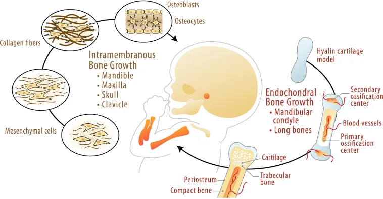
During embryogenesis, the skeleton forms by either a direct or indirect ossification process. In the case of the mandible and the maxilla, mesenchymal progenitor cells condensate and undergo direct differentiation into osteoblasts, a process known as intramembranous osteogenesis. In contrast, in the mandibular condyle, the long bones and vertebrae form initially through a cartilage template, which serves as an anlage that is gradually replaced by bone. The cartilage-dependent bone formation and growth process is known as endochondral osteogenesis (Ranly, 2000) (Fig. 1.1).
Fig. 1.1 During intramembranous osteogenesis, an ossification center develops through mesenchymal condensation. As the collagen-rich extracellular matrix develops and matures, osteoprogenitor cells undergo further osteoblastic differentiation. A subpopulation of osteoblasts becomes embedded in the mineralizing matrix and gives rise to the osteocyte lacunocanalicular network. Within the craniofacial complex, most bones develop and grow through this mechanism. Onthe other hand, long bones within the skeleton and the mandibular condyle are initially developed through the formation of a cartilaginous template that mineralizes and is later resorbed by osteoclasts and replaced by bone that is laid down afterward. The endochondral bone growth process leads to the formation of primary and secondary ossification centers that are separated by a cartilaginous structure known as the growth plate. As bone develops and matures through these two processes, structural distinct areas of compact bone and trabecular bone are formed and maintained through similar bone remodeling mechanisms.
c01f001
Alveolar bone lost as a result of an injury, disease, or trauma undergoes a repair process that is essentially a combination of endochondral and intramembranous complementary osteogenic processes (Rabie et al., 1996; Virolainen et al., 1995). A similar process occurs in most of the bone-related implant site development techniques, where osteoconduction, osteoinduction, and osteogenesis are exploited.
Bone Cells
Within bone, different cellular components can be identified. The distinct cell populations include osteogenic precursor cells, osteoblasts, osteoclasts, osteocytes, and hematopoietic elements of bone marrow. The content of this chapter will focus on the three main functional cells that are ultimately responsible for the proper skeletal homeostasis.
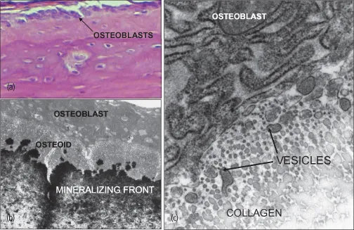
Osteoblasts are ultimately the cells responsible for bone formation. They synthesize the organic matrix components and mediate the mineralization of the matrix (Fig. 1.2). Osteoblasts are located on bone surfaces exhibiting active matrix deposition, and as their bone forming activity nears completion, some osteoblasts further differentiate into osteocytes, while others remain in the periosteal or endosteal surface of bone as lining cells. Bone lining cells are elongated cells that cover a surface of bone tissue and exhibit no synthetic activity (Rodan, 1992).
Fig. 1.2 Osteoblasts are derived from bone marrow osteoprogenitor cells and are responsible for the synthesis of the immature bone matrix known as osteoid. (A) The arrow depicts a group of osteoblasts that are lining the mature bone that contains embedded cells within the mineralized matrix. (B) Further detail of the osteoblasts lining the mature bone is clearly visualized through transmission electron microscopy (TEM). The abundant endoplasmic reticulum and Golgi apparatus within these cells reflects their high metabolic activity. Below the osteoblast layer, a collagen-rich unmineralized matrix is clearly depicted and comprises the osteoid. As the collagen mineralization occurs, a clear mineral front develops and a number of areas of crystal nucleation becomes visible. As the mineral propagates over the collagen fibers, a stable and mature bone matrix is accentuated. (C) This panel shows a higher magnification of the extracellular matrix adjacent to osteoblasts. A cross-sectional image of the collagen fibers is shown in close proximity to the cell. The arrows are pointing to a number of higher electron-dense vesicles that have detached from the osteoblast cytoplasm. These structures have been proposed to assist in the mineralization process by facilitating mineral crystal nucleation within as it occurs in cartilage.
c01f002
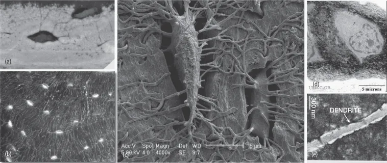
Osteocytes are stellate-shaped cells that are trapped within the mineralized bone matrix in spaces known as lacunae. They maintain a network of cytoplasmic processes known as dendrites. These osteocyte cytoplasmic projections extend through cylindrical encased compartments commonly referred to as canaliculi (Bonewald, 2007). They reach to different areas and contact blood vessels and other osteocytes. The osteocyte network is therefore an extracellular and intracellular communication channel that is sensitive at the membrane level to shear stress caused by the direction of fluid within the canaliculi space as the result of mechanical stimuli and bone deformation (Fig. 1.3). The mechanical signals are translated into biochemical mediators that will assist with the orchestration of anabolic and catabolic events within bone. This arrangement allows osteocytes to (i) participate in the regulation of blood calcium homeostasis and (ii) to sense mechanical loading and to transmit this information to other cells within the bone to further orchestrate osteoblast and osteoclast function (Burger et al., 1995; Marotti, 2000).
Fig. 1.3 The osteocyte can be defined as the orchestrator of the remodeling process within bone. (A) As bone matrix is synthesized, a number of osteoblasts become embedded within the osteoid, which later mineralize and reside in the mature matrix as osteocytes, as shown in this backscatter scanning electron microscopy (SEM) image treated with osmium to allow the visualization of the cell. (B) As shown in this confocal image, the embedded osteocytes interconnect, forming a network throughout the bone that enables these cells in the mechanosensoring capacity important in tuning up the remodeling needs. (C) The SEM image of a casted lacunocanalicular network allows the visualization of the degree of connectivity between two osteocytes and the regular diameter of the canalicular structures. (D) The high degree of mineralization of the matrix surrounding the osteocyte is clearly depicted by transmission electron microscopy. Although these cells appeared “dormant,” they are metabolically active and secrete a number of factors that allows them to modify their microenvironment. (E) A transmission electron image of a dendrite within a canaliculus allows the visualization of the space through which fluid flows and stimulates by shear stress the surface of the osteocyte cell membrane. This unique biological architectural characteristic of the osteocyte and the lacunocanalicular network represents the foundation that allows the conversion of mechanical stimuli into biochemical signals necessary for proper bone homeostasis.
c01f003
The bone formation activity is consistently coupled to bone resorption that is initiated and maintained by osteoclasts. Osteoclasts are specialized multinucleated cells that originate from the monocyte/macrophage hematopoietic lineage (Fig. 1.4). These cells have the capacity to develop and adhere to bone matrix to then secrete acid and lytic enzymes that degrade and break down the mineral and organic components of bone and calcified cartilage. The matrix degradation process results in the formation of a specialized extracellular compartment known as Howship’s lacuna (Rodan, 1992; Vaananen and Laitala-Leinonen, 2008; Vaananen et al., 2000).
Fig. 1.4 Osteoclasts are derived from cells of the macrophage/monocyte lineage and represent the bone resorbing units within the skeleton. (A) Histologically, osteoclasts can be depicted morphologically as multinucleated cells attached to bone matrix or through special staining such as the tartrate-resistant acid phosphatase (TRAP) stain highlighted in red and empha...

Índice