Clinical Pathology of the Placenta
eBook - ePub

Clinical Pathology of the Placenta

Martin Vogel, Gitta Turowski, Martin Vogel, Gitta Turowski

Compartir libro
  1. 470 páginas
  2. English
  3. ePUB (apto para móviles)
  4. Disponible en iOS y Android
eBook - ePub

Clinical Pathology of the Placenta

Martin Vogel, Gitta Turowski, Martin Vogel, Gitta Turowski

Detalles del libro
Vista previa del libro
Índice
Citas

Información del libro

This book performs a distinct introduction to the pathology of the placenta and its membranes, abortion material included, with the aim to facilitate and protect the quality of the morphological placental diagnostics by the pathologists. Seven chapters with coloured figures illustrating gross anatomy, development and maturation of the placenta explain the functional morphology in its clinical correlation of single and multiple findings for the pathologists, obstetricians and neonatologists. Moreover, the book contributes to a better understanding of pre- and perinatal investigations, maternal diseases, fetal outcomes and follow up of the newborns, as well as to the prevention of worse outcome in further pregnancies.
The atlas intends to stimulate the interest for perinatal pathology and to contribute to a better interdisciplinary understanding of pathologists and clinicians, midwives and nurses.

Preguntas frecuentes

¿Cómo cancelo mi suscripción?
Simplemente, dirígete a la sección ajustes de la cuenta y haz clic en «Cancelar suscripción». Así de sencillo. Después de cancelar tu suscripción, esta permanecerá activa el tiempo restante que hayas pagado. Obtén más información aquí.
¿Cómo descargo los libros?
Por el momento, todos nuestros libros ePub adaptables a dispositivos móviles se pueden descargar a través de la aplicación. La mayor parte de nuestros PDF también se puede descargar y ya estamos trabajando para que el resto también sea descargable. Obtén más información aquí.
¿En qué se diferencian los planes de precios?
Ambos planes te permiten acceder por completo a la biblioteca y a todas las funciones de Perlego. Las únicas diferencias son el precio y el período de suscripción: con el plan anual ahorrarás en torno a un 30 % en comparación con 12 meses de un plan mensual.
¿Qué es Perlego?
Somos un servicio de suscripción de libros de texto en línea que te permite acceder a toda una biblioteca en línea por menos de lo que cuesta un libro al mes. Con más de un millón de libros sobre más de 1000 categorías, ¡tenemos todo lo que necesitas! Obtén más información aquí.
¿Perlego ofrece la función de texto a voz?
Busca el símbolo de lectura en voz alta en tu próximo libro para ver si puedes escucharlo. La herramienta de lectura en voz alta lee el texto en voz alta por ti, resaltando el texto a medida que se lee. Puedes pausarla, acelerarla y ralentizarla. Obtén más información aquí.
¿Es Clinical Pathology of the Placenta un PDF/ePUB en línea?
Sí, puedes acceder a Clinical Pathology of the Placenta de Martin Vogel, Gitta Turowski, Martin Vogel, Gitta Turowski en formato PDF o ePUB, así como a otros libros populares de Medicine y Pathology. Tenemos más de un millón de libros disponibles en nuestro catálogo para que explores.

Información

Editorial
De Gruyter
Año
2019
ISBN
9783110451061
Edición
1
Categoría
Medicine
Categoría
Pathology

1 Normal anatomy and maturation

1.1 Anatomy and morphology

The placenta is a discoid organ formed when the chorion level, the tissue free membranes separate from the chorion frondosum in gestational week 13 to 14. It reaches a diameter between 17 and 19 cm and has a net weight of ca. 500 g prior to term. The placental net weight corresponds only to the weight of the placental disc, to which most reference values refer, whereas the gross weight includes the umbilical cord, membranes and often amniotic fluid and blood; all of which can increase the weight to 600 g or more. The maternal / basal plate area, analogous to a stylized circle, should normally reach 250 cm2 at term and can be calculated using the formula a½ × b½ × π. The placental thickness at term is between 20 and 25 mm.
The fetal / chorionic plate is the part of the placenta adjoining the amniotic sac, and the maternal / decidual basal plate lies adjacent to the uterus (Fig. 1.1). Both meet in the marginal zone. The placental tissue between the fetal / chorionic and maternal / basal plates form the grossly visible cotyledons, which are comprised of villi and the intervillous space (ca. 58 % villi and 42 % intervillous space)
Fig.1.1: Illustration. Structure of the human placenta: (1) Fetal / chorionic plate with umbilical cord vessels branching into the cotyledons, (2) cotyledons formed by villi and the intervening intervillous space, (3) maternal / basal plate with septa and interseptal spaces that enable maternal Arterial inflow (Ar) and maternal Venous outflow (Ve).

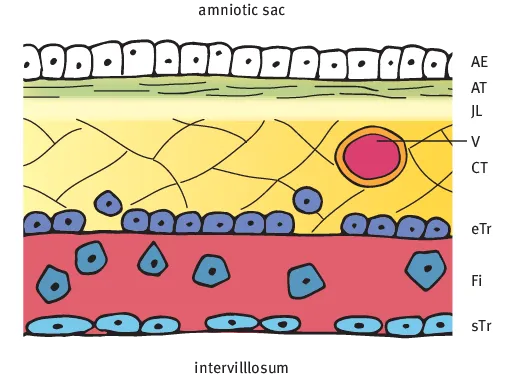
1.1.1 Chorionic plate

The chorionic plate consists of several layers that are easy to differentiate in early pregnancy.
  • The Amniotic Epithelium (AE) layer covers the chorionic plate and forms the border of the amniotic sac.
  • The Amniotic collagenous Tissue (AT) layer differs in density depending on gestational age and contains collagenous fibers.
  • The Junctional Layer (JL) is a cell and fiber poor layer between the amniotic and chorionic connective tissue layers.
  • The Chorionic connective Tissue (CT) layer is fiber rich and cell poor. Embedded here are the chorionic plate vessel branches (V) which arise from the umbilical cord vessels and supply the stem villi.
  • The subchorionic layer of the Langhans-Fibrinoid (Fi) consists mainly of matrix-type fibrinoid and invasive cells of the extravillous trophoblast. The lamellar layer close to the intervillous space consists of fibrin-type fibrinoid.
  • Focal aggregates of extravillous Trophoblast cells (eTr) are seen at the base of the chorionic connective tissue layer and in the Langhans-fibrinoid layer. Syncytiotrophoblast cells (sTr) can be focally detected at the junction to the intervillous space.
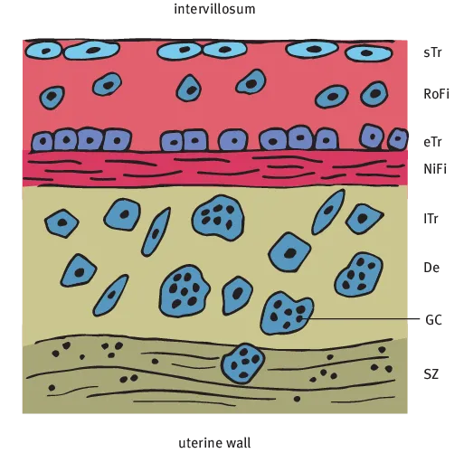
Fig.1.2: Illustration. Chorionic plate tissue layers between the amniotic sac on one side and intervillous space (intervillosum) on the other side: Amniotic Epithelium (AE), Amniotic Tissue (AT), Junctional Layer (JL), fetal vessel branches (V), Chorionic Tissue (CT), extravillous Trophoblast (eTr), Langhans-Fibrinoid (Fi) and syncytiotrophoblast (sTr).

1.1.2 Basal plate

The basal plate defines the border to the uterine wall and consists of:
  • 1.
    The extravillous trophoblast.
  • 2.
    The maternal cells of the decidua basalis, soft tissue cells and leucocytes.
  • 3.
    Fibrinoid.
Generally two types of fibrinoid can be differentiated [1,2]:
  • Fibrin-type fibrinoid originating from coagulation processes and containing fibrin rich depositions and other maternal proteins.
  • Matrix-type fibrinoid secreted from extravillous trophoblast cells and containing fetal proteins.
In early pregnancy the placenta is limited by the implantation bed and forms a zone-like structure (ch. 11). In middle pregnancy, several tissue layers form into a single basal plate of varying thickness. These different layers are not identifiable histologically.
As in the layers of the chorionic plate, syncytiotrophoblast cells (sTr) can be focally seen at the junction to the intervillous space.
  • The Rohr-Fibrinoid (RoFi) is seen next to the intervillous space and is infiltrated by non-proliferating trophoblast cells. A dense cell layer of basophilic Trophoblast cells (eTr) is seen underneath.
  • The Nitabuch-Fibrinoid (NiFi) layer is the point at which placental separation occurs at birth. This layer has a heterogeneous appearance and is comprised of connective tissue fibers, endometrial stromal cells and macrophages.
  • The Decidua basalis (De) contains stromal cells, macrophages, leucocytes and multinucleated Giant Cells (GC) of the extravillous and Intermediate trophoblast (Itr) types. Rudimentary fetal cells of the connecting stalk trophoblasts can also be found.
  • The Solution Zone (SZ) demarcates the uterus from the basal plate and contains the mucous decidual membrane that remains in the uterus following birth.
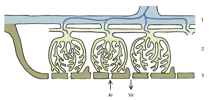
  • Vessel branches of the Spiral arteries (SP) transverse and branch within the basal plate and end in the intervillous space. One artery can have several fountain like openings in the central part of the fetal cotyledon [3]. Veins are extremely thin walled and often show sinusoidal dilatation.
Fig.1.3: Illustration. Maternal / basal plate structure between the intervillous space (intervillosum) on one side and the uterine wall on the other side: Syncytiotrophoblast (sTr), Rohr-Fibrinoid (RoFi), extravillous Trophoblast (eTr), Nitabuch-Fibrinoid (NiFi), Decidua basalis (De) containing Intermediate Trophoblasts (ITr) and Giant Cells (GC), and Solution Zone (SZ).

1.1.3 Septa and islands

The ...

Índice