I. Structure of the Head
Humans and vertebrates both share in common a regional division of the body into head, trunk, and extremities. Both also exhibit branchomery, that is, the arrangement of branchial arches, as well as metamery, that is, segmental organization of the trunk wall.
The structure of the head differs from that of the trunk in that there is an absence of segmental organization. The development of the organs of the head has resulted in the disappearance of the cranial somites and their being taken up into the structure of the cranial base. The dorso-caudal part of the cranium is thus the only part still of metameric origin. The occipital vertebra (proatlas) anterior to the atlas forms the posterior main condyle and the superior joint facets of the first cervical vertebra. These differentiations become evident in cases of developmental disorders, as, for example, manifestations of the occipital vertebra or atlas assimilation.
The rostro-dorsal segment of the skull, on the other hand, consists of unsegmented head mesectoderm and prechordal mesoderm. The structural material of the rostroventral cranium stems from the first two branchial arches.
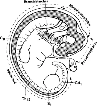
The structural arrangement originates in regionally specific inductors under the influence of the central nervous system. It is presumed that the head possesses two induction centers and the trunk one (Figure 1.1).
Figure 1.1 Organization of the head and trunk in the human embryo. (After Starck, D: Embryologie, 3rd ed., G. Thieme, Stuttgart, 1975. With permission of Georg Thieme Verlag, Stuttgart.)
The induction center of the anterior head region for the nose, the eyes, and anterior skull base is subject to the influence of the frontal brain, the prosencephalon. The posterior head region where the labyrinth, ultimobranchial bodies, and posterior skull base develop are influenced by the rhombic brain, the rhombencephalon, and the trunk by the caudal section of the neural tube.
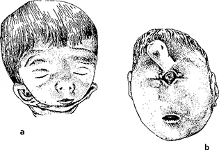
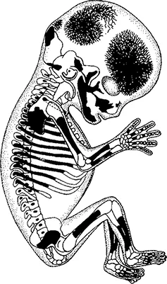
The existence of two skull formation centers is substantiated by cyclopic and otocephalic malformation patterns (Figure 1.2).
Figure 1.2 Craniofacial dysplasias: a, cyclopia; b, otocephaly. (After Schumacher, G-H: Anatomie, Lehr-buch und Atlas, Edition Zahnheilkunde. J.A. Barth, Leipzig, 1991, Vol.1. With permission.)
II. Skeletal Morphogenesis
Bone formation can ensue in two ways, either through desmal ossification, that is, directly from the mesenchyme, or indirectly through a hyaline cartilage stage, that is, through chondral ossification. Both cases first result in woven bone. The activities of osteoblasts and osteoclasts are crucial to the formation of bone as well as to its constant remodeling and regenerative capabilities.
A. Osteoblasts and Osteoclasts
Osteoblasts differentiate from mesenchymal cells. Their ultrastructure exhibits the properties of cells with high synthetic capabilities. Their initial task is to produce noncalcified osteoid, consisting of basic material (proteoglycans, glycoproteins) and collagen fibres. Osteoid production is omnidirectional, walling in the osteoblasts which then turn into osteocytes.
Osteoclasts are 30 to 100 Jim multinucleate giant cells which use enzymes (primarily proteases and phosphatases) to break down bone. They are equipped with microvilli on the side facing the bone. Their emission of lactic acid increases the pH and thus dissolves the minerals. Osteoclasts are usually found in small pockets of the broken down bone, the Howship’s lacunae. They can move in amebic fashion.
B. Desmal and Chondral Ossification
Desmal ossification begins in centers of mesenchyme cell concentration, where they become differentiated into osteoblasts. The osteoblasts synthesize the osteoid in which hydroxylapatite crystals are formed by calcium and phosphate ion enrichment. Bone trabeculae are initially laid down and converge to eventually form a network. The osteoblasts and osteoclasts mold the bone trabeculae, resulting in lamellar bone arising from the woven bone. Growth of the desmal bone follows by apposition.
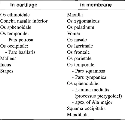
The bones of the skull vault and of the facial skull as well as the lower jaw and the diaphysis of the clavicle arise through desmal ossification (Table 1.1).
Table 1.1 The Mode of Ossification of the Bones of the Skull
Chondral ossification is the mode of ossification of the base of the skull, the vertebrae, the ribs, as well as of all the tubular bones (Figure 1.3). Cartilage here represents the first stage in bone development. Initially, a process of perichondral ossification results in the formation of a bone collar around the cartilage. Then the cartilage cells in the interior of the cartilage change into hypertrophic cartilage. Some of the cells are destroyed and the basic substance takes up calcium salts. Through endochondral ossification, mesenchyme cells from outside invade the cartilage and commence the formation of bone from the inside out. Concurrently, white blood cells (monocytes) become differentiated into chondroclasts which break down the cartilage, and mesenchyme cells become differentiated into osteoblasts which deposit bone matrix on the remaining cartilage.
Growth in diameter starts with the periosteum and is thus appositional. Longitudinal growth ensues endochondrally on a base of remaining cartilage and is thus interstitial. When there is no more cartilage remaining, bone formation and longitudinal growth ceases.
III. Skull Morphogenesis
As described above, the anlage of the skull consists of cranial somites, nonsegmented head mesectoderm, prechordal mesoderm, and the first two branchial arches.
The mesenchyme becomes concentrated into an envelope around the brain vesicles from which the skull bones arise partly through chondral and partly through desmal ossification. The cartilaginous anlage of the skull is called the chondrocranium and that arising from connective tissue, the desmocranium.
A. Chondrocranium
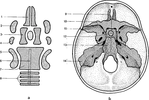
The chondrocranium arises from the fusion of several cartilage elements. In the second embryonic month, the first cartilage deposits begin to form in the basal part of the mesenchymal envelope at the anterior end of the notochord (Figure 1.4). The cartilaginous ear capsules surrounding the labyrinth arise independently of this, as do the nose capsules surrounding the olfactory organs.
Figure 1.3 Ossification centers in a 5 cm embryo. (After Schumacher G-H and Christ, B: Embryonale Entwicklung und Fehlbildungen des Menschen, Anot-omie undKlinik, 10th ed., Ullstein Mosby, Berlin, 1993. With permission.)
Figure 1.4 The anlage of the chondrocranium. a, The cartilaginous laminae; b, Their derivatives; 1, trabecular cartilage; 2, ala orbitalis; 3, hypophyseal cartilage; 4, ala temporalis; 5, labyrinthine capsule; 6, notochord; 7, parachordal cartilage; 8, occipital cartilage; 9 ethmoid bone, 10, lesser wing of sphenoid; 11, greater wing of sphenoid; 12, body of sphenoid; 13, basilar part of occipital bone; 14, petrous temporal bone. (Modified from Clara, M: Entwicklungsgeschichte des Menschen, 6 Aufl., Georg Thieme, Leipzig, 1966. With permission of Georg Thieme Verlag, Stuttgart.)
The mesenchyme situated at the cranial end of the notochord differentiates into the parachordal cartilage which then connects with the cartilage formed from the cranial somites. The trabecular and hypophyseal cartilages form anterior to the cranial chordal extremity, i.e., prechordally. The above-mentioned cartilage deposits fuse to form a long plate reaching from the nose region as far as the back of the head. The center of this cartilaginous plate contains the hypophyseal pouch (Rathke’s pouch) from whose epithelium the anterior lobe of the hypophysis arises. On both sides two further cartilaginous plates, the ala orbitalis and the ala temporalis arise. In the third month the notochord extends further caudally.
The bones of the base of the skull originate essentially in the chondrocranium (Table 1.1). The chondrocranium persists as the nasal capsule.
The auditory cartilage apparatus of the malleus, the incus, and the stapes as well as the processus styloideus of the temporal bone originate in the cartilaginous bar of the first...