1
THE NATURE OF BONES AND TEETH
THE HUMAN SKELETON
There are more than two hundred separate bones in the adult human skeleton (Figure 1.1, Table 1.1). They may be divided into three classes according to their basic shape. Long-bones are found in the limbs and take the form of hollow tubes closed at both ends. Flat bones tend to take the form of broad bony plates (for example, the bones which make up the skull vault), and irregular bones, as their name suggests, fit into neither of the above categories on account of their irregular shape. Examples of irregular bones are the vertebrae and the bones of the skull base.
THE COMPOSITION OF BONE
Bone is a composite material formed from an organic and an inorganic (mineral) part. By weight, dry bone is about 70 per cent mineral and 30 per cent organics. The great majority of the organic component is collagen, a protein which forms long fibres. The small crystals which make up the mineral portion of bone are embedded in a matrix of collagen fibres. Bone mineral is chiefly hydroxyapatite, a form of calcium phosphate whose composition approximates to the chemical formula Ca10(PO4)6(OH)2. The mineral component gives bone its rigidity, and the organic component lends it a slight ‘give’ or resilience which gives it its strength. The organic part degrades after death, which accounts for the rather brittle nature of most archaeological bone.
THE GROSS STRUCTURE OF BONE
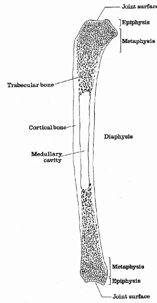
In terms of gross structure, there are two different types of bone tissue: cortical and trabecular bone. Cortical bone is the solid, dense part that forms the outer layer of the bones; it is thickest in the diaphyses (shafts) of the long-bones (Figure 1.2). It forms a thin layer around the long-bone ends and around the flat and irregular bones. Trabecular bone is less dense than cortical bone and has a honeycomb structure. Trabecular bone is located within the ends of long-bones (Figure 1.2) and in the interior of the flat and irregular bones.
In living bone, most of the outer surface is surrounded by a thin membrane, the periosteum.
The internal walls of the medullary cavities of long-bones are lined with another membrane, the endosteum, which also lines the network of tiny cavities in trabecular bone.
THE FORM AND FUNCTIONS OF THE SKELETON
Bones provide a general framework to support the body. They bear ridges for attachment for muscles and tendons, and surfaces which form joints. They also protect vital organs, provide a store of fats and blood-forming marrow, and act as a bank of mineral salts. The form of the bones can be understood in terms of these functions. This point is illustrated below with reference to the general structure of long-bones (Figure 1.2).
The tubular form of the long-bones maximises strength and minimises weight; the medullary cavity within the shaft also provides a space for blood-forming cells and fat storage. The flared ends of the shaft (the metaphyses) support the epiphyses which bear the joint surfaces by which the long-bone articulates with its neighbours. Trabecular bone is located beneath the joint surfaces. It is less rigid than cortical bone so is well adapted for the dissipation of mechanical stresses at the joints. It also forms a site of red blood cell production in the growing skeleton, and its large surface area facilitates the exchange of minerals to and from bone. The cortical bone which forms the walls of the tube provides strength and rigidity.
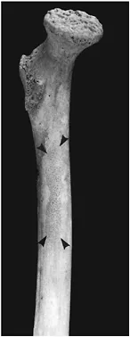
On excavation, bones have the appearance of solid, inanimate objects. This is deceptive regarding the nature of living bone. Like most other tissues in the body, bone is living tissue. It is permeated with nerves and blood vessels and, in common with most other tissues, is continually being formed and broken down. The fact that bone tissue is constantly being renewed in this way means that it can repair itself following disease or injury. It also means that, within limits, it can adapt its form according to the strains put on it. This last has been recognised since the nineteenth century, when a German anatomist, Julius Wolff, described the response of bone to mechanical forces in a manner which has become known as Wolff’s Law. He stated that ‘the form of the bone being given, the bone elements place or displace themselves in the direction of the functional pressure and increase or decrease their mass to reflect the amount of functional pressure’ (Wolff 1892, quoted in Kennedy 1989: 134). In other words, bone is sensitive to mechanical forces such as weight bearing or muscular tension, and these directly influence bone form to a certain degree. An increase in mechanical stress tends to produce bones which are more robust, i.e. are thicker and stronger, with more marked ridges for muscle attachment; a reduction in mechanical forces tends to have the opposite effect (Scott 1957). For example, exercise increases bone strength. This is illustrated by a well-known X-ray study (Jones et al. 1977) of the arm bones of professional tennis-players, which found that the thickness of the cortical bone of the humerus in the racket arm increased to the extent that it was on average about 30 per cent greater than that in the non-playing arm (Figure 1.3). Conversely, bone mineral is lost when the physical strains imposed on the bones are lessened. So, for example, patients who are bed-ridden for some time lose bone mineral (Donaldson et al. 1970), but regain it when normal activity resumes.
THE MICROSCOPIC STRUCTURE OF BONE
Bone is initially laid down as woven or primary bone; woven bone is a temporary tissue which is gradually replaced by mature, or lamellar bone. Woven bone forms the foetal skeleton, but by the time an infant is a year old it has almost entirely been replaced by lamellar bone. Woven bone may also be produced in the adult skeleton in response to disease or injury: it may form under the periosteum in response to inflammation, and is produced by some bone tumours, and during fracture repair. Woven bone is coarser and more porous than lamellar bone. The two can be distinguished with the naked eye (Figure 1.4).
Lamellar bone is composed of a series of microscopic layers (lamellae) about 4–12 microns thick (1 micron (
m) = one thousandth of a millimetre). It is stronger than woven bone. In the adult, both trabecular and cortical bone have a lamellar structure, but they differ in the way they are organised.
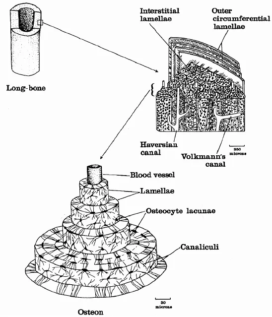
The microstructure of cortical bone is shown in Figures 1.5 and 1.6. Cortical bone is permeated by innumerable, interconnected channels, the Haversian system, upon which it relies for its blood supply. Bony lamellae are arranged concentrically around Haversian canals. There are
normally about 4–20 lamellae around each Haversian canal; this unit of bone organisation is called an osteon. Haversian canals run parallel to the long axis of the bone, and interconnect via transverse or oblique channels called Volkmann’s canals. There are also some lamellae between the osteons (interstitial lamellae), and some, termed circumferential lamellae, encircle the entire outer and inner surfaces of the bone. Trabecular bone does not have a Haversian system – its fine honeycomb structure allows it to receive sufficient nutrients from blood vessels which meander through it.
BONE CELLS
There are three main types of bone cells. Osteoblasts are responsible for formation of new bone, osteocytes are involved with the maintenance of bone as a living tissue, and osteoclasts are responsible for resorption (removal) of b...