![]()
CHAPTER 1
Carbon Nanostructures: Drug Delivery and Beyond
Agnieszka Gajewska a , Akcan Istif a , Jasra Gul a , b , Michele Chironi a , Andrea Faidiga a , Marco Rocco a , Ketty Slavec a , Teresa Gianferrara a and Tatiana Da Ros*a
a Department of Chemical and Pharmaceutical Sciences, University of Trieste, Trieste, Italy,
b International Center for Chemical and Biological Sciences (ICCBS), University of Karachi, Karachi, Pakistan
*E-mail:
[email protected] Carbon nanostructures, such as nanotubes, nanodiamonds, graphene quantum dots and carbon dots, are studied in depth as interesting materials in many different applications. In the biomedical field there are many possible uses but the area most explored, so far, is their application as drug delivery systems considering their biocompatibility and versatility. Herein we propose an analysis of some of the most recent literature related to drug delivery with carbon nanotubes, nanodiamonds, graphene quantum dots and carbon dots and some other appealing possibilities.
1.1 Introduction
Since the discovery of fullerenes in 1985, the family of carbon nanostructures has widened a lot and nowadays it includes nanotubes, nanodiamonds, nanoonions, nanoribbons, nanocones, nanohorns, graphene and its direct derivatives, as graphene quantum dots, and carbon dots. Despite the fact that they all present interesting characteristics, in the last few years the most studied, from a biological point of view, are carbon nanotubes (CNTs), graphene oxide (GO), graphene quantum dots (GQDs), carbon quantum dots (CQDs) and carbon nanodiamonds (NDs). In fact, lately the interest for fullerenes has dramatically decreased, affecting the number of studies on their properties, but there are still brilliant examples of their biological potential as reported by various authors. 1
One of the most explored bio-applications of carbon nanostructures is related to their capability to load different payloads and to cross the cellular membrane, allowing the intracellular delivery of drugs into cells. Here we will discuss some of the most recent developments in this field, except for fullerenes, which have been recently widely reviewed. 2
1.2 Carbon Nanotubes
CNTs consist of one or several concentric graphene sheets rolled up in a cylindrical shape with a range in diameter from 0.4 nm up to 100 nm. They have been studied from many different aspects.
The biological applications of CNTs are strongly dependent on all of the health hazards that this new material can cause. For several years, the effects of CNTs in cells and tissues have been explored by in vivo and toxicological studies. However, it turned out that many parameters of CNTs, (as contaminants, surface chemistry, processing methods, agglomerate states, length, diameter and more) can have various toxic effects.
As was reported, the toxic effect of CNTs can arise not directly from them, but from the residues produced during the synthetic process as nickel, cobalt or iron nanoparticles, which can remain in the CNTs and generate reactive oxygen species (ROS) in a biological environment. It turns out that ROS cause inflammatory symptoms and induce mitochondrial membrane degradation, depletion of antioxidant agents, rise in inflammatory biomarkers, and decreases cell viability. It has been demonstrated that 30% of iron in SWCNTs is able to generate free radicals within 15 min of exposure to epidermal keratinocytes in the presence of DMPO (5,5-dimethyl-1-pyrroli- ne-1-oxide). 3 In a later study it was shown that higher amounts of catalyst generate higher concentrations of free radicals and increase inflammatory responses. 4 Additionally, nickel alters the expression of the gene encoding the protein HIF1A, a factor of transcript involved in the regulation of inflammatory genes and apoptosis. 5 All studies on the toxicity of CNTs must than take into account the nature of the metallic catalyst and its percentage/quantity. It is difficult to obtain pure CNTs by removing all traces of catalysts and several methods can be employed to decrease residual catalysts including centrifugation, high-temperature annealing 6 and oxidation treatment by acid reflux. 7
Toxicity can be also influenced by the modification on the CNTs' surface. Only with acid-treatment on the CNTs' surface is it possible to introduce a number of defect sites along the CNTs' surface (as mentioned earlier). Muller et al. changed the number of defect sites on MWCNTs by mechanical grinding and annealing at high temperature and demonstrated that acute pulmonary toxicity and genotoxicity increased after intratracheal administration of MWCNTs with a larger number of defect sites. 8 However, another study by Kagan et al. showed that oxidized SWCNTs can be biodegraded more easily by myeloperoxidase enzyme, found in neutrophils and macrophages. The enzyme interacts with carboxylic sites on the nanotubes' surface 9 and oxidized CNTs may be more biocompatible than pristine CNTs from this point of view. Following this pathway, Sayes and co-workers examined cell viability in the presence of oxidized and phenylated tubes. They discovered that the phenylated tubes exhibit lower toxicity, and this can be due to the hindering of the defect sides of the tubes. 10 Dumortier at al. have examined the toxicity of CNTs functionalized by 1,3-dipolar cycloaddition. They concluded that CNTs, fully soluble in aqueous culture media, did not modify primary immune cells viability in vitro. 11 Also, the CNTs' surface area and their hydrophobic nature have an impact on the toxicity. The tubes, in fact, can potentially interact with several molecules like proteins, RNA, DNA and enzymes with toxic effects on the biological environment. 12 Dutta and co-workers found that the bovine or human serum albumin adsorption onto the CNT surface resulted in inflammatory responses after uptake by macrophage cells. Normally that effect occurs only when albumin adopts structural changes or becomes damaged. 13 The effect of functionalization of the tubes highlights the importance of assessing the toxicity profile for every type of new CNT modification.
Finally, the CNTs' length cannot be ignored. CNT sizes have an important effect on clearance. The length can range from nanometers up to millimeters. The exposure to long fiber-like material can induce dangerous damages in DNA and genetic mutations over a period of exposure, causing an extremely malignant form of cancer, mesothelioma. Symptoms of these bio-persistent fibers are the granulomas, which are the signs of oxidative stress, causing excessive fibrous tissue. 14
Macrophage are the cells responsible for the removal of foreign material like CNTs from living organisms. Fibers with a length exceeding 20 µm are extremely resistant to phagocytosis 15 and Poland et al. have shown that CNTs as spherical or stellate shaped agglomerates with less than 20 µm have no significant damaging reactions compared to samples with individualized MWCNTs, agglomerates, and ropes of MWCNTs with lengths exceeding 20 µm. 14 CNTs can be toxic, but many solutions exist, to moderate or eliminate adverse effects arising from the mentioned problems. In conclusion, CNTs should be used without the presence of metal catalysts, have an appropriate functionalization for the planned purpose with low surface oxidation, present a covered surface to effectively escape bio-interactions and be short to avoid long retention times.
To approach their potential application and effect on humans, pharmacokinetics and biodistribution in animal models have been and are currently being explored, and they depend on numerous factors such as chemical-physical characteristics, solubility, surface functionalization, aggregation of the derivatives themselves, 16 while CNT excretion is mainly renal. 17
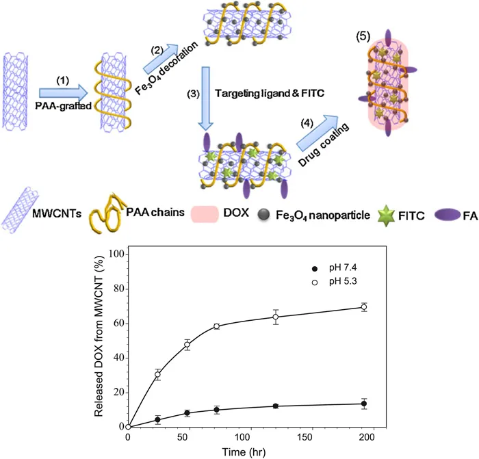
The possible biomedical applications of CNTs span many fields. One example is regenerative medicines due to their biocompatibility, resistance, and possible functionalization of their surface with biomolecules as collagen fibers to form a nanomaterial capable of acting as a scaffold in tissue regeneration, as already reported many years ago. 18 Another potentiality, not properly unraveled up to now, is their supposed antioxidant activity. If this function could be confirmed, they could also be used in the prevention of aging and preservation of food. 19 However, the most promising and studied application for CNTs is delivery systems. In fact, they can be loaded with different pharmaceutical ingredients and be delivered at specific sites, with prolonged accumulation into targeted tissues and reduced prospective systemic toxic effects. 20 The latter aspect is of particular importance in the case of anticancer drugs, the mechanism of action of which is mainly related to their toxic effects (i.e. alkylation, DNA intercalation, tubulin aggregation/disaggregation equilibrium unbalance among others). In this context, CNTs propose an opportunity to capsulate anticancer drugs, thus reducing their toxicity for an organism and enhancing local accumulation in the targeted site. 21 Moreover, it is important to remember that, with the proper modification, it is possible to stimulate the drug release from the drug-CNT complex only in a tumor environment, for example, by lowering the pH under the physiological value (Figure 1.1). 22
Figure 1.1 Top: Preparation of Fe3O4 magnetic nanoparticles decorated poly(acrylic acid)-grafted MWCNTs, covalently linked to folic acid and fluorescein (steps 1–3) and embedding doxorubicin by non-covalent interactions. Bottom: doxorubicin release profile at acid and neutral pH at 37 °C. Adapted from ref. 22 with permission from ...