![]()
The Variants are Here
The UK variant (B.1.1.7)
The first reports that a new variant, more dangerous than D614G might exist, came from news reports from the United Kingdom. Despite lockdowns, new infections were rising rapidly in Southern England during the fall of 2020. Then came the dramatic announcement from the Prime Minister's office. A unique and potentially dangerous variant was preferentially spreading in those areas experiencing a dramatic uptick in infection (Figure 18, 19, 20). In record time, a preprint was available detailing changes in the viral genome. Fast forward to February 2021: the variant has been detected in more than 50 countries, has jumped from 3.3% to 14% of cases in France in less than a month, and is projected by experts to become dominant in US hotspots in a matter of weeks. A preprint published February 3 also verified that more people are dying of this variant than ones prior-- increasing the chances of death for those infected by about 35 percent. Whether it is more lethal than the last dominant strain or simply reaching a greater proportion of vulnerable populations remains to be seen.
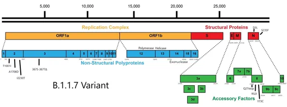
The genome sequence of the new variant revealed not one but 23 changes as compared to early strains, 20 of which alter protein structures in addition to D614G.
The collective shock of that paper, felt by government leaders, public health officials, and scientists alike, is still with us.
Figure 18. Estimate of true positive rates for classification of B.1.1.7 infection. The color gradient shows the probability of sampling a B.1.1.7 sequence conditional on sampling any sequence with 69-70del.
Source: Volz et al., 2021
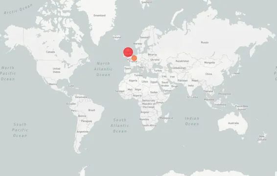
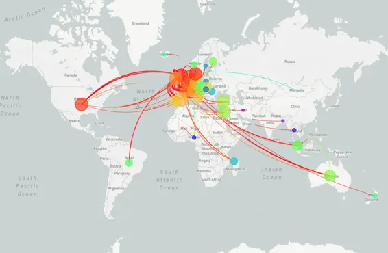
Figure 19. List of countries that have reported B.1.1.7 detection, October vs December 2020.
Source: GISAID
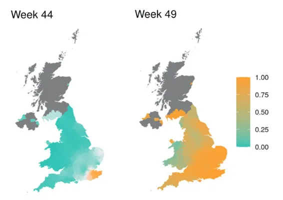
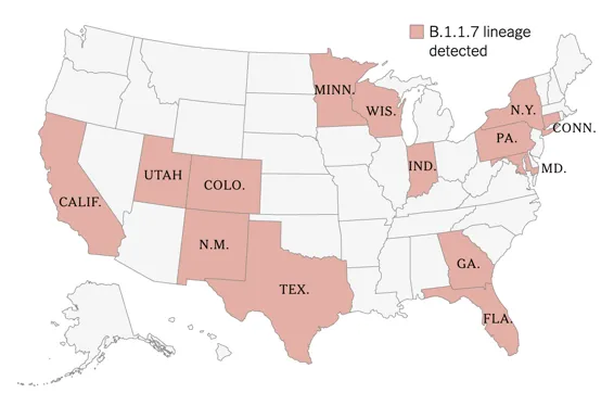
Figure 20. Map of states where local transmission of B.1.1.7 has been detected as of January 18, 2021.
Source: Corum & Zimmer, 2021
Why shock? The genome sequence of the new variant revealed not one but 21 changes as compared to early strains, 18 of which alter protein structures in addition to D614G. The new variant is now designated B.1.1.7 (aka the UK variant). Most of the 18 differences result in a change of one amino acid for another. Two are small deletions of one and two amino acids. Several changes in the nucleotide sequence of the virus do not result in any amino acid change. Eight of the changes are in the spike protein. Others are scattered about the genome, potentially altering the function of the replicative enzyme complex (orf1ab), the M protein (a virus particle surface protein), one in orf3b and several in orf8. Almost all work to date has focused on the S protein as this is the preferred vaccine target. Variants such as B.1.1.7 are known to be more contagious but have shown us they can change still further. The B.1.1.7 variant, for example, recently acquired the immune-evasive E484K mutation.
Figure 21. A linear overview of the B.1.1.7 (UK) genome.
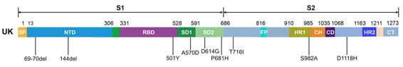
Figure 22. Mutations in the viral spike identified in B.1.1.7 (UK) in addition to D614G.
Source: Wang et al., preprint
I will describe each change as it is relevant to understanding the UK variant. As it happens, other variants now popping up around the world contain some amino acid changes identical to the UK variant as well as changes unique onto themselves.
Figure 21 is a linear display of the B.1.1.7 genome, while Figure 22 zooms in on the S protein. The functional regions of the protein in Figure 22 are highlighted in different colors, which I refer to in the following sections.
The signal peptide sequence
The initial amino acids 1-12(3) are called the signal peptide sequence. This short region directs the S protein to exit the cell membrane. The virus buds through the cell membrane, surrounding the core of the virus with a membrane decorated with the spike protein. The signal peptide is often cleaved from the mature protein as it exits the cell.
The N-terminal domain
The next region (blue) is the N-terminal domain. In the mature spike, this region folds up into a structure near the top of the spike aside the receptor-binding domain. The function of the N-terminal domain is unknown.
The receptor-binding domain
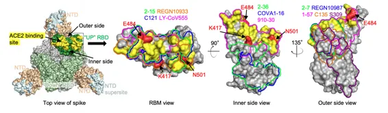
The receptor-binding domain, amino acids 331-528 (purple) folds up into a compact structure that sits atop the spike. When open, it presents a flat surface that binds tightly to the ACE2 receptor. The precise fit at the atomic level between the surface receptor-binding domain of the ACE2 protein and electrostatic attraction between oppositely charged amino acids on two surfaces determines how tightly the two proteins stick to one another (Figures 23, 24, 25). This region is the primary target of protective antibodies that block the virus's ability to bind the receptor. No receptor binding, no infection.
Think of pieces of a puzzle, each of which carries a magnetic charge. Not only must there be an exact match in the shape to the two opposing pieces, but the magnetic poles must also be complementary, positives opposite negatives (Figure 26). The puzzle pieces may fit loosely or tightly depending on the precise details of the shape. Even if the pieces fit, the magnetic poles must also match, otherwise even pieces with an exact match would repel one another. This is a very good analogy for the necessity of complementary shapes and charges that determine how well the receptor binding domain binds to the ACE2 receptor. Each amino acid has its own shape and charge characteristics. Any change in the shape or charge of the amino acid substitution will affect binding (Figures 27, 28).
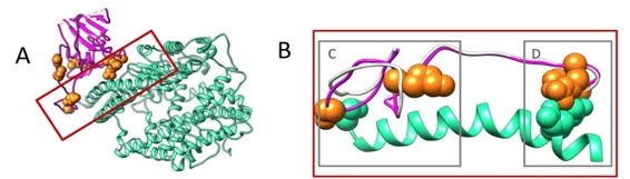
Figure 23. Left panel, top view of SARS-COV-2 spike with one RBD in the “up” conformation (pdb: 6zgg). RBD and NTD are colored green and peach, respectively. The positions of ‘inner’ and ‘outer’ sides are indicated on the “up” RBD with the ACE2-binding site colored yellow. The three panels to the right show the antibody footprints on RBD.
Source: Wang et al., preprint
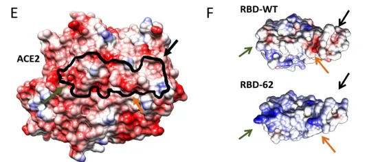
Figure 24. Global comparison between RBD-WT and RBD-62 shows overall similarity. Please note that the orientation of the receptor-binding domain is reversed compared to other figures below, with the ACE2 receptor on top.
Source: Zahradnik et al., preprint
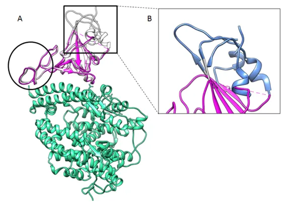
Figure 25. Cartoon representation of the Cryo-EM structure of ACE2 (cyan) in complex with RBD-62 RBM (magenta) with the RBD-62 mutations resolved in the density (orange spheres).
Source: Zahradnik et al., preprint
These bits of the virus are like puzzle pieces made of individual atoms. One atom out of place will decrease how well the pieces fit together.
Figure 26. These bits of the virus are like puzzle pieces made of individual atoms. One atom out of place will decrease how well the pieces fit together. The blue repr...