eBook - ePub
Intraoperative and Interventional Echocardiography
Atlas of Transesophageal Imaging E-Book
Donald Oxorn, Catherine M. Otto
This is a test
- 448 páginas
- English
- ePUB (apto para móviles)
- Disponible en iOS y Android
eBook - ePub
Intraoperative and Interventional Echocardiography
Atlas of Transesophageal Imaging E-Book
Donald Oxorn, Catherine M. Otto
Detalles del libro
Vista previa del libro
Índice
Citas
Información del libro
Case-based and heavily illustrated, Intraoperative and Interventional Echocardiography: Atlas of Transesophageal Imaging, 2nd Edition covers virtually every clinical scenario in which you're likely to use TEE. Drs. Donald C. Oxorn and Catherine M. Otto provide practical, how-to guidance on transesophageal echocardiography, including new approaches and state-of-the-art technologies. More than 1, 500 images sharpen your image acquisition and analysis skills and help you master this challenging technique.
- Real-world cases and abundant, detailed figures and tables show you exactly how to proceed, step by step, and get the best results.
- Each case begins with a brief presentation and discussion, and integrates clinical echocardiography, surgical pathology, and other imaging data.
- Clear descriptions of preoperative pathology guide you in choosing the best approach to common problems.
- The practice-based learning approach with expert commentary for each case helps you retain complex information and apply it in your daily practice.
- Every chapter has been thoroughly revised, with discussions of new technology and new techniques, including several techniques that are on the verge of becoming mainstream.
- New chapters cover current transcatheter valve therapies and device closures.
Preguntas frecuentes
¿Cómo cancelo mi suscripción?
¿Cómo descargo los libros?
Por el momento, todos nuestros libros ePub adaptables a dispositivos móviles se pueden descargar a través de la aplicación. La mayor parte de nuestros PDF también se puede descargar y ya estamos trabajando para que el resto también sea descargable. Obtén más información aquí.
¿En qué se diferencian los planes de precios?
Ambos planes te permiten acceder por completo a la biblioteca y a todas las funciones de Perlego. Las únicas diferencias son el precio y el período de suscripción: con el plan anual ahorrarás en torno a un 30 % en comparación con 12 meses de un plan mensual.
¿Qué es Perlego?
Somos un servicio de suscripción de libros de texto en línea que te permite acceder a toda una biblioteca en línea por menos de lo que cuesta un libro al mes. Con más de un millón de libros sobre más de 1000 categorías, ¡tenemos todo lo que necesitas! Obtén más información aquí.
¿Perlego ofrece la función de texto a voz?
Busca el símbolo de lectura en voz alta en tu próximo libro para ver si puedes escucharlo. La herramienta de lectura en voz alta lee el texto en voz alta por ti, resaltando el texto a medida que se lee. Puedes pausarla, acelerarla y ralentizarla. Obtén más información aquí.
¿Es Intraoperative and Interventional Echocardiography un PDF/ePUB en línea?
Sí, puedes acceder a Intraoperative and Interventional Echocardiography de Donald Oxorn, Catherine M. Otto en formato PDF o ePUB, así como a otros libros populares de Medicina y Cardiología. Tenemos más de un millón de libros disponibles en nuestro catálogo para que explores.
Información
Chapter 1
Coronary artery disease
Visualization of the coronary arteries and regional wall motion
CASE 1-1 Normal coronary arteries
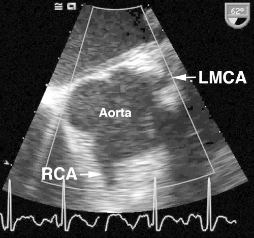
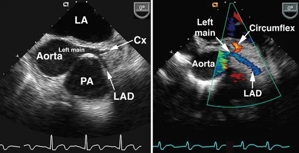
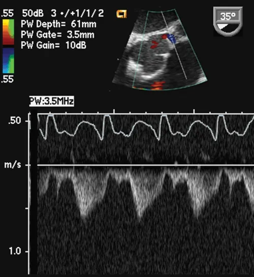
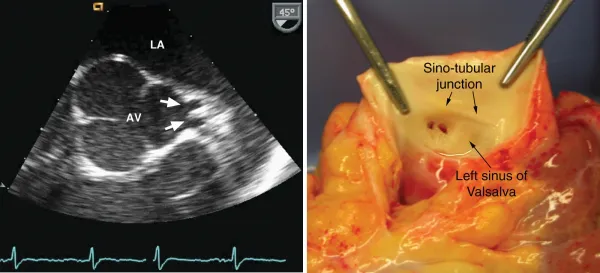
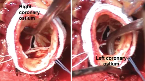
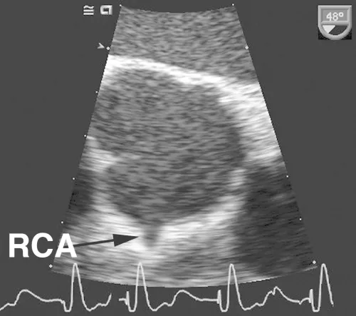
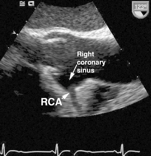
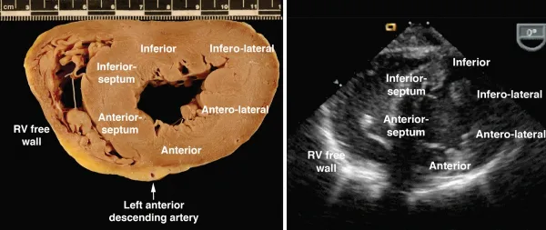
Comments
As shown in these examples, the proximal coronary arteries can often be visualized on TEE. The left main coronary artery arises from the left coronary sinus of Valsalva, is easily visualized in over 85% of patients and has a normal diameter of 4.2 ± 0.7 mm, with a slightly smaller average diameter in women (3.5 mm) compared with men (4.3 mm). The left main coronary artery bifurcates into the left anterior descendin...
Índice
Estilos de citas para Intraoperative and Interventional Echocardiography
APA 6 Citation
Oxorn, D., & Otto, C. (2016). Intraoperative and Interventional Echocardiography (2nd ed.). Elsevier Health Sciences. Retrieved from https://www.perlego.com/book/2938098/intraoperative-and-interventional-echocardiography-atlas-of-transesophageal-imaging-ebook-pdf (Original work published 2016)
Chicago Citation
Oxorn, Donald, and Catherine Otto. (2016) 2016. Intraoperative and Interventional Echocardiography. 2nd ed. Elsevier Health Sciences. https://www.perlego.com/book/2938098/intraoperative-and-interventional-echocardiography-atlas-of-transesophageal-imaging-ebook-pdf.
Harvard Citation
Oxorn, D. and Otto, C. (2016) Intraoperative and Interventional Echocardiography. 2nd edn. Elsevier Health Sciences. Available at: https://www.perlego.com/book/2938098/intraoperative-and-interventional-echocardiography-atlas-of-transesophageal-imaging-ebook-pdf (Accessed: 15 October 2022).
MLA 7 Citation
Oxorn, Donald, and Catherine Otto. Intraoperative and Interventional Echocardiography. 2nd ed. Elsevier Health Sciences, 2016. Web. 15 Oct. 2022.