![]()
CHAPTER one The Radiograph
Competent radiologic practice presupposes the availability of good-quality radiographs. Familiarity with the basic principles underlying the production of radiographs is a prerequisite for the radiologist. Accurate positioning of the animal under investigation, correct exposure factors, the use of grids and other ancillary aids, and good processing technique all influence the quality of a radiograph. The use of a technique chart is essential for consistent results. Consistency is important, particularly when studies have to be repeated over time to assess the progress of a particular case. If the radiographs in such studies are not comparable, errors of interpretation are likely to occur. Radiographs may be of poor quality because of improper positioning, improper exposure technique, or poor darkroom technique. It is hazardous to attempt to interpret such radiographs.
Radiographic technique is discussed in this book only insofar as is necessary for a proper understanding of points of interpretation. The necessary detailed information on technique can be found in any of the several works devoted to this topic.
A radiograph is a composite shadow of structures and objects in the path of an x-ray beam recorded on film. Because a radiograph is, in essence, a shadowgraph, the geometric rules applicable to the formation of shadows are also valid for radiographs. Thus, the nearer the object under examination is to the film, the sharper will be its outline. Distance of an object from the film causes magnification of the resulting shadow and some distortion and blurring. The nearer the object is to the source of radiation, the greater will be the degree of magnification. The area being studied, therefore, should be placed as near to the film as possible and at a standard acceptable distance from the source of radiation, usually 100 cm (36 to 40 inches). Because the radiograph (being a shadowgraph) outlines an object in only two planes, at least two views, made at right angles to one another (orthogonal views), are required to demonstrate the object in a three-dimensional representation. Shadows are cast not only of the outline of the body, but also of structures within it (Figure 1-1).
The radiograph is not a simple shadowgraph: some of the x-rays pass directly through the body being examined. These are the useful rays because they affect the film and produce the image. Some of the incident radiation is absorbed within the body, and some is scattered. Scattered radiation reaching the film is undesirable because it causes fogging and blurring, or “unsharpness.” Fogging gives a radiograph a cloudy or hazy appearance. Structure margins are indistinct. Grids are used to reduce scatter. As rule they should be used when the part under examination exceeds 10 cm in thickness.
Fast film/screen combinations reduce exposure times and minimize movement blur. A radiograph shows not only the outline of an organ within the body but also other body structures superimposed on it and on one another.
Not all structures allow x-rays to pass through them in the same way. Dense substances, such as bone, inhibit the passage of radiation, whereas substances that are less dense, such as gases, allow the rays to pass through them virtually unchanged. In between there are substances, such as the soft tissues, that permit more radiation to reach the film than is permitted by bone but not as much as is permitted by gases. It is this differential absorption of x-rays that enables one structure to be distinguished from another. Fluoroscopy is imaging of structures in real time using x-rays and an image intensifier. There is an increased hazard with this technique. It should not replace conventional radiography.
DENSITY AND OPACITY
A radiograph is an image made up of shadows of different opacities. Subject density is the weight per given volume of a body tissue or other object. Bone is more dense than muscle, and muscle is more dense than fat. The denser an object is, the more it inhibits the passage of radiation. Radiographic opacity is a measure of the capacity of a tissue or structure to block x-rays. Where x-rays readily reach the film, the film appears black after processing. If the x-rays are prevented from reaching part of the film, the unaffected area will appear white on the processed film. Between these two extremes, various combinations of light, dark, and gray areas are produced. Radiographic opacity therefore depends on subject density; the greater the subject density, the less radiation reaches the film.
Increased opacity denotes a whiter shadow on the radiograph than would normally be expected. The term thus refers to increased subject density as reflected on the radiograph. Decreased opacity denotes a darker shadow on the radiograph than would normally be anticipated. The decreased subject density allows more radiation to reach the film, causing a greater degree of blackening.
All objects inhibit, to some extent, the passage of radiation. Structures that absorb little of the incident radiation are said to be radiolucent. X-rays readily pass through them, and they appear dark on a radiograph. Structures that inhibit the passage of most of the incident radiation are said to be radiopaque.
Increased radiolucency represents decreased subject density; increased radiopacity represents increased subject density. A radiolucent defect is an abnormal area of decreased radiographic opacity and hence of subject density within a structure.
Five radiographic opacities can be recognized:
• Metal
• Bone or mineral
• Fluid or soft tissue
• Gas (air)
• Fat
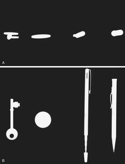
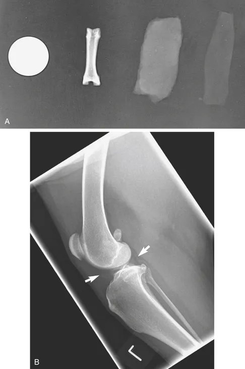
Metallic substances are very dense, and they inhibit the passage of virtually all incident radiation. Areas of film covered by such material appear white (radiopaque) on a radiograph.
Bone is not as dense as a metallic substance. It allows little radiation to pass through it compared with other body tissues. Areas of film that have been covered by bone appear almost white on a radiograph.
Fluid inhibits the passage of more of the incident radiation than gas but not as much as bone does. A fluid opacity lies between the whiteness of a bone opacity and the blackness of a gas opacity. Fluid opacities appear gray on a radiograph. Because soft tissues consist, for the most part, of fluid, soft tissue opacity and fluid opacity appear similar. All fluid opacities appear the same. It is not possible, consequently, to distinguish radiographically among blood, chyle, transudates, and exudates.
Fat opacity falls between fluid and gas opacities. Fat may help to outline structures that would not otherwise be seen; for example, perirenal fat may outline the kidneys by providing a contrasting opacity to the kidney tissues.
Gases, including air, allow x-rays to pass freely through them. Areas of film covered by gas-containing organs, such as the lungs, appear dark (radiolucent) on a radiograph.
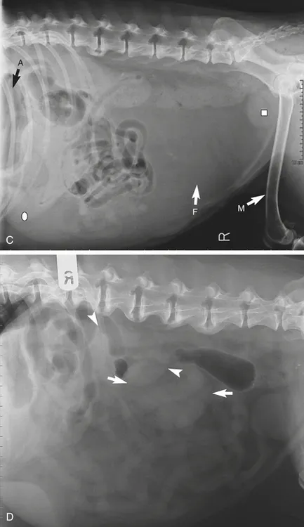
Bone, fluid, fat, and gas occur normally within the body and are said to have biologic densities. Metallic densities are introduced into the body as contrast media (explained later in this chapter), surgical implants, or foreign bodies (Figure 1-2, A to C).