
eBook - ePub
Pulmonary Pathology E-Book
A Volume in Foundations in Diagnostic Pathology Series
Dani S. Zander, Carol F. Farver
This is a test
Compartir libro
- 884 páginas
- English
- ePUB (apto para móviles)
- Disponible en iOS y Android
eBook - ePub
Pulmonary Pathology E-Book
A Volume in Foundations in Diagnostic Pathology Series
Dani S. Zander, Carol F. Farver
Detalles del libro
Vista previa del libro
Índice
Citas
Información del libro
Now fully revised to include recent advances in the field, the second edition of Pulmonary Pathology, a volume in the Foundations in Diagnostic Pathology series, is an essential foundation text for residents and pathologists. The popular template format makes it easy to use, and new information throughout brings you up to date with what's new in pulmonary pathology and pulmonary medicine, including molecular genetics and personalized medicine therapies. Practical and affordable, this resource by Drs. Dani S. Zander and Carol F. Farver is ideal for study and review as well as everyday clinical practice.
- Coverage of both common and rare neoplastic and non-neoplastic diseases of the lung and pleura.
- A focus primarily on diagnosis, with correlations to clinical and radiographic characteristics.
- Clinical and Pathologic Features summarized in quick-reference boxes for fast retrieval of information.
- Hundreds of photomicrographs and gross photographs – most in full color – depict important pathologic features, enabling you to form a differential diagnosis and compare your findings with actual cases.
- Contributions from internationally recognized pathologists, keeping you up to date with the latest information in the field.
- Consult this title on your favorite e-reader, conduct rapid searches, and adjust font sizes for optimal readability.
- Virtual Microscope slides now available online.
- Molecular genetics and personalized medicine therapies included throughout.
- New classification and approaches to diagnosis and management of pediatric diffuse lung diseases.
- 9/11-related lung disease and other recently described environmental lung diseases.
- Information on susceptibility genes for individual diseases.
- Viral linkage and new therapies for idiopathic pulmonary fibrosis, and well as information on endobronchial ultrasound-guided needle aspiration.
Preguntas frecuentes
¿Cómo cancelo mi suscripción?
¿Cómo descargo los libros?
Por el momento, todos nuestros libros ePub adaptables a dispositivos móviles se pueden descargar a través de la aplicación. La mayor parte de nuestros PDF también se puede descargar y ya estamos trabajando para que el resto también sea descargable. Obtén más información aquí.
¿En qué se diferencian los planes de precios?
Ambos planes te permiten acceder por completo a la biblioteca y a todas las funciones de Perlego. Las únicas diferencias son el precio y el período de suscripción: con el plan anual ahorrarás en torno a un 30 % en comparación con 12 meses de un plan mensual.
¿Qué es Perlego?
Somos un servicio de suscripción de libros de texto en línea que te permite acceder a toda una biblioteca en línea por menos de lo que cuesta un libro al mes. Con más de un millón de libros sobre más de 1000 categorías, ¡tenemos todo lo que necesitas! Obtén más información aquí.
¿Perlego ofrece la función de texto a voz?
Busca el símbolo de lectura en voz alta en tu próximo libro para ver si puedes escucharlo. La herramienta de lectura en voz alta lee el texto en voz alta por ti, resaltando el texto a medida que se lee. Puedes pausarla, acelerarla y ralentizarla. Obtén más información aquí.
¿Es Pulmonary Pathology E-Book un PDF/ePUB en línea?
Sí, puedes acceder a Pulmonary Pathology E-Book de Dani S. Zander, Carol F. Farver en formato PDF o ePUB, así como a otros libros populares de Medicine y Pathology. Tenemos más de un millón de libros disponibles en nuestro catálogo para que explores.
1
Normal Anatomy, Tissue Artifacts, and Incidental Structures
Douglas B. Flieder
Normal Anatomy
The lungs occupy most of the volume of the thoracic cavity. The average weights of male and female lungs are approximately 850 grams and 750 grams, respectively. The right lung is composed of ten distinct segments comprising three lobes (upper, middle, and lower), and the left lung has ten segments organized into two lobes (upper and lower). Each lobe is covered with pleura (visceral pleura) and separated from the other lobes by fissures. At the microscopic level, the lungs feature distinct yet integrated components, including conducting airways, distal airspaces, blood vessels and lymphatics, and other cellular constituents (Table 1.1).
Airways
Not only do conducting airways form the passageways through which air enters and exits the lungs, but they also warm, humidify, and aid in sterilizing incoming air. The trachea bifurcates into the left and right mainstem bronchi, which bifurcate into additional bronchi that undergo further bifurcations into smaller bronchi and then bronchioles. Airways in adult lungs usually undergo 23 divisions to finally merge with the gas exchange units, the alveoli.
Airways are classified as either bronchi or bronchioles. Bronchi have cartilaginous walls and measure more than 0.1 cm in diameter, whereas bronchioles measure less than 0.1 cm in diameter and lack cartilage. In the mainstem bronchi, hyaline cartilage is C-shaped, but as the airways enter the lung tissue, the cartilage becomes discontinuous. As the bronchial diameter decreases, the cartilage plates become smaller. Unlike bronchioles, bronchi also have submucosal salivary-type glands with both serous and mucous cells (Fig. 1.1).
Terminal bronchioles are the smallest pure conducting airways; about 30,000 terminal bronchioles are found within the lungs. The terminal bronchioles bifurcate into respiratory bronchioles, whose walls consist partially of alveoli (Fig. 1.2). Bronchioles also give rise to alveolar ducts, which terminate in alveolar sacs.
Airways are composed of mucosa, submucosa, muscularis propria, and adventitia. Bronchial epithelium lines the airway lumen and includes pseudostratified ciliated columnar epithelial cells, interspersed goblet cells and neuroendocrine cells, and underlying basal cells. The ciliated respiratory epithelial cells and goblet cells are specialized cells that function in mucociliary clearance mechanisms. Goblet cells secrete mucus, which is important for trapping inhaled particles, and the cilia propel the mucus and entrapped particles toward the pharynx, where they can be eliminated. Bronchi also feature basal cells, pluripotential reserve cells that can regenerate a damaged bronchial mucosa. Scattered neuroendocrine cells are also interspersed in the respiratory epithelium. Clusters of neuroendocrine cells can occasionally be found at airway bifurcations and are termed neuroepithelial bodies. Neuroendocrine cells may not be recognizable in routine hematoxylin and eosin-stained tissue sections but can be highlighted by immunohistochemical staining using antibodies directed against chromogranin or synaptophysin antigens. Neuroendocrine cells may play a role in lung development and/or ventilation/perfusion regulation.
In bronchioles, goblet cells are replaced by nonciliated columnar cells with prominent apical cytoplasm (Clara cells). Clara cells produce surfactant-like material, accumulate and detoxify inhaled toxins, and serve as progenitor cells for regeneration of damaged bronchiolar epithelium.
All airways feature a basement membrane composed of type III collagen and underlying elastic fibers and smooth muscle bundles. Airways are richly innervated by parasympathetic and sympathetic nerves. Blood vessels and lymphatics also course through the airway submucosa.
TABLE 1.1
Structural and Cellular Components of the Lungs
Bronchi
Epithelium
Ciliated columnar epithelial cells
Goblet cells
Basal cells
Neuroendocrine cells
Subepithelial connective tissue
Submucosal serous and mucinous acini with myoepithelial cells
Smooth muscle
Hyaline cartilage
Autonomic nervous system components
Vasculature and lymphatics
Bronchioles
Epithelium
Ciliated columnar epithelial cells
Clara cells
Subepithelial connective tissue
Smooth muscle
Autonomic nervous system components
Vasculature and lymphatics
Alveoli
Epithelium
Type I pneumocytes
Type II pneumocytes
Alveolar macrophages
Interstitium
Fibroblasts
Myofibroblasts
Monocytes/macrophages
Mast cells
Collagen and elastic fibers
Alveolar capillaries
Endothelial cells
Pericytes
Interlobular septa
Connective tissue
Veins and lymphatics
Visceral pleura
Mesothelial cells
Connective tissue with blood vessels and lymphatics
Gas Exchange Units
An average lung from a man contains approximately 300 million alveoli and 140 m2 of gas-exchanging alveolar surface. Several terminal bronchioles and associated airspaces form each pulmonary lobule, which is bounded by a fibrous septum (Fig. 1.3). The lobules function semiautonomously, with neural controls to regulate air and blood flow. Lobules consist of up to 30 individual gas exchange compartments termed acini. An acinus is an anatomic unit that consists of multiple respiratory bronchioles, alveolar ducts, and alveoli that are supplied by a single terminal bronchiole.
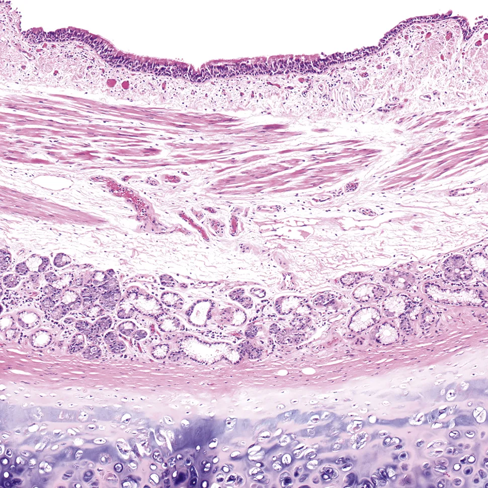
FIG. 1.1 Bronchus. The bronchial wall features pseudostratified ciliated columnar epithelium with goblet cells, submucosal seromucinous glands, bronchial vessels and lymphatics, smooth muscle, and hyaline cartilage.
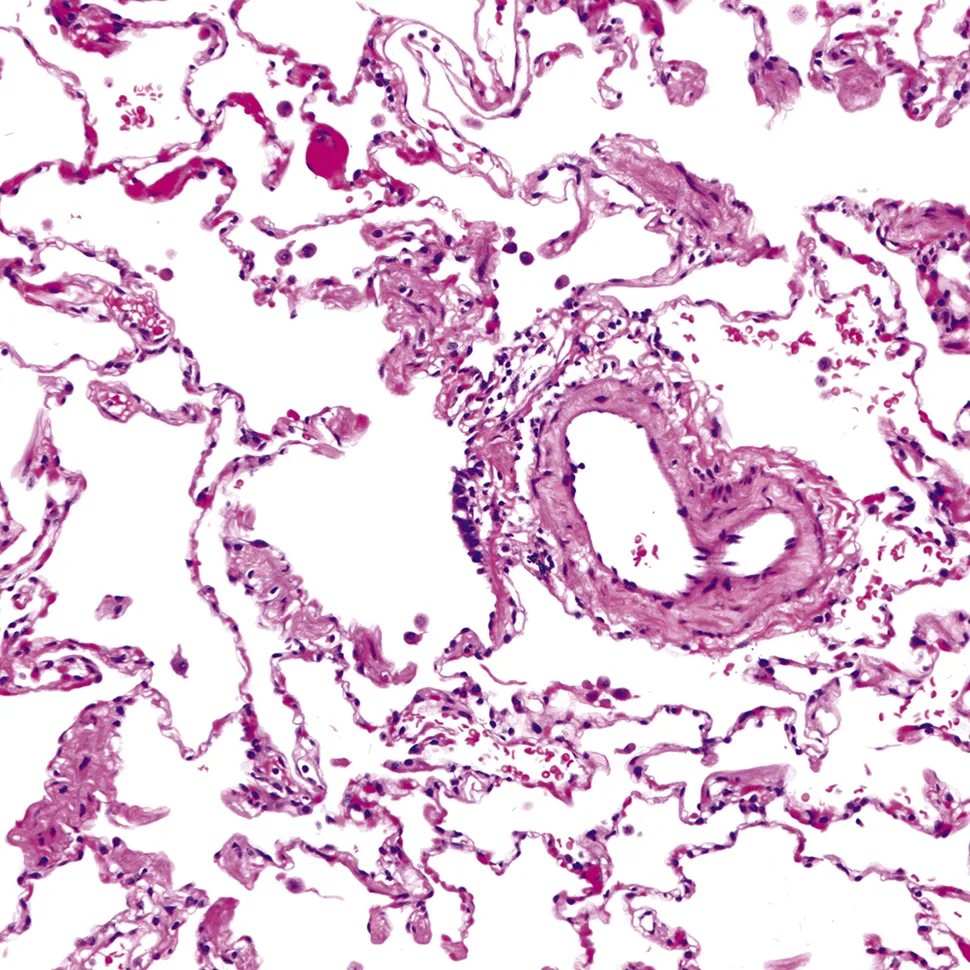
FIG. 1.2 Respiratory bronchiole and peribronchiolar structures. The respiratory bronchiole travels with a small branch of the pulmonary artery. This airway opens into an alveolar duct, as well as individual alveolar sacs. Scattered intraalveolar macrophages are a common finding and may be increased in smokers.
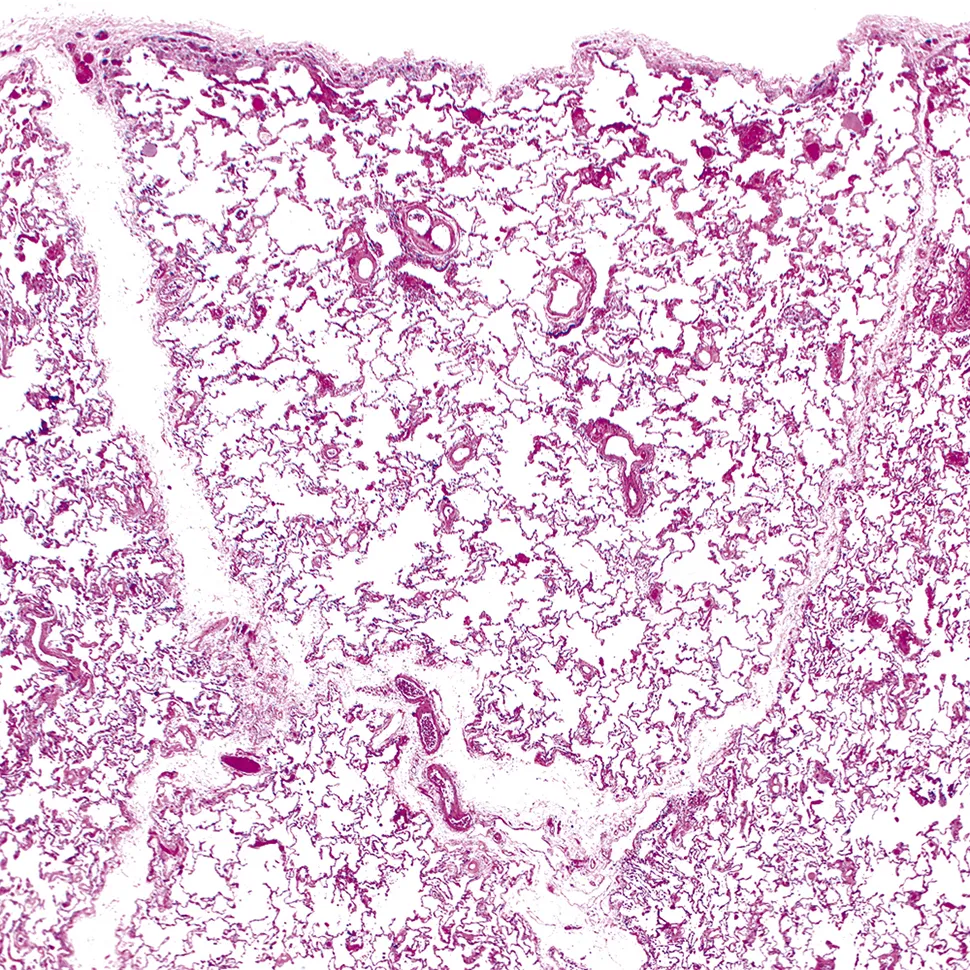
FIG....