An indispensable introduction to using digital technology in dentistry
Digital Dentistry: A Step-by-Step Guide and Case Atlas provides basic information on the use of digital resources to find a diagnosis, create a treatment plan, and execute that strategy within different dental specialisms.
This manual includes the science behind all procedures that use digital technology and provides a clinical step-by-step guide toward the use of these developments for every dental specialty area. Users will find a wide range of areas covered, from prosthodontics, restorative dentistry, and endodontics to oral and maxillofacial surgery and public health. This book also includes:
A guide to all current basic digital imaging and CAD-CAM procedures, with an emphasis on the most popular systems and software programs.
An atlas of multidisciplinary cases that were treated with digital dentistry, from diagnosis and treatment planning to execution and follow-up, in order of complexity
Assessment of the scientific basis for using digital dentistry in each category
A presentation of clinical cases to support the use of digital methodologies in all relevant scenarios
An exploration of the role of digital dentistry in dental public health, preventive dentistry, and dental education
Ideal for dental clinicians—general practitioners and specialists—as well as all other dental professionals, such as dental technologists, dental hygienists, and dental students, Digital Dentistry: A Step-by-Step Guide and Case Atlas is an essential tool and reference work to help dental practitioners streamline and update their practice with the most up-to-date technologies.
Preguntas frecuentes
¿Cómo cancelo mi suscripción?
Simplemente, dirígete a la sección ajustes de la cuenta y haz clic en «Cancelar suscripción». Así de sencillo. Después de cancelar tu suscripción, esta permanecerá activa el tiempo restante que hayas pagado. Obtén más información aquí.
¿Cómo descargo los libros?
Por el momento, todos nuestros libros ePub adaptables a dispositivos móviles se pueden descargar a través de la aplicación. La mayor parte de nuestros PDF también se puede descargar y ya estamos trabajando para que el resto también sea descargable. Obtén más información aquí.
¿En qué se diferencian los planes de precios?
Ambos planes te permiten acceder por completo a la biblioteca y a todas las funciones de Perlego. Las únicas diferencias son el precio y el período de suscripción: con el plan anual ahorrarás en torno a un 30 % en comparación con 12 meses de un plan mensual.
¿Qué es Perlego?
Somos un servicio de suscripción de libros de texto en línea que te permite acceder a toda una biblioteca en línea por menos de lo que cuesta un libro al mes. Con más de un millón de libros sobre más de 1000 categorías, ¡tenemos todo lo que necesitas! Obtén más información aquí.
¿Perlego ofrece la función de texto a voz?
Busca el símbolo de lectura en voz alta en tu próximo libro para ver si puedes escucharlo. La herramienta de lectura en voz alta lee el texto en voz alta por ti, resaltando el texto a medida que se lee. Puedes pausarla, acelerarla y ralentizarla. Obtén más información aquí.
¿Es Digital Dentistry un PDF/ePUB en línea?
Sí, puedes acceder a Digital Dentistry de Arthur R. G. Cortes, Arthur R. G. Cortes en formato PDF o ePUB, así como a otros libros populares de Medicine y Dentistry. Tenemos más de un millón de libros disponibles en nuestro catálogo para que explores.
Renan L.B. da Silva, Jun Ho Kim, Roberto A. Markarian, Rui Falacho, Djalma N. Cortes, Alan J.M. Costa, and Arthur R.G. Cortes
SUMMARY
This chapter will discuss all the terms and definitions that the dental professional needs to know to understand the procedures discussed in the following chapters. Such definitions include abbreviations and general concepts of digital imaging and digital workflow. The chapter also presents a history of the use of CAD‐CAM in dentistry in the last two decades, and the basic knowledge required plus ideas and alternatives to start with digital dentistry.
1.1 Definitions
Digital dentistry is the term used to describe the different modalities of dental treatment workflow that are mostly performed with the use of digital technologies. Several digital methods have been incorporated to dental practice to replace conventional methods and techniques in order to enhance treatment planning and predictability of execution. Nowadays, digital dentistry is considered a whole field of study within dentistry. As with any other field of study, digital dentistry involves a learning curve to be mastered and used in the clinical routine. Ultimately, the dental professional is responsible for using existing digital tools appropriately for patient treatment. In other words, the basic theories of dentistry are still the same and should be very well known by the professional, who will be able to use these new digital tools to enhance predictability in executing the treatment plan.
In order to become familiar with digital dentistry and take advantage of its benefits, it is required to learn a series of important concepts and abbreviations. The most important of these are discussed below.
1.1.1 Three‐Dimensional Imaging
Conventional two‐dimensional (2D) imaging modalities usually have several limitations such as image distortion, magnification, superimposition of anatomical structures, and lack of three‐dimensional (3D) information for diagnosis and planning. In this context, 3D imaging modalities such as cone beam computed tomography (CBCT), intraoral and facial scanning systems provide 3D digital images for dentistry [1–3]. CBCT imaging allows for visualization and assessment of bone structures with high diagnostic accuracy and precision. For CBCT images, the professional needs to understand image acquisition parameters, since the quality of the image affects the quality of the work in digital dentistry. There are several CBCT acquisition parameters, such as field of view size (FOV), peak kilovoltage (kVp), milliamperage (mA), and voxel size. Each of these parameters has an influence on CBCT quality [2–5].
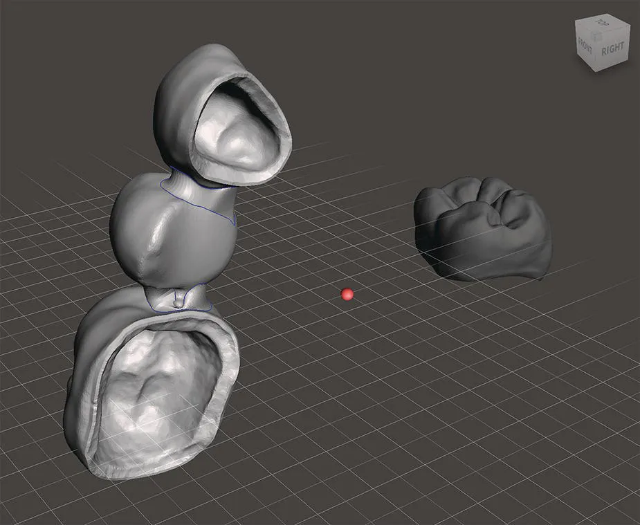
Intraoral and facial scanning can capture 3D patient images that can be used for digital treatment planning systems (Figure 1.1). The software will then develop a digital representation of the 3D object surfaces available, which will be automatically converted into 3D images composed by wireframe models.
Any 3D images can be rendered and edited in the 3D space, before being converted and saved in a specific file format [5]. As discussed in the next chapter, three file formats are commonly used in digital dentistry: OBJ, STL, and PLY. These files are based on the geometric reconstruction of objects by vectors, triangles or polygons, considering their positioning in a 3D space. After all data is ready, it is possible to store the shape of a model and other details such as color or texture.
Figure 1.1 Three‐dimensional objects imported in different coordinates of the 3D space (screen capture of MeshMixer software, Autodesk). Note that the fixed bridge is closer to the screen than the molar crown. The dynamic grid is used to orientate the spatial disposition of the 3D objects.
Three‐dimensional images can be manipulated in various ways, depending on the characteristics of the software. For example, with DICOM and STL files, using the CAD software one can plan and perform digital surgery of dental implants and wax‐up of future prostheses. After digital planning, the implant surgery guide, temporary crowns, and definitive crowns can be printed with additive manufacturing devices or milled by subtractive manufacturing devices [5, 6].
1.1.2 Coordinates and Planes
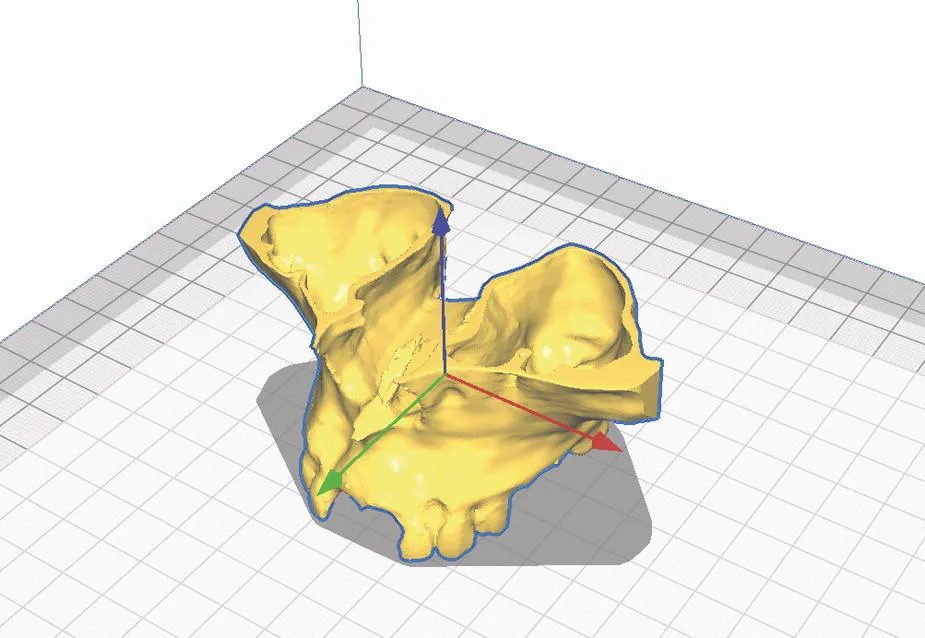
All 3D images are created or rendered in a virtual space of coordinates and planes. Any objects that are digitally designed within the 3D coordinates can be fully edited in the virtual space, before being manufactured. The coordinate system is a method of assigning numbers to points. In three dimensions, three numbers are required to specify a point. Plain 2D images have numbers related to only two coordinates (x and y). The coordinate that represents the third dimension is usually an axis called z. The z‐axis is perpendicular to both the x‐axis and the y‐axis (Figure 1.2).
The coordinates and the respective planes provide references for the location, size, and volume of the 3D images. All 3D objects have their coordinates fixed in a virtual plane of the imaging software. It is important to make sure that multiple 3D objects to be manipulated or aligned are positioned in the same spatial coordinates, which can be used as spatial references. Therefore, 3D files from different imaging methods should be in the same 3D coordinates in order to be superimposed or combined with the aim of creating a virtual patient, as explained further in this chapter.
Figure 1.2 A 3D object (reconstructed model of a max...