Atlas of Canine and Feline Urinalysis
eBook - ePub

Atlas of Canine and Feline Urinalysis

Theresa E. Rizzi, Amy C. Valenciano, Mary Bowles, Rick L. Cowell, Ronald Tyler, Dennis B. DeNicola

Compartir libro
  1. English
  2. ePUB (apto para móviles)
  3. Disponible en iOS y Android
eBook - ePub

Atlas of Canine and Feline Urinalysis

Theresa E. Rizzi, Amy C. Valenciano, Mary Bowles, Rick L. Cowell, Ronald Tyler, Dennis B. DeNicola

Detalles del libro
Vista previa del libro
Índice
Citas

Información del libro

Atlas of Canine and Feline Urinalysis

Atlas of Canine and Feline Urinalysis offers an image-based reference for performing canine and feline urinalyses. Hundreds of full-color images depict techniques, physical characteristics, urine chemistry, and microscopic characteristics of urine sediment in dogs and cats. Written by leading experts, this highly illustrated resource acts as an aid to accurately identifying cytological features and interpreting both chemical and sediment findings.

Logically organized for easy navigation, the book covers urine collection and specimen handling, initial assessment, urine chemistry, and microscopic findings, including casts, crystals, cells, organisms, and artifacts. Atlas of Canine and Feline Urinalysis is an important diagnostic tool for veterinary undergraduate and graduate students, veterinary technicians, general practitioners, veterinary clinical pathologists, and specialists in other disciplines.

Key features

  • Presents hundreds of full-color images for reference and picture-matching while using urinalysis as a diagnostic tool
  • Provides a complete guide to properly performing a urinalysis exam in the veterinary practice
  • Emphasizes collection techniques, physical assessment, urine chemistry, and the microscopic sediment exam
  • Covers casts, crystals, cells, organisms, and artefacts
  • Offers a practical, visual resource for incorporating urinalysis into the clinic

Preguntas frecuentes

¿Cómo cancelo mi suscripción?
Simplemente, dirígete a la sección ajustes de la cuenta y haz clic en «Cancelar suscripción». Así de sencillo. Después de cancelar tu suscripción, esta permanecerá activa el tiempo restante que hayas pagado. Obtén más información aquí.
¿Cómo descargo los libros?
Por el momento, todos nuestros libros ePub adaptables a dispositivos móviles se pueden descargar a través de la aplicación. La mayor parte de nuestros PDF también se puede descargar y ya estamos trabajando para que el resto también sea descargable. Obtén más información aquí.
¿En qué se diferencian los planes de precios?
Ambos planes te permiten acceder por completo a la biblioteca y a todas las funciones de Perlego. Las únicas diferencias son el precio y el período de suscripción: con el plan anual ahorrarás en torno a un 30 % en comparación con 12 meses de un plan mensual.
¿Qué es Perlego?
Somos un servicio de suscripción de libros de texto en línea que te permite acceder a toda una biblioteca en línea por menos de lo que cuesta un libro al mes. Con más de un millón de libros sobre más de 1000 categorías, ¡tenemos todo lo que necesitas! Obtén más información aquí.
¿Perlego ofrece la función de texto a voz?
Busca el símbolo de lectura en voz alta en tu próximo libro para ver si puedes escucharlo. La herramienta de lectura en voz alta lee el texto en voz alta por ti, resaltando el texto a medida que se lee. Puedes pausarla, acelerarla y ralentizarla. Obtén más información aquí.
¿Es Atlas of Canine and Feline Urinalysis un PDF/ePUB en línea?
Sí, puedes acceder a Atlas of Canine and Feline Urinalysis de Theresa E. Rizzi, Amy C. Valenciano, Mary Bowles, Rick L. Cowell, Ronald Tyler, Dennis B. DeNicola en formato PDF o ePUB, así como a otros libros populares de Medicine y Veterinary Medicine. Tenemos más de un millón de libros disponibles en nuestro catálogo para que explores.

Información

Año
2017
ISBN
9781119110415
Edición
1
Categoría
Medicine

1
Sample Collection and Handling

The method of urine collection and the subsequent handling of the sample can influence the interpretation of results. The following discussion is a review of urine collection and handling techniques.

Collection of Urine Samples

The three techniques for urine collection are: free-catch, catheterization, and cystocentesis. Each of these methods of collection and their associated advantages and disadvantages will be discussed. General considerations related to urine sample collection, handling, and submission, regardless of collection method, are listed in Box 1.1 (Figures 1.11.4).

Box 1.1 General considerations for urine sample collection, handling, and submission.

Collection

  1. Observe principles of aseptic technique as much as possible
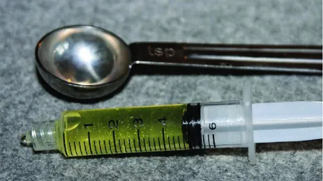
  2. Collect adequate volume (minimum of 5 mL recommended) (Figure 1.1)
  3. Clean and/or sterile container provided by veterinarian preferred for collection for UA
  4. Sterile container preferred for collection of sample to be cultured
  5. When possible, withhold drugs and fluid administration prior to sample collection

Handling

  1. Ideally UA should be performed within 60 minutes of sample collection and no longer than 6 hours following collection
  2. Refrigerate and cap the sample when UA cannot be performed within 60 minutes of collection (if possible, perform the dipstick portion prior to refrigeration)
  3. Refrigerated samples should be brought to room temperature prior to performing UA

Submission

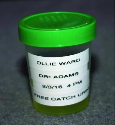
  1. Container submitted should identify sample as urine, be capped, and have adequate patient identification (Figure 1.2)
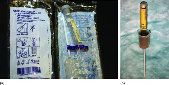
  2. Consider using preservative tube* for storage and submission of sample for culture
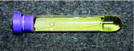
  3. Sample for cytologic evaluation should include one or more air-dried slides of urine sediment prepared by the blood smear or squash preparation technique along with a urine sample placed in an EDTA tube (Figure 1.3)
  4. Pertinent information should be provided for samples submitted for evaluation to an outside laboratory, including patient signalment, history, and method of urine collection
  5. Fasted samples submitted for evaluation frequently have increased specific gravity values, more cells and casts, and decreased pH values compared to non-fasted samples
UA=urinalysis
*e.g. BD Vacutainer®Culture and Sensitivity Preservative Tube. The BD 4-mL urine culture preservative tube kit includes a urine transfer straw which can be used to facilitate aspiration of urine into the tube (Figure 1.4). Use of the transfer straw is optional. Although the manufacturer recommends adding a minimum of 3 mL of patient urine for an optimum preservative to urine ratio, obtaining that amount for urine from canine and feline patients can be challenging. It has been the authors' experience that the BD 4-mL urine culture preservative tube can be employed successfully for culture using a much smaller volume (≥0.5 mL) of urine, when necessary.
Image described by caption.
Figure 1.1 The minimum recommended volume for routine urinalysis is 5 mL or 1 teaspoon.
Image described by caption.
Figure 1.2 Urine submitted for analysis should be submitted in a suitable, capped container with appropriate labeling.
Image described by caption.
Figure 1.3 Cytologic evaluation of urine can be enhanced by including a urine sample submitted in an EDTA tube.
Image described by caption.
Figure 1.4 (a) BD Vacutainer® 4-mL urine culture preservative tube kit. (b) Culture preservative tube attached to urine transfer straw.

Free-Catch Urine Collection

The free-catch method of urine collection (Box 1.2 and Figures 1.51.9) is often easy to perform but is dependent upon the cooperation of the patient and may be difficult to accomplish in patients with conditions producing urge incontinence. Samples are usually collected during normal voiding or by manual external compression of the urinary bladder. Normal voiding free-catch urine sampling can often be performed by the owners and does not pose a risk to the pet. The manual compression of a distended urinary bladder (Box 1.3 and Figures 1.101.14) may be at the convenience of the collector; however, drawbacks include sample contamination, urinary bladder trauma, and reflux of infected urine into the ureters, kidney, and prostate. Furthermore, this technique cannot be used following a cystotomy operation and may be unpleasant in other postoperative laparotomy patients.
Collecting a midstream urine sample is preferred to minimize sample contamination; however, some contamination with cells, bacteria, and debris from the distal urethra, genital tract, and external skin and hair coat is unavoidable. Obtaining an optimal free-catch sample can be facilitated by using one container to collect the beginning of the urine stream and then changing to a second collecting container as the urine stream continues. The urine in the second container should be more representative of a true midstream sample. In some cases, a free-catch sample containing white cells, bacteria, and/or protein may be an indication to collect a subsequent patient urine sample via cystocentesis or catheterization in order to help establish the source of the abnormalities identified in the voided sample.
Sometimes a satisfactory free-catch sample cannot be obtained either during normal voiding or via manual expression, most commonly due to the pet's behavior or urge incontinence. Manual expression can be especially problematic in male cats as a result of resistance to handling and difficulty in initiating voiding due to the small diameter of the male feline urethra. Collecting urine by catheterization or cystocentesis are alternative options but not readily accomplished in all patients. Consequently, analysis of post-voided urine collected from a variety of surfaces may be necessary in select cases (Box 1.4 and Figure 1.15). Voided cat urine can sometimes be collected from a clean litter pan to which nonabsorbable plastic beads (e.g. Uri-Void™) or hydrophobic sand (Kit4Cat™) has been added. Cat owners may also use clean glass aquarium beads, straws which have been cut up, or plastic craft beads as litter substitutes. In some cases urine can be successfully collected from a litter pan after a feline patient has voided on top of clinging plastic wrap that has been placed over the cat's usual litter or, in the case of outdoor cats, over a layer of dirt (Figure 1.16). At times the only available urine for analysis is a sample that has been voided onto a floor, tabletop, or other contaminated surface. This contamination factor must be taken into consideration when evaluating such a sample. Prompt examination of the collected urine sample should decrease the level of artifacts encountered. As would be expected, the less contaminated the collection surface, the more reliable the urinalysis results obtained. However, disinfectants used in cleaning the surface from which a urine sample is subsequently obtained also have the potential to alter the urinalysis results, particularly when performing dipstick colorimetric tests. Post-voided urine samples obtained from a surface are generally unsatisfactory for accurate identification of infectious agents, especially if there is a time delay in analysis. At minimum, specific gravity of the urine specimen can usually be determined with reasonable accuracy.

Box 1.2 Techniques for obtaining free catch urine samples – normal voiding.

Canine Technique

  1. If the dog's hair coat around the vulva or prepuce is notably dirty, clean the area and pat dry
  2. Walk the dog on a leash early in the morning, after feeding, or at another time that the dog is accustomed to urinating
  3. Observe the dog for initiation of micturition and be prepared to collect a sample at the beginning of urination. If desired, latex gloves can be worn by the collector for his/her protection. The collector can plan on positioning the urine container with his/her hands or with a device designed to hold and position the container. Commercial collection devices are available (e.g. Olympic Clean-Catch™) or can be made at home. One such homemade device consists of a yardstick or pole/broom-type handle taped to the handle of a ladle or suitable plastic measuring cup (Figure 1.5)). Have one or two suitable containers available for urine collection. The container(s) should be clean and dry and appropriate for the size of the dog. Container(s) may be provided by the veterinarian or a suitable clean, dry household plastic or glass container may be used for urine collection. Smaller dogs may require a flatter collecting receptacle such as a shallow plastic tray, a Styrofoam plate with a raised rim, or a metal, disposable pie plate (Figure 1.6)
  4. As soon as micturition is initiated or a micturition posture is assumed, place the collection container as unobtrusively as possible under the vulva, immediately anterior and ventral to the prepuce, or directly in the urine stream produced. If possible, obtain at least 5–10 mL (1–2 tsp) for urinalysis. If the collector is able, a second container (midstream sample) should be positioned for urine collection provided the dog is continuing to urinate after a sample of the initial urine stream has been obtained in the fir...

Índice