Abdominal X-rays for Medical Students
eBook - ePub

Abdominal X-rays for Medical Students

Christopher Clarke, Anthony Dux

Compartir libro
  1. English
  2. ePUB (apto para móviles)
  3. Disponible en iOS y Android
eBook - ePub

Abdominal X-rays for Medical Students

Christopher Clarke, Anthony Dux

Detalles del libro
Vista previa del libro
Índice
Citas

Información del libro

Highly Commended at the British Medical Association Book Awards 2016 Abdominal X-rays for Medical Students is a comprehensive resource offering guidance on reading, presenting and interpreting abdominal radiographs. Suitable for medical students, junior doctors, nurses and trainee radiographers, this brand new title is clearly illustrated using a unique colour overlay system to present the main pathologies and to highlight the abnormalities in abdomen x-rays.

Abdominal X-rays for Medical Students:

  • Covers the key knowledge and skills necessary for practical use
  • Provides an effective and memorable way to analyse and present abdominal radiographs - the unique 'ABCDE' system as developed by the authors
  • Presents each radiograph twice, side by side: the first as seen in the clinical setting, and the second with the pathology clearly highlighted
  • Includes self-assessment to test knowledge and presentation technique

With a systematic approach covering both the analysis of radiographs and next steps mirroring the clinical setting and context, Abdominal X-rays for Medical Students is a succinct and up-to-date overview of the principles and practice of this important topic.

Preguntas frecuentes

¿Cómo cancelo mi suscripción?
Simplemente, dirígete a la sección ajustes de la cuenta y haz clic en «Cancelar suscripción». Así de sencillo. Después de cancelar tu suscripción, esta permanecerá activa el tiempo restante que hayas pagado. Obtén más información aquí.
¿Cómo descargo los libros?
Por el momento, todos nuestros libros ePub adaptables a dispositivos móviles se pueden descargar a través de la aplicación. La mayor parte de nuestros PDF también se puede descargar y ya estamos trabajando para que el resto también sea descargable. Obtén más información aquí.
¿En qué se diferencian los planes de precios?
Ambos planes te permiten acceder por completo a la biblioteca y a todas las funciones de Perlego. Las únicas diferencias son el precio y el período de suscripción: con el plan anual ahorrarás en torno a un 30 % en comparación con 12 meses de un plan mensual.
¿Qué es Perlego?
Somos un servicio de suscripción de libros de texto en línea que te permite acceder a toda una biblioteca en línea por menos de lo que cuesta un libro al mes. Con más de un millón de libros sobre más de 1000 categorías, ¡tenemos todo lo que necesitas! Obtén más información aquí.
¿Perlego ofrece la función de texto a voz?
Busca el símbolo de lectura en voz alta en tu próximo libro para ver si puedes escucharlo. La herramienta de lectura en voz alta lee el texto en voz alta por ti, resaltando el texto a medida que se lee. Puedes pausarla, acelerarla y ralentizarla. Obtén más información aquí.
¿Es Abdominal X-rays for Medical Students un PDF/ePUB en línea?
Sí, puedes acceder a Abdominal X-rays for Medical Students de Christopher Clarke, Anthony Dux en formato PDF o ePUB, así como a otros libros populares de Medicine y Radiology, Radiotherapy & Nuclear Medicine. Tenemos más de un millón de libros disponibles en nuestro catálogo para que explores.

Información

Año
2015
ISBN
9781118600542

About X-rays

What are X-rays?

X-rays are a form of ionising radiation. They are part of the electromagnetic spectrum and have sufficient energy to cause ionisations. They contain more energy than ultraviolet (UV) waves but less energy than gamma rays.
c1-fig-0001
Figure 1: The electromagnetic spectrum (Freq is short for frequency).
Radiation is the transfer of energy in the form of particles or waves.
Ionising radiation is the radiation with sufficient energy to cause ionisations, which is a process whereby radiation removes an outer shell electron from an atom. Thus ionising radiation is able to cause changes on a molecular level in biologically important molecules (e.g. DNA).
Uses of ionising radiation include conventional X-rays (plain radiographs), contrast studies, computed tomography (CT), nuclear medicine and positron emission tomography (PET).

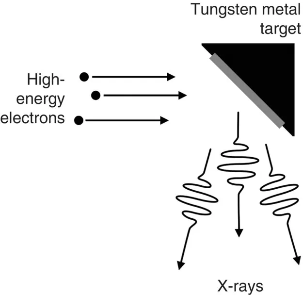
How are X-rays produced?

X-rays are produced by focusing a high-energy beam of electrons onto a metal target (e.g. tungsten). The electrons hit the metal target and some will have enough energy to knock out another electron from the inner shell of one of the metal atoms. As a result, electrons from higher energy levels then fill up this vacancy and X-rays are emitted in the process. Producing X-rays this way is extremely inefficient (~0.1%), so most of the energy is wasted as heat. This is why X-ray tubes need to have advanced cooling mechanisms. The X-rays produced then pass through the patient and onto a detector mechanism which produces an image.
c1-fig-0002
Figure 2: X-ray production.

How do X-rays make an image?

Main points include the following:
  1. The resulting image on the X-ray detector is a two-dimensional (2D) representation of a three-dimensional (3D) structure.
  2. While passing through a patient the X-ray beam is absorbed in proportion to the cube of the atomic number of the various tissues through which it passes. By convention, the greater the amount of radiation hitting a detector, the darker the image will be. Therefore, the less “dense” a material is, the more X-rays get through and the darker the image. Conversely the more “dense” a material is, the more X-rays are absorbed and the image appears whiter. Materials of low “density” appear darker than those of high “density”.
  3. Structures can only be seen if there is sufficient contrast with surrounding tissues (contrast is the difference in absorption between one tissue and another).
c1-fig-0003
Figure 3: The spectrum of tissues of different densities as seen on a conventional radiograph. The radiograph example shows the left lumbar region of a patient who swallowed a battery.

How are X-ray images (radiographs) stored?

In some hospitals radiographs are printed onto X-ray film, but most places now use a computer-based digital radiograph storage system for storing X-ray images, thereby eliminating the need for film.
This system is known as Picture Archiving and Communication System (PACS). Doctors and other healthcare professionals are able to view the images (radiographs) on a computer screen, making it easy to manipulate the image (e.g. changing the contrast, zooming in/out, etc).
The advantages are ease of access, both locally and internationally, cost saving and no more lo...

Índice