Biological Sciences
Aneuploidy
Aneuploidy is a genetic condition characterized by an abnormal number of chromosomes in a cell, often resulting from errors in cell division. This can lead to developmental abnormalities and is associated with conditions such as Down syndrome and Turner syndrome. Aneuploidy can occur in both somatic cells and gametes, impacting the health and viability of an organism.
Written by Perlego with AI-assistance
Related key terms
1 of 5
11 Key excerpts on "Aneuploidy"
- Joshua Copel, Mary E. D'Alton, Helen Feltovich, Eduard Gratacos, Anthony O. Odibo, Lawrence D. Platt, Boris Tutschek, Lawrence Platt, Mary E. D'Alton, Helen Feltovich, Eduard Gratacos, Anthony O. Odibo, Lawrence Platt, Boris Tutschek(Authors)
- 2017(Publication Date)
- Elsevier(Publisher)
Aneuploid conditions are often suspected by prenatal ultrasound (US) when multiple, and sometimes severe, structural anomalies are seen. Different aneuploid conditions are typically associated with specific constellations of US findings. US on its own, however, is an insufficient screening tool for Aneuploidy. Other methods, including serum biomarkers and noninvasive prenatal screening, should also be a part of a comprehensive prenatal screening program.Disorder
Definition
Aneuploidy refers to an abnormal copy number of one or more chromosomes. Aneuploid conditions have a subtraction (monosomy) or an addition (trisomy) of chromosomal material in one or more of the chromosome pairs.Polyploidy refers to an abnormal copy number of all of the chromosomes. The normal human cell is diploid, meaning it contains two copies of genetic material, with one copy coming from each parent. A triploid cell has 69 chromosomes (69, XXX, 69 XXY, 69, XYY) and thus three copies of genetic material. Triploidy is most commonly the result of ovum fertilization by two spermatozoa. It is not compatible with life and usually ends in pregnancy loss in the first trimester. The rare fetuses born alive usually die shortly after birth.Prevalence and Epidemiology
Aneuploidy is the most common genetic abnormality detected by prenatal diagnosis. Maternal age is directly related to the incidence of Aneuploidy. For example, as maternal age increases, the combined risk of delivering a child affected by any of the three most common trisomies (trisomy 13, 18, or 21) increases from about 1 : 1065 at age 25 to 1 : 300 at age 35 and 1 : 28 at age 45.Prenatal screening approaches use one or a combination of tests, including maternal serum biomarkers, cell-free fetal deoxyribonucleic acid, nuchal translucency, and detailed anatomic US. Each of these tests is associated with their own detection rates for the various aneuploid conditions, but none of them are considered diagnostic modalities. Chorionic villus sampling (CVS) and amniocentesis remain the mainstays for definitive prenatal diagnosis of Aneuploidy. While percutaneous umbilical blood sampling can also be used, it is not commonly performed for Aneuploidy detection in the absence of another indication for the procedure (e.g., planned fetal transfusion). eBook - PDF
Chromosomal Abnormalities
A Hallmark Manifestation of Genomic Instability
- Marcelo L. Larramendy, Sonia Soloneski, Marcelo L. Larramendy, Sonia Soloneski(Authors)
- 2017(Publication Date)
- IntechOpen(Publisher)
An aneuploid cell can have one or more extra chromosomes, called hyperploid, or it could have lost one or more chromosomes, which is called hypoploid. Following this defini -tion of Aneuploidy, a cell that has doubled its complete genome without dividing is called tetraploid and not aneuploid, because a balanced genome is still present. © 2017 The Author(s). Licensee InTech. This chapter is distributed under the terms of the Creative Commons Attribution License (http://creativecommons.org/licenses/by/3.0), which permits unrestricted use, distribution, and reproduction in any medium, provided the original work is properly cited. Aneuploidy is well known from cancer and systemic trisomies such as Down syndrome. Indeed, at least two out of three cancers exhibit Aneuploidy [1–3]. Although it has been shown that Aneuploidy causes stress and reduces cellular fitness [ 4–7], cancer cells have somehow found a way to cope with Aneuploidy and manage to proliferate despite the detri-mental consequences of Aneuploidy. This is known as the Aneuploidy paradox [6]. Perhaps by selecting numerical chromosomal abnormalities that promote tumor progression in addi-tion to other structural genomic rearrangements, cancer cells can survive and keep grow-ing [8, 9 ]. The profound effect that Aneuploidy has on healthy cells is emphasized by the fact that, besides sex-chromosome abnormalities, in humans, only three systemic autosomal trisomies are compatible with life: trisomy 21 causing Down syndrome, trisomy 13 causing Patau’s syndrome and trisomy 18 causing Edward’s syndrome [10–12]. The viability of these systemic aneuploidies can probably be explained by the fact that these three chromosomes contain the lowest number of genes of all human autosomes. Even though these trisomies can be compatible with life, the majority of such trisomic pregnancies end with a miscarriage, and the children that do survive until birth suffer from severe cognitive and developmental defects [13]. eBook - PDF
Introducing Genetics
From Mendel to Molecules
- Alison Thomas(Author)
- 2014(Publication Date)
- Garland Science(Publisher)
Any organism or cell with a chromosome number that is not an exact multiple of the haploid number of chromosomes is described as aneuploid (Figure 8.1). 8.2 Aneuploidy The most frequent examples of Aneuploidy are cases when a single chromosome is either lost from or added to a normal diploid set (i.e. monosomy or trisomy ). The extra or lacking chromosome can be an autosome or a sex chromosome. Aneuploidy usually results from non-disjunction during meiosis, when either a pair of homologous chromosomes fails to segregate during anaphase I or a EUPLOIDY diploidy 2 n (two copies of each homolog) haploidy n (one copy of each homolog) polyploidy (more than two chromosome sets) triploidy 3 n (three chromosome sets) tetraploidy 4 n (four chromosome sets) Aneuploidy monosomy 2 n –1 (one chromosome missing) trisomy 2 n +1 (one extra chromosome) Figure 8.1 Terminology associated with variation in chromosome number, illustrated by considering three homologous chromosome pairs. 114 CHAPTER 8 Variation in Chromosomal Number and Structure pair of chromatids does not separate during anaphase II. Figure 8.2 illustrates this process. Normally a gamete should have one copy of every chromosome. If non-disjunction occurs during the formation of a gamete it becomes unbalanced – lacking or containing two copies of a particular chromosome. Following fer-tilization of such gametes by a normal haploid gamete, monosomic and trisomic zygotes result (Figure 8.2). Non-disjunction can also occur during mitotic division if sister chromatids fail to separate during anaphase. This results in somatic clones of monosomic or trisomic cells. If mitotic non-disjunction occurs during early embryonic devel-opment it produces a mosaic individual who has patches of cells with different chromosome numbers. 8.3 Monosomy The phenotypic consequences to the individual who develops from an aneu-ploid zygote varies depending upon the chromosome involved, but may be severe. eBook - ePub
Anatomy and Physiology for Midwives E-Book
Anatomy and Physiology for Midwives E-Book
- Jane Coad, Kevin Pedley, Melvyn Dunstall(Authors)
- 2019(Publication Date)
- Elsevier(Publisher)
Box 7.11 ). If the changes can be seen by light microscopy such as a marked structural abnormality or an atypical number of chromosomes, they are termed ‘gross aberrations’ and can be detected from an examination of the karyotype. Chromosomal abnormalities can be classified as numerical or structural, affecting either the autosomes or the sex chromosomes. These types of abnormality are easier than a single-gene abnormality to detect.BOX 7.11 Incidence of Chromosomal Abnormalities- • Incidence of major chromosomal abnormality
-
- • About 1 in 200 live births
- • About 1 in 20 perinatal deaths (stillbirths and early neonatal deaths)
- • About 1 in 2 early spontaneous abortions
- • About 1 in 100 births: single-gene (unifactorial) disorder
- • About 1 in 50 births: + major congenital abnormality
Numerical Abnormalities
The loss or gain of one or more chromosomes is described as Aneuploidy (wrong number of chromosomes), whereas cells with the correct number of chromosomes are euploidic. It is estimated that 10–25% of all human fetuses are aneuploidal, predominantly due to nondisjunction in maternal meiosis, although trisomy 18 most often results from nondisjunction in meiosis II. Aneuploidy appears to occur more frequently in humans than in other species, possibly because of deliberate or occupational exposure to environmental factors such as tobacco smoke, alcohol, oral contraception use, radiation exposure and industrial chemicals, which is probably one of the reasons for a high rate of miscarriage in humans.Aneuploidy is usually due to nondisjunction in the formation of the gametes resulting in a zygote that does not have 46 chromosomes. Monosomy describes the loss of a complete chromosome and trisomy describes the addition of a single chromosome, as in Down syndrome (trisomy 21 or T21 caused by an additional copy of chromosome 21) (see Table 7.1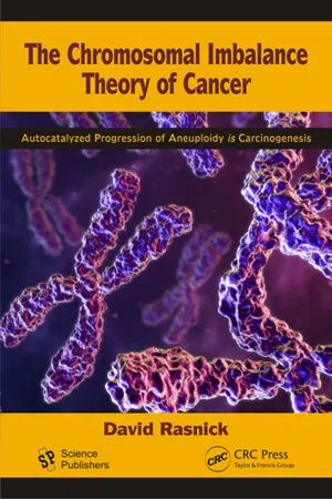 eBook - PDF
The Chromosomal Imbalance Theory of Cancer
The Autocatalyzed Progression of Aneuploidy is Carcinogenesis
- David Rasnick(Author)
- 2016(Publication Date)
- CRC Press(Publisher)
This section introduces the mathematical tools needed to investigate how quantitative changes to the genome caused by chromosomal imbalance affect the overall metabolic activity (hence phenotype) of a cell. The chromosomal imbalance theory asserts that the multiplicity of structural and functional cancer-specific phenotypes are the direct consequence of Aneuploidy. Supporting this view, Lindsley et al. showed that Aneuploidy readily produced numerous complex phenotypes of Drosophila, including reduced survival, small size, and a variety of morphological abnormalities such as rough eyes, abnormal wings and bristle patterns, and a misshapen abdomen. The authors concluded that, “The primary point of theoretical interest to emerge from these studies is that the deleterious effects of Aneuploidy are, in the main, caused by the additive effects of genes that slightly reduce viability and not by the individual effects of a few aneuploid-lethal genes among a large array of dosage insensitive loci” (Hodgkin 2005, Lindsley et al. 1972, Sandler and Hecht 1973). Recent “experimental data have pointed to the conclusion that Aneuploidy is a main driving force for the observed adaptive evolution” in yeast (Rancati et al. 2008) and that fitness ranking between euploid and aneuploid cells is dependent on context and karyotype, providing the basis for the notion that Aneuploidy can directly underlie phenotypic evolution and cellular adaptation (Pavelka et al. 2010a). “Changes in karyotype and the transcriptome can mediate the gene expression changes necessary for phenotypic determination” (Gao et al. 2007). Aneuploidy, alters the expression of many genes by a small extent (direct effect) and a few genes by a large extent (indirect effect) and can thus be viewed as a large-effect mutation (Rancati et al. 2008, Springer et al. 2010, Upender et al. 2004).
- Cheryl Natividad(Author)
- 2019(Publication Date)
- Delve Publishing(Publisher)
Abnormalities in the chromosomes may involve a deviation in their number or structure. These may occur accidentally during cell division like when sister chromatids failed to separate during the formation of egg or sperm or when a chromosome undergoes rearrangement during the early development stages of the embryo. The age of the mother (i.e. older women have a higher risk of giving birth to babies with chromosome abnormalities than younger women) and certain environmental factors may increase the chances of occurrence of these erroneous events (Genetic Alliance, 2009; Nonal Human Genome Research Institute, 2016). Abnormalities in chromosome number Errors during meiosis may result to in cells that have more or less than the normal number of chromosomes. If there is a failure of separation or non-disjunction of homologous chromosomes during meiosis I or of sister chromatids during meiosis II, then one pole will be receiving more chromosomes while the other pole will have missing chromosomes. This eventually produces gametes with an abnormal chromosome number. Non-disjunction during mitosis will also produce cells with excessive or lacking chromosome number (Cross and Wolstenholme, 2001). There are two types of aberrant chromosome number: euploidy and Aneuploidy. Aberrant euploidy refers to having less or more than the normal set of chromosomes. The abnormal chromosome number is a multiple of the haploid chromosome number. On the other hand, Aneuploidy is having less or more than the normal chromosome number but the number of excess or missing chromosomes is less than the haploid set. Aberrant euploidy Sexually-reproducing animals have diploid chromosome number as a result of combining the haploid sets contributed by the paternal and maternal gametes during fertilization. The haploid chromosome number is normally eBook - PDF
Biochemical Evolution
The Pursuit of Perfection
- Athel Cornish-Bowden(Author)
- 2016(Publication Date)
- Garland Science(Publisher)
So, even the best of biological theories usually have to contend with one or two inconsistencies that have to be left to future biologists to clarify. There remains the question of timing. If Aneuploidy is the consequence of cancer, then we should expect to find some cells that are clearly cancerous but which have not yet become aneuploid and vice versa: If Aneuploidy is the cause, we should expect to find noncancerous aneuploid cells. The latter observation is easy to make: After all, any individual with Down syndrome or another trisomy is an entire organism of 10 14 aneuploid 9 The building next to the one where I work was remodeled last year, and I was surprised to see wall panels being installed over an insulating material that looks exactly like the asbestos fiber that was used everywhere 40 years ago. Apparently it is amorphous silica fiber , but if the carcinogenicity of asbestos is not chemical in origin, who is to say that chemically different substances with similar physical properties will not be carcinogenic? I wonder how thoroughly the asbestos substitutes have been checked for carcinogenicity. 218 13. Aspects of Cancer but noncancerous cells. Moreover, although such trisomic individuals do not necessarily develop cancer, they do have a high probability of developing cancer. A person with Down syndrome, for example, has about 30 times the average probability of developing leukemia. Even such a small degree of Aneuploidy is also sufficient to produce around 80 characteristic symptoms of the syndrome, implying that it has many biochemical effects. Does Aneuploidy necessarily lead to cancer? What about the converse observation: Can we find cancerous cells that are not aneuploid? Duesberg and his collaborators studied this question by ex-amining the effects of the carcinogenic chemical dimethylbenzanthracene on Chinese hamster embryo cells. eBook - PDF
Biology and Pathology of the Oocyte
Role in Fertility, Medicine and Nuclear Reprograming
- Alan Trounson, Roger Gosden, Ursula Eichenlaub-Ritter(Authors)
- 2013(Publication Date)
- Cambridge University Press(Publisher)
Section 5 Pathology Chapter 29 Alterations in the gene expression of aneuploid oocytes and associated cumulus cells Dagan Wells The importance of Aneuploidy in human reproductive failure Human reproduction is a remarkably inefficient pro- cess. On average, fertile couples attempting to conceive only succeed in achieving a clinical pregnancy one month out of every five. For infertile patients undergo- ing in vitro fertilization (IVF) pregnancy rates are sim- ilarly low. More than 80% of the embryos transferred to the uterus during IVF treatment fail to implant and two-thirds of cycles do not produce a child [1]. As a result, most IVF patients require two or more rounds of treatment to achieve a pregnancy. There are many potential reasons why an embryo might not estab- lish a pregnancy; however, it is clear that one of the most important is chromosome abnormality. This is particularly true for embryos derived from women of advanced reproductive age. While it is not unusual for half of the blastocyst stage embryos produced by women in their early thirties to be chromosomally abnormal, this figure increases dramatically with age, such that an Aneuploidy rate exceeding 75% is typical for blastocysts from women over the age of 40 [2]. The high prevalence of Aneuploidy, coupled with its detri- mental impact on development, explains the majority of embryo implantation failures and miscarriages. Evi- dence for the lethality of Aneuploidy comes from the detection of chromosome imbalances in the majority of miscarriages [3, 4] and from blinded studies where embryos, later revealed to be chromosomally abnor- mal, had been transferred to patients [5]. Aneuploidy detected in human preimplantation embryos can be derived from sperm or arise due to errors occurring in the mitotic divisions follow- ing fertilization. However, the vast majority of such anomalies originate in the oocyte.
- William A. Stini(Author)
- 2011(Publication Date)
- De Gruyter Mouton(Publisher)
Polyploidy in Humans and Other Vertebrates FATHI ABDEL-HAMEED Each species has a characteristic chromosomal number and morphology. In higher plants and animals, the nuclei of the somatic tissues contain two similar sets, or a diploid number (2n) of chromosomes. One set is donated by a haploid male gamete and represents the paternal genome, while the other represents the maternal genome. A degree of variability in the karyotypes of members of a given species is common in natural popula-tions. If variability involves deviation from normal structure for certain chromosomes, it is described as a structural aberration, while changes in chromosome number are described as numerical aberrations. Under normal conditions, various somatic tissues show the same ploidy level, i.e., homogeneous for diploidy. Infrequently, however, an individual may possess more than one cell, an occurrence known as mosaicism. In tissue cultures, especially long-term cultures, a progressive alteration in chromosome constitution over time can result in heteroploid cell lines, i.e., cell lines showing great variation in chromosomal number. The chromosome number of a single somatic cell can differ from diploidy in two ways: (1) in the case of Aneuploidy, the chromosome number is anything other than an exact multiple of the basic number ( Λ :) or of the haploid number (1«) of a true diploid; and (2) in the case of true polyploidy or euploidy, the chromosome number is exactly three or more times the gametic number of a true diploid or that of a hypothetical diploid (3 n, An,. . . etc.). Various forms of Aneuploidy, including mono-somy (In — 1), nullisomy (2 η — 2), trisomy (2 η + 1), double trisomy (2 η + 1 + 1), and tetrasomy (2η + 2), have been observed in several plant and animal groups. Since the discovery of the first known aneuploid condition in man, trisomy 21 (Lejeune et al. 1959), several other autosomal and sex-chromosome Aneuploidy conditions have been described, and it has eBook - PDF
- Zuzana Storchova(Author)
- 2012(Publication Date)
- IntechOpen(Publisher)
155, No. 1, pp. 113-119, ISSN 1552-4833 Yang, AH.; Kaushal, D.; Rehen, SK.; Kriedt, K.; Kingsbury, MA.; McConnell, MJ.& Chun, J. (2003). Chromosome segregation defects contribute to Aneuploidy in normal neural progenitor cells. The Journal of Neuroscience , Vol. 23, No. 32, 10454-10462, ISSN 0270-6474 Yoon, PW.; Freeman, SB.; Sherman, SL.; Taft, LF.; Gu, Y.; Pettay, D.; Flanders, W.; Khoury, MJ. & Hassold, TJ. (1996). Advanced maternal age and the risk of Down syndrome characterized by the meiotic stage of chromosomal error: a population-based study. The American Journal of Human Genetics , Vol. 58, No. 3, pp.628-633, ISSN 0002-9297 Aneuploidy in Health and Disease 122 Yurov, YB.; Iourov, IY.; Monakhov, VV.; Soloviev, IV.; Vostrikov, VM. & Vorsanova, SG. (2005). The variation of Aneuploidy frequency in the developing and adult human brain revealed by an interphase FISH study. Journal of Histochemistry & Cytochemistry , Vol. 53, No. 3, pp. 385-390, ISSN 0022-1554 7 Sex Chromosome Aneuploidies Eliona Demaliaj 1 , Albana Cerekja 2 and Juan Piazze 3 1 Department of Obstetric-Gynecology, Faculty of Medicine, University of Tirana Hospital Mbreteresha Geraldine, Tirane 2 Gynecology and Obstetrics Ultrasound Division, ASL Roma B, Rome 3 Ultrasound Division, Ospedale di Ceprano/Ospedale SS. Trinità di Sora, Frosinone 1 Albania 2,3 Italy 1. Introduction Sex chromosome Aneuploidy is defined as a numeric abnormality of an X or Y chromosome, with addition or loss of an entire X or Y chromosome. Sex chromosome mosaicism, in which one or more populations of cells have lost or gained a sex chromosome, also is common. The most commonly occurring sex chromosome mosaic karyotypes include 45,X/46XX, 46XX/47,XXX, and 46,XY/47,XXY. Less frequent are those sex chromosome abnormalities where addition of more than one sex chromosome or a structural variant of an X or Y chromosome occur. eBook - PDF
Chromosome Engineering in Plants
Genetics, Breeding, Evolution
- P.K. Gupta, T. Tsuchiya(Authors)
- 1991(Publication Date)
- Elsevier Science(Publisher)
Altenburg (1957, p. 289) writes that an aneuploid has two sets and part of a third and that it is therefore sometimes referred to as a heteroploid. Sinnott et al. (1958, p. 196) again state that heteroploidy implies changes involving the numbers of chromosomes in a set. In the same way, Begg (1959, p. 128) speaks of 'those changes which involve gains or losses of whole chromosomes {heteroploidy), or of whole sets of chromosomes {polyploidy or haploidy) Hovanitz (1953, p. 412) represents a new variant in the use of these terms when writing in the following way: Ά term heteroploidy has been used by a number of authors in the past to cover all phases of whole chromosome variation in the nucleus although this term has also been restricted to synonymy with polyploidy'. The latter part of the statement is surprising but demonstrates that some writers consider heteroploid to mean polyploid, whereas the more frequent misuse is to consider heteroploid synonymous with aneuploid. As pointed out previously heteroploidy comprises polyploidy as well as Aneuploidy. Dobzhansky in his well-known book Genetics and the Origin of Species (1951, p. 231) uses the terms orthoploid and heteroploid in quite a new and special sense when speaking about the gametes formed by a translocation heterozygote. The balanced gametes are said to be orthoploid in contrast to the unbalanced gametes with duplicated and missing segments which are said to be heteroploid. Terminology of chromosome numbers 21 Rieger and Michaelis (1958, pp. 398, 24) in their comprehensive dictionary of genetical and cytogenetical terms consider orthoploid chromosome numbers to be 2x, x, 6x, etc., whereas anorthoploid should be Ix, 3x, 5x, etc. The same statement is made by Kappert (1953, p. 230). As pointed out above several times, this is certainly not the meaning of Winkler's terms.
Index pages curate the most relevant extracts from our library of academic textbooks. They’ve been created using an in-house natural language model (NLM), each adding context and meaning to key research topics.