Chemistry
Nuclear Spin Resonance
Nuclear spin resonance is a phenomenon in which atomic nuclei absorb and re-emit electromagnetic radiation at specific frequencies when placed in a magnetic field. This technique is widely used in chemistry to study the structure and dynamics of molecules, as well as to determine the chemical environment of atoms within a molecule. Nuclear spin resonance is the basis for nuclear magnetic resonance (NMR) spectroscopy.
Written by Perlego with AI-assistance
Related key terms
1 of 5
10 Key excerpts on "Nuclear Spin Resonance"
 eBook - PDF
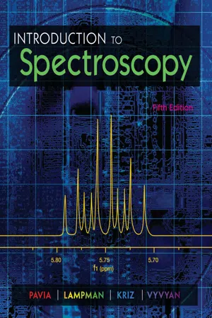
- Donald Pavia, Gary Lampman, George Kriz, James Vyvyan, Donald Pavia, Gary Lampman, George Kriz, James Vyvyan(Authors)
- 2014(Publication Date)
- Cengage Learning EMEA(Publisher)
215 C H A P T E R 5 NUCLEAR MAGNETIC RESONANCE SPECTROSCOPY Part One: Basic Concepts N uclear magnetic resonance (NMR) is a spectroscopic method that is even more important to the organic chemist than infrared spectroscopy. Many nuclei may be studied by NMR techniques, but hydrogen and carbon are most commonly available. Whereas infrared (IR) spectroscopy reveals the types of functional groups present in a molecule, NMR gives information about the number of magnetically distinct atoms of the type being studied. When hydrogen nuclei (protons) are studied, for instance, one can determine the number of each of the distinct types of hydrogen nuclei as well as obtain information regarding the nature of the immediate environment of each type. Similar information can be determined for the carbon nuclei. The combination of IR and NMR data is often sufficient to determine completely the structure of an unknown molecule. 5.1 NUCLEAR SPIN STATES Many atomic nuclei have a property called spin: the nuclei behave as if they were spinning. In fact, any atomic nucleus that possesses either odd mass, odd atomic number, or both has a quantized spin angular momentum and a magnetic moment. The more common nuclei that possess spin include 1 1 H, 2 1 H, 13 6 C, 14 7 N, 17 8 O, and 19 9 F. Notice that the nuclei of the ordinary (most abundant) isotopes of car-bon and oxygen, 12 6 C and 16 8 O, are not included among those with the spin property. However, the nu-cleus of the ordinary hydrogen atom, the proton, does have spin. For each nucleus with spin, the number of allowed spin states it may adopt is quantized and is determined by its nuclear spin quan-tum number I . For each nucleus, the number I is a physical constant, and there are 2 I + 1 allowed spin states with integral differences ranging from + I to − I . The individual spin states fit into the sequence + I , ( I − 1), .
- Denis Rousseau(Author)
- 2012(Publication Date)
- Academic Press(Publisher)
In this spectrometer, a 10-ppm chemical-shift range corre-sponds to a frequency spread of 3000 Hz; thus, the resonance frequency of two Ή nuclei in different chemical environments will differ from each other by, at most, — 3000 Hz. In the same spectrometer, 1 3 C nuclei will resonate at — 75 MHz; the difference in the resonance frequencies of different carbons will be less than ~ 20 kHz. 1. NUCLEAR MAGNETIC RESONANCE 19 E. Spin-Spin Interactions Each spin generates a magnetic field due to its magnetic dipole moment. This field is experienced by all other spins and causes two major spin-spin interactions. These are the scalar and the dipole-dipole couplings. In N M R spectra of liquids the most common manifestation of scalar interac-tions is the multiplet structure observed from coupled nuclei. On the other hand, at least in liquids, dipole-dipole coupling is usually the major source of relaxation of the spins. 1. Scalar or J Coupling The scalar coupling is an interaction between two magnetic nuclei in the same molecule and is mediated by electrons in the chemical bonds. Con-sider a simple case with two magnetic nuclei, A and B, with two electrons in the bonding orbital between them. The electron spins are paired so that the net electron spin vanishes. Therefore, in first order, there is no interaction between the electron spins and the nuclear spins. But since A has a magnetic nucleus, the electron closer to A will tend to orient itself in the magnetic field of the nuclear dipole; this in turn would induce an orientational preference for the electron near Β and, consequently, for the nuclear spin of B. Thus, the net effect is a coupling between A and Β nuclei which depends on the relative orientation of the nuclear spins alone and not on the orientation of the molecule as a whole with respect to the external field. eBook - PDF
- Rose Marie O. Mendoza(Author)
- 2019(Publication Date)
- Arcler Press(Publisher)
Figure 5.1: Schematic representation of NMR. Source: https://chembam.com/techniques/nmr/ The NMR phenomenon can usually be described in the nutshell as follows. If the sample is placed in the magnetic field (MF) and is given RF radiation at a suitable frequency, nuclei in the sample are able to absorb the energy. The frequency of radiation essential for the absorption of energy is dependent on three main things. At first, comes the characteristic of kind of the nucleus (e.g., 13 C or 1 H). Second, the frequency is dependent on the chemical environment of the nucleus. For instance, the hydroxyl and methyl protons of the methanol absorb at the different frequencies and amide protons of the two different tryptophan deposits in the native protein absorb at the different frequencies as they are in the different chemical environments. The Nuclear Magnetic Resonance Spectroscopy (NMR) 143 NMR frequency is also dependent on the spatial location in the MF if that specific field isn’t uniform everywhere. This last variable gives the basis for the MRI (magnetic resonance imaging), for the self-diffusion coefficient measurements and for the coherence selection. For imaging and for diffusion coefficient measurements, the MF varies linearly over the sample. However, for the spectroscopic purposes, the MF is wished to be homogeneous over the sample. The homogeneity necessities for the NMR spectroscopy are rather severe; the MF should not vary more than ten ppb (parts per billion) over the volume of sample. After absorption of the energy by nuclei, length of the time and manner in which nuclei disperse that energy can be utilized to reveal the information regarding the variety of the dynamic processes. 5.2. MAGNETIC RESONANCE 5.2.1. Nuclear Spins Nuclei have +ve charge. Many nuclei act as they were spinning. Anything which possesses charge and moves has the magnetic moment and yields the MF. eBook - ePub
- Rui Yang(Author)
- 2018(Publication Date)
- CRC Press(Publisher)
5 Nuclear Magnetic Resonance SpectroscopyIn an external magnetic field, the nucleus of an atom may exist in various spin states; thus, it can have different energies. When this nucleus is irradiated with the electromagnetic waves of an appropriate frequency, it absorbs the energy and jumps from the ground state to the excited state, producing a nuclear magnetic resonance (NMR) signal. Similar to ultraviolet–visible (UV–Vis) and infrared (IR) spectroscopy, NMR spectroscopy is also a type of absorption spectroscopy. The only difference is that in NMR spectroscopy, nuclei absorb radiofrequency (RF) energy and exhibit spin transitions; in UV–Vis spectroscopy, electrons absorb UV–Vis light and exhibit electronic transitions; while in infrared (IR) spectroscopy, molecules absorb IR light and exhibit vibrational and rotational transitions.
In NMR spectroscopy, nuclei with nonzero magnetic moments are studied.5.1 Basic Principle of NMR5.1.1 Magnetic Moment of NucleusA spinning nucleus possesses angular momentum P . As the nucleus has a positive charge, a magnetic field with a magnetic moment μ, is produced, because of spinning. The angular momentum and magnetic moment are related to each other as follows:μ = γ P .5.15.1Here, γ is the nuclear magnetogyric ratio , an intrinsic character of a specific nucleus. μ is expressed in units of the nuclear magneton β, where β is a constant (5.05 × 10⁻²⁷ J/T). Table 5.1
- SachchidaNand Shukla(Author)
- 2019(Publication Date)
- Arcler Press(Publisher)
Nuclear Magnetic Resonance Spectroscopy (NMR) 5 CONTENTS 5.1. Introduction ................................................................................... 134 5.2. Overview of Concepts ................................................................... 134 5.3. Quantum Mechanical Description ................................................. 137 5.4. Description of The Nuclear Quantum Number ............................... 138 5.5. The Population of The Energy Levels .............................................. 139 5.6. Nmr Spectra of Several Nuclei ....................................................... 142 5.7. Fine Structure of NMR Spectrum .................................................... 147 5.8. Nuclear Relaxation ........................................................................ 151 5.9. The Noe Phenomenon ................................................................... 153 5.10. Use Of Nuclear Magnetic Resonance To Monitor the Rate Processes ....................................................................... 156 5.11. Miscellaneous Uses ..................................................................... 158 References ............................................................................................. 163 Introduction to Modern Instrumentation Methods and Techniques 134 5.1. INTRODUCTION Nuclear magnetic resonance (NMR) spectroscopy was discovered after World War II and from then the applications of NMR spectroscopy to chemistry have been expanding continuously. It was quite natural then that Nuclear magnetic resonance took a vital part in undergraduate chemistry education. In recent years, applications of Nuclear magnetic resonance have been stretched to medicine and biology (Tompa et al., 1996; Bilgic et al., 2015). The fundamental principles are normally covered in physical chemistry course and every undergraduate physical chemistry course textbook includes a chapter on it. eBook - PDF
Physical Chemistry
A Modern Introduction, Second Edition
- William M. Davis(Author)
- 2011(Publication Date)
- CRC Press(Publisher)
371 12 Magnetic Resonance Spectroscopy Electronsandcertainatomicnucleipossessintrinsicmagneticmomentsthatgiveriseto aninteractionenergywithanexternalmagneticfield�Thedifferencebetweentheinterac-tionenergiesofthedifferentstatescanbeprobedbymagneticresonancespectroscopy, an immensely powerful technique now used for qualitative analysis, determination of molecularstructure,andmeasurementofreactionratesanddynamics�Itsuseextendsto imagingmacroscopicobjects,andmedicalapplicationsfordiagnosticworkhavebecome widespread�Thischapterprovidesthequantummechanicalfoundationformultinuclear magneticresonancespectroscopy� 12.1 Nuclear Spin States Atomicnucleiconsistofneutronsandprotonsandhaveastructurethatisasrichastheelec-tronicstructureofatomsandmolecules�Inchemistry,though,nuclearstructureisoften unimportantsinceitdoesnotchangeinthecourseofachemicalreaction�Itisfrequently appropriatetoconsidernucleitobesimplypositivelychargedpointmasses,aswehave donetothispoint�However,therearesomeimportantmanifestationsofnuclearstructure thathavebeenexploitedindevelopingpowerfultypesofmolecularspectroscopies� Protons and neutrons have an intrinsic spin and an intrinsic magnetic moment� Experimentshaverevealedthattherearejusttwopossibleorientationsoftheirspinvec-torswithrespecttoa z -axisdefinedbyanexternalfield�Thismeansthattheirintrinsic spinquantumnumberis1/2;theyarefermions�Whereastheletter S iscommonlyused for the electron spin quantum number, the letter I is commonly used for nuclear spin quantumnumbers�Thus, I =1/2foraproton,andtheallowedvaluesforthequantum numbergivingthenuclearspinangularmomentumprojectiononthe z -axis, M I ,are+1/2 and−1/2� Theangularmomentumcouplingoftheintrinsicprotonandneutronspinsinaheavy eBook - PDF
Chemical Analysis
Modern Instrumentation Methods and Techniques
- Francis Rouessac, Annick Rouessac, John Towey(Authors)
- 2022(Publication Date)
- Wiley(Publisher)
Chemical Analysis: Modern Instrumentation Methods and Techniques , Third Edition. Francis Rouessac and Annick Rouessac, translated by John Towey. © 2022 John Wiley & Sons Ltd. Published 2022 by John Wiley & Sons Ltd. Companion Website: www.wiley.com/go/Rouessac/Analysis3e Nuclear Magnetic Resonance Spectroscopy Chapter 15 N uclear magnetic resonance (NMR) is a spectroscopic method based on the study of the nuclei of certain elements. It has become irreplaceable in a variety of areas, such as chemistry, biology, the food industry or medical imaging (MRI). It is one of the most powerful methods to obtain structural information on organic or inorganic compounds. A succession of technological advances has led to a growing number of applications for it, whether analytical or not, in industry, ranging Chapter 15: Nuclear Magnetic Resonance Spectroscopy 388 from oil exploration to the identification of food products. This chapter is limited to a simplified study of NMR in solution in the field of organic compounds. 15.1 GENERAL INTRODUCTION Nuclear magnetic resonance refers to a property of matter that has given its name to powerful method of materials investigation, in particular to identify the structure of organic or inorganic compounds. NMR spectrometers are therefore often found in research laboratories, although there are routine applications based on this property, using simpler and more robust dedicated instruments (see Figure 15.31). Organic chemists quickly understood the interest of NMR, which has accelerated its development and favoured the appearance of many technical improvements. eBook - PDF
Nuclear Magnetic Resonance
Volume 1
- R K Harris(Author)
- 2007(Publication Date)
- Royal Society of Chemistry(Publisher)
A Specialist Periodical Report Nuclear Magnetic Resonance Volume 1 A Review of Recent Published Literature up to June 1971 Senior Reporter R. K. Harris, School of Chemical Sciences, University of East Anglia Reporters N. Boden, University of Leeds P. Diehl, University of Basel, Switzerland E. G. Finer, Unilever Research Ltd., Welwyn M. 1. Foreman, University of Strathclyde D. G. Gillies, Royal Holloway College, University of London R. Grinter, University of East Anglia P. M. Henrichs, University of Basel, Switzerland R. G. Jones, University of &sex W. T. Raynes, Shemeld University D. Shaw, Varian Associates Ltd., Walfon-on-Thames @ Copyright 1972 The Chemical Society Burlington House, London, WIV OBN ISBN : 0 85186 252 7 Library of Congress Catalog Card No. 72-78527 Printed in Great Britain by Alden & Mowbray Ltd at the Alden Press, Oxford Foreword This volume of Specialist Periodical Reports is the first in a series covering the topic of Nuclear Magnetic Resonance. It largely follows the practice set by other Specialist Periodical Reports, but it should be obvious from the list of contents that on the whole this volume is intended to be oriented towards a discussion of the phenomenon of n.m.r. itself rather than towards a summary of applications of n.m.r. in given areas of chemistry. This is more readily done for some aspects of n.m.r. than for others; thus Chapter 3, Relaxation, is heavily phenomenon-orientated, whereas Chapter 8, Macromolecules and Solids, necessarily has some degree of chemical orientation. However, a de- liberate attempt has been made to exclude chapter-headings based on com- pound-type or on a particular n.m.r.-active nucleus (e.g. ‘l 3C Resonance’). Clearly, the division of n.m.r. into the areas given by the chapter-headings is arbitrary. In some instances there is overlap of material; it is hoped that such overlap is helpful and not excessive. eBook - PDF
Spin Dynamics
Basics of Nuclear Magnetic Resonance
- Malcolm H. Levitt(Author)
- 2008(Publication Date)
- Wiley(Publisher)
The twin challenges of sensitivity and resolution are the keynotes of NMR instrumentation. The NMR signal is weak for two reasons. First, the individual nuclear magnetic moments are very small compared with those of electrons. Second, the distibution of nuclear magnetic moments is nearly isotropic. As described in Chapter 2, macroscopic nuclear magnetism depends on the very slight thermal imbalance of the distribution of magnetic moments. In ordinary circumstances, this imbalance represents only about 1 part in 10 5 . Detection of the weak nuclear magnetism is a considerable instrumental challenge. The problem of resolution has been mastered, and a great deal of progress has been made on the problem of sensitivity. Nevertheless, sensitivity remains a great limitation of the NMR technique, at least in its conventional form. Rather large amounts of sample are required, compared with other spectroscopies. Around 10 14 nuclear spins are typically required to obtain a usable NMR signal. 1 In the following sections, I outline the operation of a typical solution-state NMR spectrometer. Solid- state NMR spectrometers, and NMR imaging instruments, are based on similar principles, although with technical differences that are largely restricted to the probe and magnet. 4.1 The Magnet Most NMR experiments require a magnetic field which is homogeneous (i.e. independent of position) within at least 1 part in 10 9 . This extremely high magnetic field homogeneity must be maintained over the entire volume of the sample, i.e. around a cubic centimetre in solution NMR and many hundreds of cubic cen- timetres in NMR imaging experiments. The magnetic field must also be extremely stable with respect to time. A perfectly uniform magnetic field avoids inhomogeneous broadening of the NMR signal (see Section 3.6) and allows the resolution of small differences in the nuclear Larmor frequency, due to chemical shifts, spin–spin couplings, or other molecular interactions.
- Jay A. Glasel, Murray P. Deutscher(Authors)
- 1995(Publication Date)
- Academic Press(Publisher)
But the result was still a one-dimensional spectrum, and the problem of resolution in proton spectra of biopolymers remained. During the late 1970s, two-dimen- sional (2D) NMR methods were developed (Ernst et al., 1987) that revolution- ized the use of NMR as a structure determination tool for biopolymers, particu- larly proteins (Bax and Lerner, 1986). These methods greatly improve resolution by dispersing NMR spectra along two frequency axes rather than one, and allow the simultaneous measurement of NMR parameters and inter- actions that can only be determined by individual experiments with 1D meth- Chapter 7 NuclearMagnetic Resonance 349 ods. The 2D methods have been extended to higher dimensions, and 3D and 4D experiments have allowed further improvements in resolution so that larger molecules can be profitably studied by NMR. The great proliferation of NMR experiments has been accompanied by an elaborate jargon to describe the methods and purposes of the experiments, and many fanciful acronyms have been devised. These terms may be amusing, confusing, or annoying, but they cannot be avoided; those used here are explained in the glossary, or in the text as they arise. A. Two-Dimensional Nuclear Magnetic Resonance As a review, in a typical homonuclear one-dimensional NMR experiment, the magnetizations of all the spins, which at equilibrium are initially aligned along B0, are simultaneously flipped into the x,y plane by a 90 ~ observation pulse. This is called the preparation period of the experiment. The magnetization in the x,y plane is measured over a detection period (t 2) as a signal that varies with time, s(t 2), called the FID, by being sampled at discrete points in time, digitized, and stored in a computer. The time variable, t2, is used here to allow our notation for this one-dimensional experiment to agree with two-dimensional NMR terminology; t 2 is just the detection period discussed previously.
Index pages curate the most relevant extracts from our library of academic textbooks. They’ve been created using an in-house natural language model (NLM), each adding context and meaning to key research topics.