Psychology
CAT and PET Scan
CAT (Computerized Axial Tomography) and PET (Positron Emission Tomography) scans are imaging techniques used in psychology to study brain structure and function. CAT scans provide detailed images of the brain's structure, while PET scans show brain activity by measuring blood flow and glucose metabolism. These scans help psychologists understand the relationship between brain structure, function, and psychological processes.
Written by Perlego with AI-assistance
Related key terms
1 of 5
12 Key excerpts on "CAT and PET Scan"
 eBook - PDF
- Marie T. Banich, Rebecca J. Compton(Authors)
- 2018(Publication Date)
- Cambridge University Press(Publisher)
The invention of this method enabled standardized brain atlases, which could be used to co-register damage across patients and link damage in a given area across individuals to simi- larities in behavioral deficits. • Positron emission tomography (PET) uses the decay of a radioactive isotope introduced into the brain, typically either glucose or oxygen, to examine brain metabolism. Areas of the brain that are particularly active will take up more of the isotope and provide a greater signal. The use of PET allowed researchers for the first time to record activ- ity from specific brain regions in response to cognitive and emotional demands, thus showing the neurologically nor- mal brain in action. The Twenty-First Century: The Brain Imaging Revolution • Magnetic resonance imaging (MRI) provides the ability to examine both brain anatomy and brain function without the use of ionizing radiation, which vastly expanded the questions and populations that could be examined with brain imaging. Moreover, it provided greater anatomical resolution than CAT scans and greater temporal resolution than PET studies, and as such has become a mainstay of cognitive neuroscience research.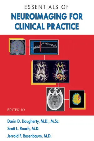
- Darin D. Dougherty, Scott L. Rauch, Jerrold F. Rosenbaum, Darin D. Dougherty, Scott L. Rauch, Jerrold F. Rosenbaum(Authors)
- 2008(Publication Date)
- American Psychiatric Association Publishing(Publisher)
75 3 Positron Emission Tomography and Single Photon Emission Computed Tomography Darin D. Dougherty, M.D., M.Sc. Scott L. Rauch, M.D. Alan J. Fischman, M.D., Ph.D. Whereas computed tomography (CT; see Chapter 1 in this volume) and magnetic resonance imaging (MRI; see Chapter 2 in this volume) provide structural im- ages of the brain, positron emission tomography (PET) and single photon emission computed tomography (SPECT) are radiological technologies that are used to measure numerous aspects of brain function. PET and SPECT, along with functional magnetic resonance im- aging (fMRI; see Chapter 4 in this volume), are power- ful tools for neuroscience research. Although PET and SPECT are still primarily research tools in the field of psychiatry, there is growing clinical utility for these methodologies. We begin this chapter by briefly de- scribing the principles that underlie these methods. We then discuss the use of PET and SPECT in both the clin- ical psychiatry and neuroscience research environ- ments. Finally, we propose future directions for the use of PET and SPECT in psychiatry. Principles of PET and SPECT Positron Emission Tomography Positron Emission PET measures radioactive decay in order to form im- ages of biological tissue function. Specifically, unstable 76 ESSENTIALS OF NEUROIMAGING FOR CLINICAL PRACTICE nuclides are introduced into the organism being stud- ied, and the PET camera detects the resulting radioac- tive decay and uses these data to construct functional images. Commonly used positron-emitting nuclides in PET studies include 11-carbon ( 11 C), 15-oxygen ( 15 O), 18-fluorine ( 18 F), and 13-nitrogen ( 13 N) (Table 3– 1). These nuclides are incorporated into the desired molecules, resulting in a radiopharmaceutical (see subsection titled “Radiopharmaceuticals” later in this chapter). eBook - ePub
- Barry Reisberg(Author)
- 2008(Publication Date)
- Free Press(Publisher)
A formidable array of impressive new technologies are currently being applied to the solution of diagnostic questions in the area of dementia. The most exciting of these technologies have already been mentioned earlier in our story of brain failure. Virtually all utilize a computer to process a vast amount of information which gives us a more accurate picture of brain structure or function. These technologies include computerized axial tomographic scanning of the brain substance (“CT scans,” or “CAT scans”), positron emission tomographic imaging of the brain (”PET scans”), and radioisotopic measurements of regional cerebral blood flow (rCBF). Computer analyses have also considerably increased the sophistication of traditional measures of brain electrical activity, such as electroencephalography (EEG). This has resulted in such complex but apparently very useful technologies as quantitative pharmaco-EEG (QPEEG) and “neurometrics.’’CT scans are computer-produced pictures of successive “slices” of brain substance. The shading of the picture is determined by the density of the substance visualized. Hence areas of the brain which contain mostly fluid can be distinguished from areas which contain tissue. CT scans also appear to be capable of distinguishing areas of the brain which contain predominately neuronal cells, or “gray matter,” from areas of the brain which contain mostly axonal fibers, “white matter.”1When first introduced in the mid-1970s, CT scans appeared to hold out considerable promise of enabling clinicians to accurately diagnose senile dementia. Since decay of particular portions of the outermost layers of the brain substance is one of the features observed when autopsies are conducted on the brains of persons with Alzheimer’s disease, it was hoped that the extent of brain decay as visualized in CT images would have diagnostic significance. Also, since multi-infarct dementia is characterized by the frequent presence of substantial hemorrhagic areas within the brain—what are referred to at autopsy as “cerebral softenings”—it was hoped that CT scans would be of diagnostic value in this area as well. eBook - PDF
Positron Emission Tomography
Recent Developments in Instrumentation, Research and Clinical Oncological Practice
- Sandro Misciagna(Author)
- 2013(Publication Date)
- IntechOpen(Publisher)
Section 2 PET Applications in Neurological and Behavioral Research Chapter 4 Functional Imaging Studies of Human Cognition Using Positron Emission Tomography Sandro Misciagna Additional information is available at the end of the chapter http://dx.doi.org/10.5772/57124 1. Introduction In past times behavioural neurologists have shown how discrete brain lesions provoked different types of cognitive disorders such as language, praxic, gnostic, spatial or memory domain. They interpreted these anatomo-clinical associations conjecturing that the normal brain function (impaired by brain damage) was localized within the lesioned region (lesional hypothesis) and had been impaired from brain damage (Marshall & Fink, 2003). They also hypothesized that cognitive impairments could arise from lesions that spared the functional centers but disconnected them from other centers (disconnectional hypothesis). According this argument “basic psychological functions” are localized in a punctate fashion and complex psychological functions are constituted from many such basic functions joined together in distributed circuits. It follows that the symptoms may have arisen from a reconfiguration of the entire circuit in response to cerebral damage. During the late 19th century, the advent of structural brain imaging, first the computed tomography (CT) and later the magnetic resonance imaging (MRI), gave the possibility to study anatomical localization of the cognitive deficits that were manifest after brain injury. Since then neuroimaging studies have helped medical doctors in clinical practice to identify cerebral damage caused by spaced-occupying lesions, strokes or degenerative processes. eBook - ePub
- Aaron Newman(Author)
- 2019(Publication Date)
- SAGE Publications Ltd(Publisher)
11 Positron Emission Tomography (PET)Learning Objectives
After reading this chapter, you should be able to:- Explain the origins of the PET signal at the subatomic level.
- Explain how PET data is acquired and reconstructed.
- Define PERs and list the names and uses of several.
- Describe the potential risks of PET and how they are mitigated.
- Explain the constraints that PET imposes on experimental design in cognitive neuroscience.
- List several use cases for PET over fMRI in cognitive neuroscience research.
- Discuss the value of multimodal PET.
Introduction
Positron emission tomography (PET) is a unique imaging modality in several ways. Firstly, although it is very safe, PET is nominally more invasive than other imaging techniques because it involves introducing radioactive tracers into the body. Secondly, although – like fMRI – PET allows indirect identification of neural activity levels through measurements of blood flow and tracking the uptake of oxygen, PET can also be used to map the distribution of many other molecules in the brain, including receptors and neuromodulators, as well as disease markers such as amyloid (a hallmark of Alzheimer’s disease).PET was first developed in the 1970s and has steadily evolved as a technique over time. The basis of PET imaging is introducing a radioactive tracer with a very short half-life into the body (through injection or inhalation), and then recording how it gets distributed in the tissue of interest. Because the radioactivity is very short-lived and used in low concentrations, PET is a safe technique to use – although precautions need to be followed concerning the handling of the radioactive materials, and the number of scans that an individual can undergo in a specified period of time. As such, whether PET is truly a ‘non-invasive’ technique can be debated, but at worst it is minimally invasive and it is certainly safe.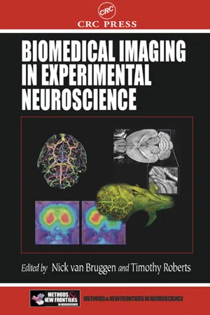
- Nick Van Bruggen, Timothy P.L. Roberts, Nick Van Bruggen, Timothy P.L. Roberts(Authors)
- 2002(Publication Date)
- CRC Press(Publisher)
Positron emission tomography (PET) has been used for a number of years in the study of the function and neurochemistry in the human and nonhuman primate brain. 1 PET is a noninvasive and extremely safe imaging modality that allows for repeated observations in an individual. The adaptation of PET to repeatedly and noninvasively image small animals represents a unique opportunity to explore neu-roplasticity and neuropathology in living rodents. This chapter will describe the basic physical principles underlying PET imaging, review some of the work that has successfully utilized PET in rodent brain imaging, and will indicate future directions for rodent research with PET. 9.2 BASIC PRINCIPLES OF POSITRON EMISSION TOMOGRAPHY IMAGING PET is a radiotracer imaging technique that utilizes small amounts of a compound or biomolecule of interest labeled with radioactive atoms. The radiation emitted as the radioactive label decays is picked up by a series of external detectors and the resulting information is used to compute images that show the distribution of the Small Animal Imaging with Positron Emission Tomography 273 radioactive tracer in the subject. PET can essentially be thought of as a noninvasive version of autoradiography, with inferior spatial resolution, but with the advantages that the pharmacokinetics of the tracer can be measured in a single experiment and repeat studies can be performed on the same subject. 9.2.1 P OSITRON -E MITTING R ADIONUCLIDES AND T RACERS PET imaging makes exclusive use of radionuclides that decay by positron emission. Table 9.1 lists commonly used positron-emitting radionuclides. Some of the radio-nuclides are isotopes of biologically ubiquitous elements such as carbon, nitrogen, and oxygen, enabling radioactively labeled tracers of small organic molecules to be produced by direct isotopic substitution, for example, by replacing a stable carbon-12 atom with a positron-emitting carbon-11 atom. eBook - PDF
Synaptic Plasticity
Basic Mechanisms to Clinical Applications
- Michel Baudry, Xiaoning Bi, Steven S. Schreiber(Authors)
- 2005(Publication Date)
- CRC Press(Publisher)
Positron emission tomography (PET) has been used for a number of years in the study of the function and neurochemistry in the human and nonhuman primate brain (1). It is a noninvasive, and extremely safe imaging modality that allows for repeated observations in an individual. The adapta-tion of PET to repeatedly and noninvasively image small animals represents a unique opportunity to explore neuroplasticity and neuropathology in living rodents. This chapter describes some of the basic physical principles underlying PET imaging, review some of the work that has successfully utilized PET in rodent brain imaging and will indicate future directions for rodent research with PET. 2. PRINCIPLES OF PET This section reviews some of the fundamental principals of PET, for more detailed descriptions, see Ref. 2, among many other reviews. Positron emis-sion tomography utilizes radiolabeled nuclides to obtain images of where that tracer is concentrated within the living animal. These radionuclides 160 Kornblum et al. emit positrons as they decay. Some of these radionuclides are isotopes of elements common to all biological systems, such as carbon, nitrogen, and oxygen, enabling labeled tracers of small organic molecules to be produced by direct isotopic substitution, for example, by replacing a carbon-12 atom with a positron-emitting carbon-11 atom. These radionuclides are produced in a biomedical cyclotron (3). After the radionuclide is produced, rapid che-mical synthetic pathways are needed to produce the radiolabeled product and it must then be immediately injected into the subject, with the time between production and injection typically being no more than 4–6 half-lives. With carbon-11, this amounts to around 2 hr from production of radionuclide to injection of radiotracer. Despite these difficulties, due to its chemical advantages, carbon-11 remains the radionuclide of choice in PET for labeling receptor ligands and many drugs (4).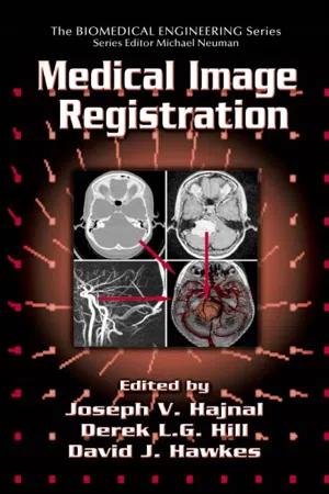 eBook - PDF
- Joseph V. Hajnal, Derek L.G. Hill, Joseph V. Hajnal, Derek L.G. Hill(Authors)
- 2001(Publication Date)
- CRC Press(Publisher)
Labeling with a radioactive nuclide allows the syn-thesis of specific tracers, which are used to determine, for example, cerebral blood flow or glucose consumption in the human brain. Hence, PET is repre-sentative of a functional imaging modality, and the primary interest in the development of PET was to quantify the three-dimensional distribution of Registration of MRI and PET Images for Clinical Applications 201 the radioactive tracer with subsequent interpretation in a framework of phys-iological models. The physical phenomenon underlying the positron emission limits the res-olution of PET principally to 2 to 3 mm. During practical applications of PET imaging, the resolution is further reduced due to the limited detector size and the smoothing applied during image reconstruction. The latter is necessary to maintain sufficient signal and limit the influence of statistical noise. For further details the reader is referred to excellent reviews (see, for example, Eriksson et al. 2 ) and to Chapter 5 of this book for a discussion on the influ-ence of scanner errors. Image registration most often relies on specific features detectable in the images, i.e., characteristic tracer uptake patterns in an organ or part of the human body. This is especially important when retrospective registration techniques are employed. PET images are stored with standard image orien-tation transverse to the patient’s head-foot axis. A pixel represents a measure-ment of radioactivity concentration at a particular position inside the field of view (i.e., the volume covered by the detector system). These pixel values can be related to a physiological variable by means of an appropriate model. Usu-ally, the radioactive tracer does not have a uniform distribution but shows a tracer-dependent-specific pattern with increased uptake, for instance, in the brain’s gray matter compared to white matter, or in the heart compared to lung tissue.
- Ana Lucia Abujamra(Author)
- 2011(Publication Date)
- IntechOpen(Publisher)
It is often used to accurately determine the stage of the Diagnostic Techniques and Surgical Management of Brain Tumors 38 brain tumour. PET images produce visual impression of detailed biochemical changes caused by brain tumours and their metabolic activities. Despite the recognized limitations of fluorodeoxyglucose positron emission tomography (FDG-PET) in brain tumour imaging due to the high background of normal gray matter, this imaging modality provides critical information for the management of patients with cerebral parenchyma tumours by providing a: Global picture of the tumour and thus guiding the appropriate site for biopsy, thereby improving accuracy of the technique and reducing the number of biopsy samples Prediction of metabolic activity and aggressiveness of the tumour, thereby enhancing the ability to provide a prognosis. Another area, which has been investigated extensively in tumour imaging, is differentiating between recurrent tumour and treatment-related changes – for example, radiation necrosis or postsurgical changes. The aggressive tumour responds with a bright signal because of its high uptake of FDG, but radiation necrosis will show no changes. FDG-PET has equally demonstrated its usefulness in differentiating lymphoma from infectious toxoplasmosis in patients with acquired immune deficiency syndrome with almost 100% accuracy, and is the investigation of choice in that setting [30]. In recent years, an increasing number of brain tumour PET studies have used other tracers, such as labeled methionine, tyrosine, thymidine, choline, fluoromisonidazole, EF5 and so on, of which positron-labeled amino acid analogues, nucleotide analogues, and the hypoxia imaging tracers are of special interest [30]. The major advantage of these radiotracers over FDG is the markedly reduced background activity in gray matter, which allows detection of even smaller lesions and low-grade tumours with high precision.
- Derek J. Chadwick, Julie Whelan, Derek J. Chadwick, Julie Whelan(Authors)
- 2008(Publication Date)
- Wiley(Publisher)
Wiley. Chichester (Ciba Foundation Symposium 163) p 76-92 Data from functional positron emission tomography (PET) neuroimaging are universally interpreted with reference to models. These models range from the physiological, as in the compartmental models of radioligand binding, to the psychological-for example, ‘cognitive subtraction’ (Petersen et a1 1988). We present a model for the combined manipulation of behaviour and neurotransmitter function which uses conjoint psychological and pharmacological challenge. The nature of functional brain systems and their associated neurotransmitter systems can be inferred from studying patients with specific behavioural deficits 76 Psychopharmacological PET studies 77 and identifying the sites of abnormal brain physiology. These inferences are based on the ‘lesion’ model of brain dysfunction and are limited, in that neither the behavioural nor the regional brain deficit is under experimental control. PET studies of regional cerebral blood flow (rCBF) activation permit experimental control of two aspects of brain function, namely the neurocognitive processes and neurotransmitter function. We suggest that combined psychological and pharmacological challenge in the same individuals provides more information than either challenge alone. To demonstrate this point we report a study which combines (or, in terms of experimental layout, crosses) a memory task with acute administration of the novel anxiolytic drug buspirone. Crossing psychological and pharmacological treatments enables the sites of interaction to be identified. An interaction in this instance is defined as a functional (psychological) increase/decrease in local neuronal activity which can be manipulated by acute drug administration. An interaction of psychological and pharmacological effects on rCBF is taken as evidence of an association between the functional system and the neurotransmitter system(s) affected by the drug. eBook - PDF
- Philip Palin Dendy, Brian Heaton(Authors)
- 2011(Publication Date)
- CRC Press(Publisher)
The desire for quantification was paramount and the application of coincidence detection of positron emissions provides the basis for the realisation of this goal today. Over the ensuing period a broad range of technological advances have occurred and this has led to the transition of PET from the research laboratory to a routine clinical imaging tool. Progress in PET has been dependent on several factors: (1) Development of the cyclotron and associated radiochemistry to routinely produce well-characterised radiopharmaceuticals targeted at particular clinical problems (2) Development of the electronic and detector technologies needed to provide the high resolution and sensitivity for positron detection in the clinical environment (3) Development of computer technology to provide the resource to process the very large data rates encountered in a timely manner (4) Development of the image reconstruction and data analysis tools to provide repro-ducible quantitative measures of radionuclide distributions and kinetics in vivo These developments alone were not sufficient to see the widespread adoption of PET imaging in a routine clinical setting. This came with the development in 2000 of two com-mercially available integrated PET and X-ray CT scanners to form the PET/CT scanner (Beyer et al. 2000). This added the advantages of a high resolution anatomical imaging modality (CT) to the highly sensitive functional imaging modality of PET, providing inherently registered and ‘fused’ images (Figure 11.1). This combination of function and structure has seen the widespread adoption of PET/CT in oncology as well as in neurologi-cal and cardiac applications. Many of the challenges of the use of positron emitters remain. The application of the methodology is inextricably linked to the production and availability of well-characterised radiopharmaceuticals targeted to specific biochemical pathways and processes.
- Stephen F. Davis, William Buskist, Stephen F. Davis, William F. Buskist(Authors)
- 2007(Publication Date)
- SAGE Publications, Inc(Publisher)
Human imaging methods brought about the opportunity to study location of function without the limitations of natural dysfunc-tion or disease. Most of these methods, however, reflect signals that are collected over seconds. Accordingly, there is strong interest in combining the location information from human imaging methods and rapid electrical activity provided by EEG recordings. The advent of human imaging methods, PET in the 1980s and fMRI in the early 1990s, provided greater opportunities to visualize activation in areas of the brain during sensory, perceptual, cognitive, social, affective, and motor processing. Furthermore, fMRI allowed the study of activation throughout the whole brain rather than in single regions at one time. The potential to image many areas at once opens up the possibility of studying how areas work together to produce complex behaviors. COUPLING BETWEEN NEURAL ACTIVITY, METABOLISM, AND BLOOD FLOW Although PET and fMRI are two different methods for studying human neural activation during task performance, these techniques rely on the assumption that increases in brain metabolism are reflections of an increase in neural activation. Therefore, to trust the relevance of the data from these methodologies requires an understanding of the relation between neural activity and brain metabolism. It may help to know first that the brain uses a substantial proportion of the energy consumed by the body. The brain uses 15 percent to 20 percent of the oxygen consumed despite making up only 2 percent of body weight (Clarke & Sokoloff, 1999; Roland, 1993). The glucose and oxygen used by the brain serves to produce ATP. This molecule is valuable because the free energy it stores is easily acces-sible. ATP is considered the currency required for the biological reactions that support life. Instead of storing ATP, the brain makes ATP as needed. This requires that neural activity stimulate increases in metabolism.
Index pages curate the most relevant extracts from our library of academic textbooks. They’ve been created using an in-house natural language model (NLM), each adding context and meaning to key research topics.