
eBook - ePub
Rheumatology, Orthopaedics and Trauma at a Glance
Catherine Swales, Christopher Bulstrode
This is a test
Condividi libro
- English
- ePUB (disponibile sull'app)
- Disponibile su iOS e Android
eBook - ePub
Rheumatology, Orthopaedics and Trauma at a Glance
Catherine Swales, Christopher Bulstrode
Dettagli del libro
Anteprima del libro
Indice dei contenuti
Citazioni
Informazioni sul libro
Rheumatology, Orthopaedics and Trauma at a Glance is the new edition of The Musculoskeletal System at a Glance. The book now includes not just basic anatomy, but also features presenting complaints and patient examination and reflects the increased coverage of rheumatology, making it relevant for students at all levels.
Rheumatology, Orthopaedics and Trauma at a Glance
- Expands its coverage of rheumatology to include all major topics on the medical student curriculum
- Includes fully illustrated chapters on examination of each part of the musculoskeletal system
- Provides self-assessment case studies to test knowledge and provide clinical context
- Consolidates all information relating to the musculoskeletal system in one title
Rheumatology, Orthopaedics and Trauma at a Glance is ideal for all medical students studying the musculoskeletal system or taking an orthopaedics or rheumatology rotation.
Domande frequenti
Come faccio ad annullare l'abbonamento?
È semplicissimo: basta accedere alla sezione Account nelle Impostazioni e cliccare su "Annulla abbonamento". Dopo la cancellazione, l'abbonamento rimarrà attivo per il periodo rimanente già pagato. Per maggiori informazioni, clicca qui
È possibile scaricare libri? Se sì, come?
Al momento è possibile scaricare tramite l'app tutti i nostri libri ePub mobile-friendly. Anche la maggior parte dei nostri PDF è scaricabile e stiamo lavorando per rendere disponibile quanto prima il download di tutti gli altri file. Per maggiori informazioni, clicca qui
Che differenza c'è tra i piani?
Entrambi i piani ti danno accesso illimitato alla libreria e a tutte le funzionalità di Perlego. Le uniche differenze sono il prezzo e il periodo di abbonamento: con il piano annuale risparmierai circa il 30% rispetto a 12 rate con quello mensile.
Cos'è Perlego?
Perlego è un servizio di abbonamento a testi accademici, che ti permette di accedere a un'intera libreria online a un prezzo inferiore rispetto a quello che pagheresti per acquistare un singolo libro al mese. Con oltre 1 milione di testi suddivisi in più di 1.000 categorie, troverai sicuramente ciò che fa per te! Per maggiori informazioni, clicca qui.
Perlego supporta la sintesi vocale?
Cerca l'icona Sintesi vocale nel prossimo libro che leggerai per verificare se è possibile riprodurre l'audio. Questo strumento permette di leggere il testo a voce alta, evidenziandolo man mano che la lettura procede. Puoi aumentare o diminuire la velocità della sintesi vocale, oppure sospendere la riproduzione. Per maggiori informazioni, clicca qui.
Rheumatology, Orthopaedics and Trauma at a Glance è disponibile online in formato PDF/ePub?
Sì, puoi accedere a Rheumatology, Orthopaedics and Trauma at a Glance di Catherine Swales, Christopher Bulstrode in formato PDF e/o ePub, così come ad altri libri molto apprezzati nelle sezioni relative a Medicine e Internal Medicine & Diagnosis. Scopri oltre 1 milione di libri disponibili nel nostro catalogo.
Informazioni
1
Musculoskeletal structure and function
The locomotor system is composed of bone, cartilage, muscle, tendons and ligaments.
Bone
Bone is essentially a mineralised connective tissue. It is comprised of two subtypes:
- 1 Woven bone is formed when bone is laid down rapidly, as in the developing foetus, healing fractures or bone-forming tumours.
- 2 Lamellar bone is laid down slowly. It is structurally strong and forms the adult skeleton. It is arranged in two forms:
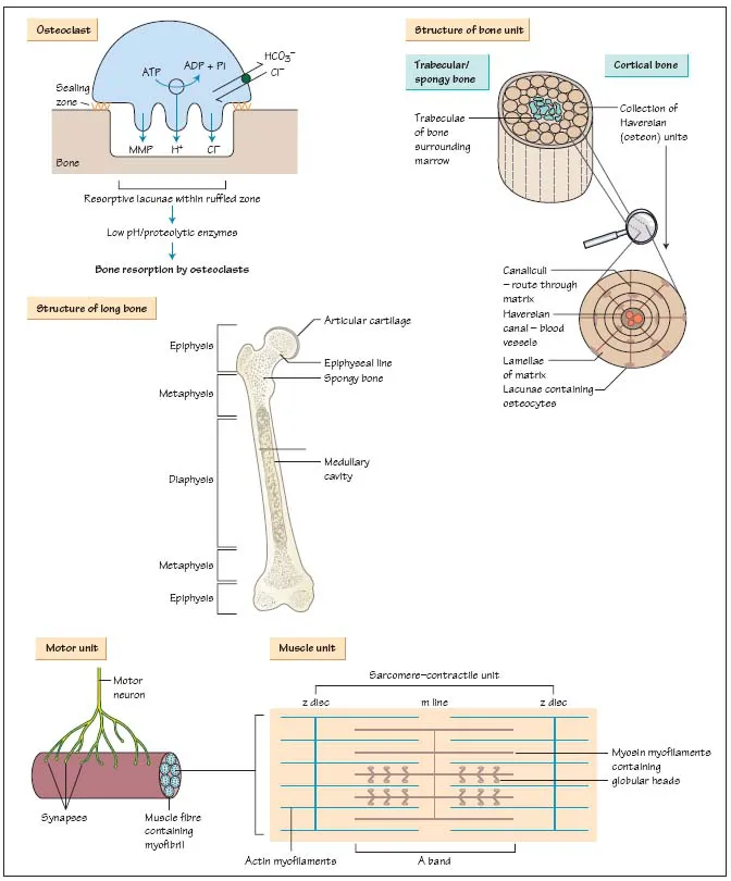
- Cortical or compact bone comprises 80% of the skeleton, accounting for most of the shafts of long bones. It is formed by Haversian systems: rings of collagen and matrix containing central blood vessels and lining cells called osteocytes.
- Trabecular or medullary bone is found in contact with bone marrow cells between the cortices, at the end of long bones and in vertebral bodies. In trabecular bone the collagen and matrix run as sheets (lamellae) parallel to the bone surface.
The three main cell types in bone are:
- 1 Osteoblasts (‘builders’) are responsible for bone formation by forming organised lamellae of mineralised matrix and collagen. Osteoblasts lie in sheets on the surface of bone trabeculae and their activity is closely coupled to osteoclasts.
- 2 Osteoclasts (‘cutters’) resorb bone. These giant multinucleated cells migrate across bone, settle on an area to be resorbed and the plasma membrane adjacent to the bone surface becomes a ‘ruffled border’. Secretion of proteolytic enzymes (e.g. matrix metalloproteinases, MMP) and hydrochloric acid onto the bone surface remove mineral and matrix simultaneously.
- 3 Osteocytes are mature, relatively inactive osteoblasts that lie in lacunae within bone.
Osteoblasts and osteoclasts are coupled into bone remodelling units that keep adult bone mass relatively constant.
See Chapter 31 ‘Disorders of Bone Metabolism’ for more details of osteoclast/osteoblast cell biology.
Bone is covered in a vascular membrane (the periosteum) which is a major source of blood supply to the bone (the other supply is derived from perforating vessels which then run up in the medulla). The periosteum is helpful when reducing fractures, as it is often partly intact and can be used to guide the broken frgments together. The periosteum is also important in fracture healing, supplying cells which ‘organise the haematoma’ around the fracture site (see below).
The protein matrix of bone consists largely of type I collagen. Osteoblasts lay down triple helices of type I collagen into organised lamellae containing unmineralised matrix (osteoid). The tensile strength of bone is increased by covalent bonds between collagen sheets; rigidity is conferred by mineralisation of bone matrix, with deposition of hydroxyapatite crystals between the lamellae.
Bone remodelling occurs throughout life to repair damaged bone. Alternating cycles of recruitment, differentiation and activation of osteoclasts and osteoblasts maintain the structural integrity of bone throughout life; with advancing age however, bone loss exceeds formation. Vigorous bone remodelling follows fracture in 5 stages:
- – Clot or haematoma formation from bleeding vessels within bone.
- – Organisation and recruitment of new populations of osteoblasts.
- – Callus formation from new osteoid and woven bone formation.
- – Modelling by osteoblasts/clasts transforms woven to lamellar bone.
- – Remodelling strengthens bone in direction of maximal stress.
Movement stimulates this process, so rigid fixing of fractures with plates prevents callus formation, and healing occurs more slowly on the background of standard bone remodelling. Bone is unique in its ability to heal without scar formation.
Bone increasese its circumference by the generation of new bone immediately under the periosteum, but length increases at epiphyseal growth plates. These are cartilage plates with their own blood supply which lie between the epiphysis (end of the bone) and the metaphysis, the part of the bone which connects with the diaphysis (shaft of the bone). These epiphyseal plates are weaker than the surrounding bone and therefore fractures in growing skeletons tend to occur at this site. If the fracture affects the blood supply or the anatomy of the growth plate then development may be affected.
Cartilage
Cartilage is composed of chondrocytes and chondroblasts, which create a matrix of type II collagen, and proteoglycans to bind water. Adult cartilage consists of four layers – the superficial, middle, deep and calcified zones, which differ in pattern of collagen fibre deposition, and water and cell content. Articular (hyaline) cartilage is an avascular and aneural shock absorber. It covers articular surfaces and allows friction-free movement of joints. Fibrocartilage forms the menisci and intervertebral discs.
Cartilage is lost either through mechanical degeneration at points of load-bearing (in osteoarthritis) or through resorption in an inflamed joint (in rheumatoid arthritis) or both. As cartilage contains no blood vessels, it heals slowly if damaged.
Muscle
Muscle is formed by fibres that differ according to their twitch rate and fatiguability.
- Type 1 muscle fibres are slow twitch (red) fibres that are highly resistant to fatigue. They have abundant mitochondria and are designed to maintain sustained contractions such as needed in posture control.
- Type 2 muscle fibres are fast twitch (white) fibres and are designed to produce greater force and rapidity of contraction but fatigue rapidly.
Fibres of similar types group together with a lower motor neuron to form a motor unit. Muscle fibres contain myofibrils formed by the contractile myofilaments actin and myosin. The myosin-binding sites on actin are covered by tropomyosin and troponin. However, when an action potential reaches a motor unit, stimulation causes calcium release into the surrounding cytoplasm (sarcoplasm). The calcium binds with troponin sites on tropomyosin, revealing the active binding sites and disinhibiting actin filaments. These cross-link with the globular heads on myosin, causing shortening of the motor unit. The muscle relaxes once calcium levels fall and the cross-links are broken.
Tendons and Iigaments
Both of these specialised connective tissues are composed of type I collagen. Tendons attach muscle to bone, while ligaments connect bones to one another, supplying support to a joint.
Nerves
Nerves are arranged in a segmental fashion, one pair at each level. The sensory nerves enter dorsally, supplying sensory information from a stereotyped strip of skin (dermatome). The motor root exits ventrally; a myotome is the motor equivalent of a dermatom...