
eBook - ePub
Atom Probe Tomography
Put Theory Into Practice
Williams Lefebvre, Francois Vurpillot, Xavier Sauvage, Williams Lefebvre, Francois Vurpillot, Xavier Sauvage
This is a test
Condividi libro
- 416 pagine
- English
- ePUB (disponibile sull'app)
- Disponibile su iOS e Android
eBook - ePub
Atom Probe Tomography
Put Theory Into Practice
Williams Lefebvre, Francois Vurpillot, Xavier Sauvage, Williams Lefebvre, Francois Vurpillot, Xavier Sauvage
Dettagli del libro
Anteprima del libro
Indice dei contenuti
Citazioni
Informazioni sul libro
Atom Probe Tomography is aimed at beginners and researchers interested in expanding their expertise in this area. It provides the theoretical background and practical information necessary to investigate how materials work using atom probe microscopy techniques, and includes detailed explanations of the fundamentals, the instrumentation, contemporary specimen preparation techniques, and experimental details, as well as an overview of the results that can be obtained. The book emphasizes processes for assessing data quality and the proper implementation of advanced data mining algorithms.
For those more experienced in the technique, this book will serve as a single comprehensive source of indispensable reference information, tables, and techniques. Both beginner and expert will value the way the book is set out in the context of materials science and engineering. In addition, its references to key research outcomes based upon the training program held at the University of Rouen—one of the leading scientific research centers exploring the various aspects of the instrument—will further enhance understanding and the learning process.
- Provides an introduction to the capabilities and limitations of atom probe tomography when analyzing materials
- Written for both experienced researchers and new users
- Includes exercises, along with corrections, for users to practice the techniques discussed
- Contains coverage of more advanced and less widespread techniques, such as correlative APT and STEM microscopy
Domande frequenti
Come faccio ad annullare l'abbonamento?
È semplicissimo: basta accedere alla sezione Account nelle Impostazioni e cliccare su "Annulla abbonamento". Dopo la cancellazione, l'abbonamento rimarrà attivo per il periodo rimanente già pagato. Per maggiori informazioni, clicca qui
È possibile scaricare libri? Se sì, come?
Al momento è possibile scaricare tramite l'app tutti i nostri libri ePub mobile-friendly. Anche la maggior parte dei nostri PDF è scaricabile e stiamo lavorando per rendere disponibile quanto prima il download di tutti gli altri file. Per maggiori informazioni, clicca qui
Che differenza c'è tra i piani?
Entrambi i piani ti danno accesso illimitato alla libreria e a tutte le funzionalità di Perlego. Le uniche differenze sono il prezzo e il periodo di abbonamento: con il piano annuale risparmierai circa il 30% rispetto a 12 rate con quello mensile.
Cos'è Perlego?
Perlego è un servizio di abbonamento a testi accademici, che ti permette di accedere a un'intera libreria online a un prezzo inferiore rispetto a quello che pagheresti per acquistare un singolo libro al mese. Con oltre 1 milione di testi suddivisi in più di 1.000 categorie, troverai sicuramente ciò che fa per te! Per maggiori informazioni, clicca qui.
Perlego supporta la sintesi vocale?
Cerca l'icona Sintesi vocale nel prossimo libro che leggerai per verificare se è possibile riprodurre l'audio. Questo strumento permette di leggere il testo a voce alta, evidenziandolo man mano che la lettura procede. Puoi aumentare o diminuire la velocità della sintesi vocale, oppure sospendere la riproduzione. Per maggiori informazioni, clicca qui.
Atom Probe Tomography è disponibile online in formato PDF/ePub?
Sì, puoi accedere a Atom Probe Tomography di Williams Lefebvre, Francois Vurpillot, Xavier Sauvage, Williams Lefebvre, Francois Vurpillot, Xavier Sauvage in formato PDF e/o ePub, così come ad altri libri molto apprezzati nelle sezioni relative a Ciencias físicas e Espectroscopia y análisis de espectro. Scopri oltre 1 milione di libri disponibili nel nostro catalogo.
Informazioni
Argomento
Ciencias físicasChapter One
Early Developments and Basic Concepts
D. Blavette, and X. Sauvage Groupe de Physique des Matériaux, University and INSA of Rouen, Normandie University, France
Abstract
Since the 1990s, atom probe tomography (APT) technique has reached a recognized position among the most important analytical microscopy techniques. APT is able to map out the spatial distribution of atomic species in three dimensions. With a unique combination of chemical sensitivity and three dimensional capabilities with a spatial resolution near the atomic scale it provides unique information about nanoscale structures. In this introduction chapter, the overall fundamentals and performances of APT, its main fields of application in material science, the historical background, and modern challenges are briefly described.
Keywords
Application; Atom probe; Concepts; Development; HistoryIntroduction
Since the 1990s, atom probe tomography (APT) technique has reached a recognized position among the most important analytical microscopy techniques. APT is able to map out the spatial distribution of atomic species in three dimensions (3D). With a unique combination of chemical sensitivity and 3D capabilities with a spatial resolution near the atomic scale it provides unique information about nanoscale structures. In this introduction chapter, the overall fundamentals and performances of APT, its main fields of application in material science, the historical background, and modern challenges are briefly described.
Atom Probe Tomography in Materials Science Today
The Unique Information Provided by Atom Probe Tomography
Imaging materials in 3D at the atomic scale has not only been a dream but also a challenge. This has been of vital importance for fundamental researches dealing with phase transformations or segregation of solute atoms to lattice defects. The characterization of microstructures at the ultimate scale is also of great importance for materials of industrial importance as it enables to better control the genesis and development of microstructures and to optimize processing routes and properties. Both functional and mechanical properties of materials depend not only on microscale features like grain sizes, precipitates, but also on nanoscale features such as clusters of solutes, chemical gradients, nanoprecipitates, and segregations on structural defects.
Scanning electron microscopes (SEM) have high imaging resolutions down to about 10 nm thanks to Field Emission Gun but their analytical capability based on X-ray emission is, however, still limited. This is the result of the scattering of electrons in matter that gives rise to a significant spread of the signal up to about 1 μm. This limitation can be easily overcome in the transmission electron microscope (TEM) or scanning-TEM where the sample thickness is typically less than 100 nm. In TEM, the limitation of the spatial resolution comes mainly from the aberrations of lenses used to focus the electron beam. During the past 10 years, the implementation of spherical aberration correctors in commercial instruments pushed the routine analytical capability of TEMs down to the atomic scale [1]. However, the chemical sensitivity of analytical TEM, based on electron energy loss spectroscopy or energy dispersive X-ray spectroscopy is typically limited to about one atomic percent in routine experiments. Besides, data and images are provided through the projection in two dimensions across the electron-transparent sample and part of the 3D information might be lost. Recent developments in TEM tomography [2] make it also possible to access part of this 3D information but hardly with a near-atomic scale resolution. Secondary ion mass spectroscopy overcomes electron microscopy techniques for the chemical sensitivity by several orders of magnitude (Fig. 1.1) but at the expense of the spatial resolution as the imaging capability is typically limited by a resolution of about 50 nm [3].
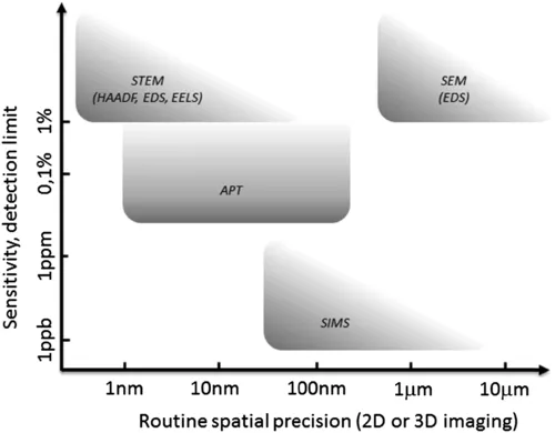
Figure 1.1 Range of routine spatial precision in two-dimensional (2D) or three-dimensional (3D) imaging mode and chemical sensitivity of major analytical microscopy techniques showing that the atom probe tomography (APT) combines near-atomic resolution with sensitivity close to few atomic parts per million. EDS, Energy dispersive X-ray spectroscopy; EELS, electron energy loss spectroscopy; SEM, scanning electron microscopy; SIMS, secondary ion mass spectroscopy; STEM, scanning transmission electron microscope.
In the landscape of analytical microscopy (Fig. 1.1), APT exhibits unique capabilities: it combines 3D information, near-atomic resolution (0.1 nm in depth <1 nm at the sample surface) with a chemical sensitivity down to a few atomic parts per million in the best cases. The typical analyzed volumes are 50 × 50 × 200 nm3 (Fig. 1.2). Compared to TEM, APT is certainly less versatile. First, it is a destructive technique. Measurements cannot be repeated on the same sample. It is also difficult to relate the chemical information to structure. Besides, the field of view or the region of interest is more limited. However, the unique capabilities of APT to investigate materials in 3D at a near-atomic scale, including semiconductors and oxides, have been leading to a growing community both in the academic world and industry.
Fields of Application
Field ion microscopy (FIM), invented by E.W. Müller in the early 1950s, can be viewed as the precursor of modern APT. Both are based on high electric fields. It has provided major contributions in surface science and physical metallurgy thanks to its atomic-scale resolution (Fig. 1.3). As early as the 1960s it has been used to image crystalline defects like grain boundaries (GBs) [4] or vacancies [5]. Phase contrast also made it possible to investigate at least qualitatively precipitation, voids, irradiation defects, segregation to lattice defects or surface diffusion, or reactions in metallic alloys [6].
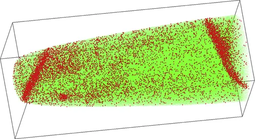
Figure 1.2 Three-dimensional reconstruction of a volume analyzed by atom probe tomography in a steel showing both the carbon segregation to grain boundaries (two are displayed) and a nanoscale carbide [Fe, green; C, red; 45 × 45 × 120 nm3].
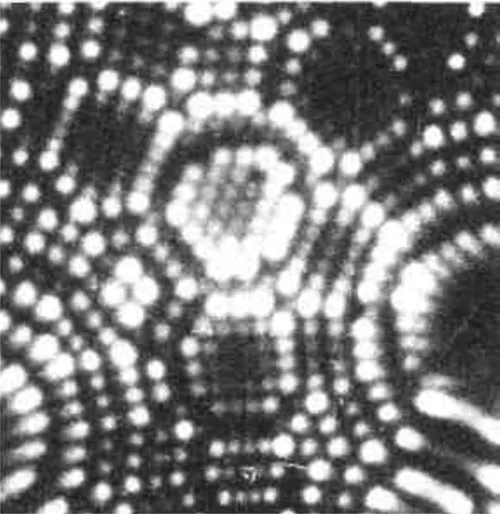
Figure 1.3 Field ion microscopy image of the surface of a PtAu alloy. Each dot is the image of a single atom. The intersection of crystalline atomic planes with the hemispherical surface of the sample gives rise to the specific ring contrast. With courtesy of C. Martin, GPM, France.
The early applications of atom probe in the 1970s were mainly limited to good conductors of electricity, that is, metallic alloys. Metallic glasses [7],...