Plant Biochemistry
eBook - ePub

Plant Biochemistry

Hans-Walter Heldt,Birgit Piechulla

Condividi libro
  1. 656 pagine
  2. English
  3. ePUB (disponibile sull'app)
  4. Disponibile su iOS e Android
eBook - ePub

Plant Biochemistry

Hans-Walter Heldt,Birgit Piechulla

Dettagli del libro
Anteprima del libro
Indice dei contenuti
Citazioni

Informazioni sul libro

The fully revised and expanded fourth edition of Plant Biochemistry presents the latest science on the molecular mechanisms of plant life. The book not only covers the basic principles of plant biology, such as photosynthesis, primary and secondary metabolism, the function of phytohormones, plant genetics, and plant biotechnology, but it also addresses the various commercial applications of plant biochemistry. Plant biochemistry is not only an important field of basic science explaining the molecular function of a plant, but is also an applied science that is in the position to contribute to the solution of agricultural and pharmaceutical problems.

Plants are the source of important industrial raw material such as fat and starch but they are also the basis for the production of pharmaceutics. It is expected that in the future, gene technology will lead to the extensive use of plants as a means of producing sustainable raw material for industrial purposes. As such, the techniques and use of genetic engineering to improve crop plants and to provide sustainable raw materials for the chemical and pharmaceutical industries are described in this edition. The latest research findings have been included, and areas of future research are identified.

  • Offers the latest research findings in a concise and understandable manner
  • Presents plant metabolism in the context of the structure and the function of plants
  • Includes more than 300 two-color diagrams and metabolic schemes
  • Covers the various commercial applications of plant biochemistry
  • Provides extensive references to the recent scientific literature

Domande frequenti

Come faccio ad annullare l'abbonamento?
È semplicissimo: basta accedere alla sezione Account nelle Impostazioni e cliccare su "Annulla abbonamento". Dopo la cancellazione, l'abbonamento rimarrà attivo per il periodo rimanente già pagato. Per maggiori informazioni, clicca qui
È possibile scaricare libri? Se sì, come?
Al momento è possibile scaricare tramite l'app tutti i nostri libri ePub mobile-friendly. Anche la maggior parte dei nostri PDF è scaricabile e stiamo lavorando per rendere disponibile quanto prima il download di tutti gli altri file. Per maggiori informazioni, clicca qui
Che differenza c'è tra i piani?
Entrambi i piani ti danno accesso illimitato alla libreria e a tutte le funzionalità di Perlego. Le uniche differenze sono il prezzo e il periodo di abbonamento: con il piano annuale risparmierai circa il 30% rispetto a 12 rate con quello mensile.
Cos'è Perlego?
Perlego è un servizio di abbonamento a testi accademici, che ti permette di accedere a un'intera libreria online a un prezzo inferiore rispetto a quello che pagheresti per acquistare un singolo libro al mese. Con oltre 1 milione di testi suddivisi in più di 1.000 categorie, troverai sicuramente ciò che fa per te! Per maggiori informazioni, clicca qui.
Perlego supporta la sintesi vocale?
Cerca l'icona Sintesi vocale nel prossimo libro che leggerai per verificare se è possibile riprodurre l'audio. Questo strumento permette di leggere il testo a voce alta, evidenziandolo man mano che la lettura procede. Puoi aumentare o diminuire la velocità della sintesi vocale, oppure sospendere la riproduzione. Per maggiori informazioni, clicca qui.
Plant Biochemistry è disponibile online in formato PDF/ePub?
Sì, puoi accedere a Plant Biochemistry di Hans-Walter Heldt,Birgit Piechulla in formato PDF e/o ePub, così come ad altri libri molto apprezzati nelle sezioni relative a Biowissenschaften e Botanik. Scopri oltre 1 milione di libri disponibili nel nostro catalogo.

Informazioni

Anno
2010
ISBN
9780123849878
Edizione
4
Categoria
Botanik

1 A leaf cell consists of several metabolic compartments
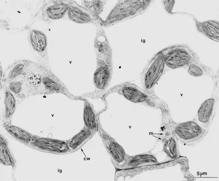
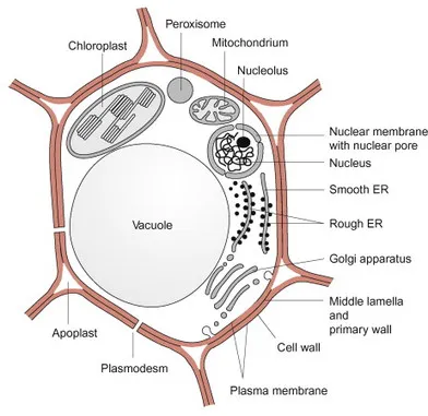
In higher plants photosynthesis occurs mainly in the mesophyll, the chloroplast-rich tissue of leaves. Figure 1.1 shows an electron micrograph of a mesophyll cell and Figure 1.2 shows a schematic presentation of the cell structure. The cellular contents are surrounded by a plasma membrane called the plasmalemma and are enclosed by a cell wall. The cell contains organelles, each with its own characteristic shape, which divide the cell into various compartments (subcellular compartments). Each compartment has specialized metabolic functions, which will be discussed in detail in the following chapters (Table 1.1). The largest organelle, the vacuole, usually fills about 80% of the total cell volume. Chloroplasts represent the next largest compartment, and the rest of the cell volume is filled with mitochondria, peroxisomes, the nucleus, the endoplasmic reticulum, the Golgi bodies, and, outside these organelles, the cell plasma, called cytosol. In addition, there are oil bodies derived from the endoplasmic reticulum. These oil bodies, which occur in seeds and some other tissues (e.g., root nodules), are storage organelles for triglycerides (see Chapter 15).
Image
Figure 1.1 Electron micrograph of mesophyll tissue from tobacco. In most cells the large central vacuole is to be seen (v). Between the cells are the intercellular gas spaces (ig), which are somewhat enlarged by the fixation process. c: chloroplast; cw: cell wall; n: nucleus; m: mitochondrion.
(By D. G. Robinson, Heidelberg.)
Image
Figure 1.2 Schematic presentation of a mesophyll cell. The black lines between the red cell walls represent the regions where adjacent cell walls are glued together by pectins.
Table 1.1 Subcellular compartments in a mesophyll cell* and some of their functions
Percent of the total cell volume Functions (incomplete)
Vacuole 79 Maintenance of cell turgor.
Store of, e.g., nitrate, glucose and storage proteins, intermediary store for secretory proteins, reaction site of lytic enzymes and waste depository
Chloroplasts 16 Photosynthesis, synthesis of starch and lipids
Cytosol 3 General metabolic compartment, synthesis of sucrose
Mitochondria 0.5 Cell respiration
Nucleus 0.3 Contains the genome of the cell. Reaction site of replication and transcription
Peroxisomes Reaction site for processes in which toxic intermediates, such as H2O2 and glyoxylate, are formed and eliminated
Endoplasmic reticulum Storage of Ca++ ions, participation in the export of proteins from the cell and in the transport of newly synthesized proteins into the vacuole and their secretion from the cell
Oil bodies (oleosomes) Storage of triacylglycerols
Golgi bodies Processing and sorting of proteins destined for export from the cells or transport into the vacuole
* Mesophyll cells of spinach; data by Winter, Robinson, and Heldt (1994).
The nucleus is surrounded by the nuclear envelope, which consists of the two membranes of the endoplasmic reticulum. The space between the two membranes is known as the perinuclear space. The nuclear envelope is interrupted by nuclear pores with a diameter of about 50 nm. The nucleus contains chromatin, consisting of DNA double strands that are stabilized by being bound to basic proteins (histones). The genes of the nucleus are collectively referred to as the nuclear genome. Within the nucleus, usually off-center, lies the nucleolus, where ribosomal subunits are formed. These ribosomal subunits and the messenger RNA formed by transcription of the DNA in the nucleus migrate through the nuclear pores to the ribosomes in the cytosol, the site of protein biosynthesis. The synthesized proteins are distributed between the different cell compartments according to their final destination.
The cell contains in its interior the cytoskeleton, which is a three-dimensional network of fiber proteins. Important elements of the cytoskeleton are the microtubuli and the microfilaments, both macromolecules formed by the aggregation of soluble (globular) proteins. Microtubuli are tubular structures composed of α and β tubuline monomers. The microtubuli are connected to a large number of different motor proteins that transport bound organelles along the microtubuli at the expense of ATP. Microfilaments are chains of polymerized actin that interact with myosin to achieve movement. Actin and myosin are the main constituents of the animal muscle. The cytoskeleton has many important cellular functions. It is involved in the spatial organization of the organelles within the cell, enables thermal stability, plays an important role in cell division, and has a function in cell-to-cell communication.
1.1 The cell wall gives the plant cell mechanical stability
The difference between plant cells and animal cells is that plant cells have a cell wall. This wall limits the volume of the plant cell. The water taken up into the cell by osmosis presses the plasma membrane against the inside of the cell wall, thus giving the cell mechanical stability. The cell walls are very complex structures; in Arabidopsis about 1,000 genes were found to be involved in its synthesis. Cell walls also protect against infections.
The cell wall consists mainly of carbohydrates and proteins
The cell wall of a higher plant is made up of about 90% carbohydrates and 10% proteins. The main carbohydrate constituent is cellulose. Cellulose is an unbranched polymer consisting of D-glucose molecules, which are connected to each other by β-1,4 glycosidic linkages (Fig. 1.3A). Each glucose unit is rotated by 180° from its neighbor, so that very long straight chains can be formed with a chain length of 2,000 to 25,000 glucose residues. About 36 cellulose chains are associated by interchain hydrogen bonds to a crystalline lattice structure known as a microfibril. These crystalline regions are impermeable to water. The microfibrils have an unusually high tensile strength, are very resistant to chemical and biological degradations, and are in fact so stable that they are very difficult to hydrolyze. However, many bacteria and fungi have cellulose-hydrolyzing enzymes (cellulases). These bacteria can be found in the digestive tract of some animals (e.g., ruminants), thus enabling them to digest grass and straw. It is interesting to note that cellulose is the most abundant organic substance on earth, representing about half of the total organically bound carbon.
Image
Image
Image
Figure 1.3 Main constituents of the cell wall. A. Cellulose; B. A hemicellulose; C. Constituent of pectin
Hemicelluloses are also important constituents of the cell wall. They are defined as those polysaccharides that can be extracted by alkaline solutions. The name is derived from an initial belief, which later turned out to be incorrect, that hemicelluloses are precursors of cellulose. Hemicelluloses consist of a variety of polysaccharides that contain, in addition to D-glucose, other carbohydrates such as the hexoses D-mannose, D-galactose, D-fucose, and the pentoses D-xylose and L-arabinose. Figure 1.3B shows xyloglycan as an example of a hemicellulose. The basic structure is a β-1,4-glucan chain to which xylose residues are bound via α-1,6 glycosidic linkages, which in part are linked to D-galactose and D-fucose. In addition to this, L-arabinose residues are linked to the 2′OH group of the glucose.
Another major constituent of the cell wall is pectin, a mixture of polymers from sugar acids, such as D-galacturonic acid, which are connected by α-1,4 glycosidic links (Fig. 1.3C). Some of the carboxyl groups are esterified by methyl groups. The free carboxyl groups of adjacent chains are linked by Ca++ and Mg++ ions (Fig. 1.4). When Mg++ and Ca++ ions are absent, pectin is a soluble compound. The Ca++/Mg++ salt of pectin forms an amorphous, deformable gel that is able to swell. Pectins function like glue in sticking neighboring cells together, but these cells can be detached again during plant growth. The food industry makes use of this property of pectin when preparing jellies and jams.
Image
Figure 1.4 Ca++ and Mg++ ions mediate electrostatic interactions between pectin strands.
The structural proteins of the cell wall are connected by glycosidic linkages to the branched polysaccharide chains and belong to the class of proteins known as glycoproteins. The carbohydrate portion of these glycoproteins varies from 50% to over 90%.
For a plant cell to grow, the very rigid cell wall has to be loosened in a precisely controlled way. This is facilitated by the protein expansin, which occurs in growing tissues of all flowering plants. It probably functions by breaking hydrogen bonds between cellulose microfibrils and cross-linking polysa...

Indice dei contenuti