
eBook - ePub
Gray's Anatomy
Henry Gray, Henry Carter
This is a test
Condividi libro
- English
- ePUB (disponibile sull'app)
- Disponibile su iOS e Android
eBook - ePub
Gray's Anatomy
Henry Gray, Henry Carter
Dettagli del libro
Anteprima del libro
Indice dei contenuti
Citazioni
Informazioni sul libro
Written in the 1850s when its author was a young doctor, Gray's Anatomy was the most comprehensive and accessible medical textbook of its time. This edition comprises an abridged version of the classic 1860 text - the last to be published during Gray's lifetime - and the masterly wood-block illustrations that ensured the runaway success of the original book.A national and international treasure, the original Gray's Anatomy is a pleasure to dip into or to consult in depth. It is essential reading for anyone with an interest in the history of medicine or in the amazingly complex machine that is the human body.
Domande frequenti
Come faccio ad annullare l'abbonamento?
È semplicissimo: basta accedere alla sezione Account nelle Impostazioni e cliccare su "Annulla abbonamento". Dopo la cancellazione, l'abbonamento rimarrà attivo per il periodo rimanente già pagato. Per maggiori informazioni, clicca qui
È possibile scaricare libri? Se sì, come?
Al momento è possibile scaricare tramite l'app tutti i nostri libri ePub mobile-friendly. Anche la maggior parte dei nostri PDF è scaricabile e stiamo lavorando per rendere disponibile quanto prima il download di tutti gli altri file. Per maggiori informazioni, clicca qui
Che differenza c'è tra i piani?
Entrambi i piani ti danno accesso illimitato alla libreria e a tutte le funzionalità di Perlego. Le uniche differenze sono il prezzo e il periodo di abbonamento: con il piano annuale risparmierai circa il 30% rispetto a 12 rate con quello mensile.
Cos'è Perlego?
Perlego è un servizio di abbonamento a testi accademici, che ti permette di accedere a un'intera libreria online a un prezzo inferiore rispetto a quello che pagheresti per acquistare un singolo libro al mese. Con oltre 1 milione di testi suddivisi in più di 1.000 categorie, troverai sicuramente ciò che fa per te! Per maggiori informazioni, clicca qui.
Perlego supporta la sintesi vocale?
Cerca l'icona Sintesi vocale nel prossimo libro che leggerai per verificare se è possibile riprodurre l'audio. Questo strumento permette di leggere il testo a voce alta, evidenziandolo man mano che la lettura procede. Puoi aumentare o diminuire la velocità della sintesi vocale, oppure sospendere la riproduzione. Per maggiori informazioni, clicca qui.
Gray's Anatomy è disponibile online in formato PDF/ePub?
Sì, puoi accedere a Gray's Anatomy di Henry Gray, Henry Carter in formato PDF e/o ePub, così come ad altri libri molto apprezzati nelle sezioni relative a History e World History. Scopri oltre 1 milione di libri disponibili nel nostro catalogo.
Informazioni
Argomento
HistoryCategoria
World HistoryTHE SKULL.
The Skull, or superior expansion of the vertebral column, is composed of four vertebræ, the elementary parts of which are specially modified in form and size, and almost immoveably connected, for the reception of the brain, and special organs of the senses. These vertebræ are the occipital, parietal, frontal, and nasal. Descriptive anatomists, however, divide the skull into two parts, the Cranium and the Face. The Cranium (κράνος, a helmet), is composed of eight bones: viz., the occipital, two parietal, frontal, two temporal, sphenoid, and ethmoid. The face is composed of fourteen bones; viz., the two nasal, two superior maxillary, two lachrymal, two malar, two palate, two inferior turbinated, vomer, inferior maxillary. The ossicula auditûs, the teeth, and Wormian bones, are not included in this enumeration.

THE OCCIPITAL BONE.
The Occipital Bone (fig. 22) is situated at the back part and base of the cranium, is trapezoid in form, curved upon itself, and presents for examination two surfaces, four borders, and four angles.
The External Surface is convex. Midway between the summit of the bone and the posterior margin of the foramen magnum is a prominent tubercle, the external occipital protuberance, for the attachment of the Ligamentum nuchæ; and descending from it, as far as the foramen, a vertical ridge, the external occipital crest. This tubercle and crest, vary in prominence in different skulls. Passing outwards from the occipital protuberance on each side are two semicircular ridges, the superior curved lines; and running parallel with these from the middle of the crest, are the two inferior curved lines. The surface of the bone above the superior curved lines is smooth on each side, and in the recent state, is covered by the Occipito-frontalis muscle, whilst the ridges, as well as the surface of the bone between them, serve for the attachment of numerous muscles. The superior curved line gives attachment internally to the Trapezius, externally to the Occipito-frontalis, and Sterno-cleido mastoid; to the extent shewn in the figure. The depressions between the curved lines to the Complexus internally, the Splenius capitis and Obliquus superior externally. The inferior curved line, and the depressions below it, afford insertion to the Rectus capitis posticus, major and minor.
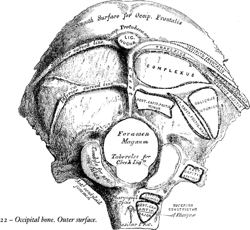
The foramen magnum is a large oval aperture, its long diameter extending from before backwards. It transmits the spinal cord and its membranes, the spinal accessory nerves, and the vertebral arteries. Its back part is wide for the transmission of the cord, and the corresponding margin rough for the attachment of the dura mater enclosing it; the fore part is narrower, being encroached upon by the condyles; it has projecting towards it from below the odontoid process, and its margins are smooth and bevelled internally to support the medulla oblongata. On each side of the foramen magnum are the condyles, for articulation with the atlas; they are convex, oblong or reniform in shape, and directed downwards and outwards; they converge in front, and encroach slightly upon the anterior segment of the foramen. On the inner border of each condyle is a rough tubercle for the attachment of the ligaments (check) which connect this bone with the odontoid process of the axis; whilst external to them is a rough tubercular prominence, the transverse or jugular process, (the representative of the transverse process of a vertebra) channelled in front by a deep notch, which forms part of the jugular foramen. The under surface of this process affords attachment to the Rectus capitis lateralis; its upper or cerebral surface presents a deep groove, which lodges part of the lateral sinus, whilst its prominent extremity is marked by a quadrilateral rough surface, covered with cartilage in the fresh state, and articulating with a similar surface on the petrous portion of the temporal bone. On the outer side of each condyle, near its fore part, is a foramen, the anterior condyloid; it is directed downwards, outwards, and forwards, and transmits the hypoglossal nerve. This foramen is sometimes double. Behind each condyle is a fossa,* perforated at the bottom by a foramen, the posterior condyloid, for the transmission of a vein to the lateral sinus. In front of the foramen magnum is a strong quadrilateral plate of bone, the basilar process, wider behind than in front; its under surface, which is rough, presenting in the median line a tubercular ridge, the pharyngeal spine, for the attachment of the tendinous raphe and Superior constrictor of the pharynx; and on each side of it, rough depressions for the attachment of the Recti capitis antici, major and minor.
The Internal or Cerebral Surface (fig. 23) is deeply concave. The posterior or occipital part is divided by a crucial ridge into four fossæ. The two superior, the smaller, receive the posterior lobes of the cerebrum, and present slight eminences and depressions corresponding to their convolutions. The two inferior, which receive the lateral lobes of the cerebellum, are larger than the former, and comparatively smooth; both are marked by slight grooves for the lodgment of arteries. At the point of meeting of the four divisions of the crucial ridge is an eminence, the internal occipital protuberance. It nearly corresponds to that on the outer surface, and is perforated by one or more large vascular foramina. From this eminence, the superior division of the crucial ridge, runs upwards to the superior angle of the bone; it presents occasionally a deep groove for the superior longitudinal sinus, the margins of which give attachment to the falx cerebri. The inferior division, the internal occipital crest, runs to the posterior margin of the foramen magnum, on the edge of which it becomes gradually lost: this ridge, which is bifurcated below, serves for the attachment of the falx cerebelli. The transverse grooves pass outwards to the lateral angles; they are deeply channelled, for the lodgment of the lateral sinuses, their prominent margins affording attachment to the tentorium cerebelli.† At the point of meeting of these grooves is a depression, the ‘Torcular Herophili’,‡ placed a little to one or the other side of the internal occipital protuberance. More anteriorly is the foramen magnum, and on each side of it, but nearer its anterior than its posterior part, the internal openings of the anterior condyloid foramina; the internal openings of the posterior condyloid foramina being a little external and posterior to them, protected by a small arch of bone. In front of the foramen magnum is the basilar process, presenting a shallow depression, the basilar groove, which slopes from behind, upwards and forwards, and supports the medulla oblongata; and on each side of the basilar process is a narrow channel, which, when united with a similar channel on the petrous portion of the temporal bone, forms a groove, which lodges the inferior petrosal sinus.

Angles. The superior angle is received into the interval between the posterior superior angles of the two parietal bones: it corresponds with that part of the skull in the fœtus which is called the posterior fontanelle. The inferior angle is represented by the square-shaped surface of the basilar process. At an early period of life, a layer of cartilage separates this part of the bone from the sphenoid; but in the adult, the union between them is osseous. The lateral angles correspond to the outer ends of the transverse grooves, and are received into the interval between the posterior inferior angles of the parietal and the mastoid portion of the temporal.
Borders. The superior extends on each side from the superior to the lateral angle, is deeply serrated for articulation with the parietal bone, and forms by this union the lambdoid suture. The inferior border extends from the lateral to the inferior angle; its upper half is rough, and articulates with the mastoid portion of the temporal, forming the masto-occipital suture: the inferior half articulates with the petrous portion of the temporal, forming the petro-occipital suture: these two portions are separated from one another by the jugular process. In front of this process is a deep notch, which, with a similar one on the petrous portion of the temporal, forms the foramen lacerum posterius. This notch is occasionally subdivided into two parts by a small process of bone, and presents an aperture at its upper part, the internal opening of the posterior condyloid foramen.
Structure. The occipital bone consists of two compact laminæ, called the outer and inner tables, having between them the diploic tissue; this bone is especially thick, at the ridges, protuberances, condyles, and anterior part of the basilar process; whilst at the bottom of the fossæ, especially the inferior, it is thin, semitransparent, and destitute of diploë.
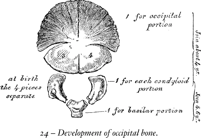
Development. (fig. 24). The occipital bone has four centres of development; one for the posterior or occipital part, which is formed in membrane, one for the basilar portion; and one for each condyloid portion, which are formed in cartilage.
The centre for the occipital portion appears about the tenth week of fœtal life; and consists, according to Blandin and Cruvelhier, of a small oblong plate which appears in the situation of the occipital protuberance.* The condyloid portions then ossify, and lastly the basilar portion. At birth, the bone consists of four parts, separate from one another, the occipital portion being fissured in the direction above indicated. At about the fourth year, the occipital and the two condyloid pieces join; and about the sixth year the bone consists of a single piece. At a later period, between the eighteenth and twenty-fifth years, the occipital and sphenoid become united, forming a single bone.
Articulations. With six bones; two parietal, two temporal, sphenoid, and atlas.
Attachment of Muscles. To the superior curved line are attached the Occipito-frontalis, Trapezius, and Sterno-cleido-mastoid. To the space between the curved lines, the Complexus, Splenius capitis, and Obliquus superior; to the inferior curved line, and the space between it and the foramen magnum, the Rectus posticus major and minor; to the transverse process, the Rectus lateralis; and to the basilar process, the Recti antici majores and minores, and Superior Constrictor of the pharynx.
THE PARIETAL BONES.
The Parietal Bones (paries, a wall), form by their union the sides and roof of the skull; each bone is of an irregular quadrilateral form, and presents for examination two surfaces, four borders, and four angles.
Surfaces. The external surface (fig. 25) is convex, smooth, and marked about its centre by an eminence, called the parietal eminence, which indicates the point where ossification commenced. Crossing the centre of the bone in an arched direction is a curved ridge, the temporal ridge, for the attachment of the temporal fascia. Above this ridge, the surface of the bone is rough and porous, and covered by the aponeurosis of the Occipito-frontalis; below it the bone is smooth, forms part of the temporal fossa, and affords attachment to the Temporal muscle. At the back part of the superior border, close to the sagittal suture, is a small foramen, the parietal foramen, which transmits a vein to the superior longitudinal sinus. Its existence is not constant, and its size varies considerably.
The internal surface (fig. 26), concave, presents eminences and depressions for lodging the convolutions of the cerebrum, and numerous furrows for the ramifications of the meningeal arteries; the latter run upwards and backwards from the anterior inferior angle, and from the central and posterior part of the lower border of the bone. Along the upper margin is part of a shallow groove, which, when joined to the opposite parietal, forms a channel for the superior longitudinal sinus, the elevated edges of which afford attachment to the falx cerebri. Near the groove are seen several depressions; they lodge the Pacchionian bodies. The internal opening of the parietal foramen is also seen when that aperture exists.

Borders. The superior, the longest and thickest, is dentated to articulate with its fellow of the opposite side, forming the sagittal suture. The inferior is divided into three parts; of these, the anterior is thin and pointed, bevelled at the expense of the outer surface, and overlapped by the tip of the great wing of the sphenoid; the middle portion is arched, bevelled at the expense of the outer surface, and overlapped by the squamous portion of the temporal; the posterior portion being thick and serrated for articulation with the mastoid portion of the temporal. The anterior border, deeply serrated, is bevelled at the expense of the outer surface above, and of the inner below; it articulates with the frontal bone, forming the coronal suture. The posterior border, deeply denticulated, articulates with the occ...