Cardiology of the Horse is a multi-author, contemporary reference on equine cardiology. The first section reviews the physiology, pathophysiology and pharmacology of the equine cardiovascular system. The second section describes diagnostic methods from basic to specialist examination skills and the third section addresses the investigation and management of common clinical problems using a problem-orientated approach. Suitable for students, general and specialist practitioners and teachers.
An up-to-date account of current clinical practice in equine cardiology covering:
recent developments in research and practice
problem-orientated approaches helpful to both general and specialist practitioners
clinical management of specific groups from foals and racehorses to geriatric patients
cardiac problems related to exercise, anaesthesia and intensive care
A superb companion DVD of clinical cases with extensive footage combining theory and clinical practice:
echocardiograms
heart sounds and murmurs
ECGs
radiography
pathology
Extensive linking of text to DVD, integrating fundamental principles and diagnostic data with information on clinical management of specific problems.
Domande frequenti
Come faccio ad annullare l'abbonamento?
È semplicissimo: basta accedere alla sezione Account nelle Impostazioni e cliccare su "Annulla abbonamento". Dopo la cancellazione, l'abbonamento rimarrà attivo per il periodo rimanente già pagato. Per maggiori informazioni, clicca qui
È possibile scaricare libri? Se sì, come?
Al momento è possibile scaricare tramite l'app tutti i nostri libri ePub mobile-friendly. Anche la maggior parte dei nostri PDF è scaricabile e stiamo lavorando per rendere disponibile quanto prima il download di tutti gli altri file. Per maggiori informazioni, clicca qui
Che differenza c'è tra i piani?
Entrambi i piani ti danno accesso illimitato alla libreria e a tutte le funzionalità di Perlego. Le uniche differenze sono il prezzo e il periodo di abbonamento: con il piano annuale risparmierai circa il 30% rispetto a 12 rate con quello mensile.
Cos'è Perlego?
Perlego è un servizio di abbonamento a testi accademici, che ti permette di accedere a un'intera libreria online a un prezzo inferiore rispetto a quello che pagheresti per acquistare un singolo libro al mese. Con oltre 1 milione di testi suddivisi in più di 1.000 categorie, troverai sicuramente ciò che fa per te! Per maggiori informazioni, clicca qui.
Perlego supporta la sintesi vocale?
Cerca l'icona Sintesi vocale nel prossimo libro che leggerai per verificare se è possibile riprodurre l'audio. Questo strumento permette di leggere il testo a voce alta, evidenziandolo man mano che la lettura procede. Puoi aumentare o diminuire la velocità della sintesi vocale, oppure sospendere la riproduzione. Per maggiori informazioni, clicca qui.
Cardiology of the Horse è disponibile online in formato PDF/ePub?
Sì, puoi accedere a Cardiology of the Horse di Celia Marr, Mark Bowen in formato PDF e/o ePub, così come ad altri libri molto apprezzati nelle sezioni relative a Medicine e Equine Veterinary Science. Scopri oltre 1 milione di libri disponibili nel nostro catalogo.
I was almost tempted to think with Fracastorius that the motion of the heart was only to be comprehended by God.
William Harvey, 16281
An appreciation of the anatomy of the heart and great vessels is central to the understanding of cardiac function and disease and for optimal interpretation of diagnostic techniques such as auscultation, echocardiography and radiography. This chapter also reviews cardiovascular physiology focusing on impulse conduction within the equine heart and on the heart as a muscular pump. Although clinically important parameters such as stroke volume, cardiac output and blood pressure are emphasized, these haemodynamic parameters are actually the ultimate functional expression of the biochemical and biophysical processes of myocyte excitation, contraction and relaxation.
ANATOMY OF THE HEART AND GREAT VESSELS(
ET)
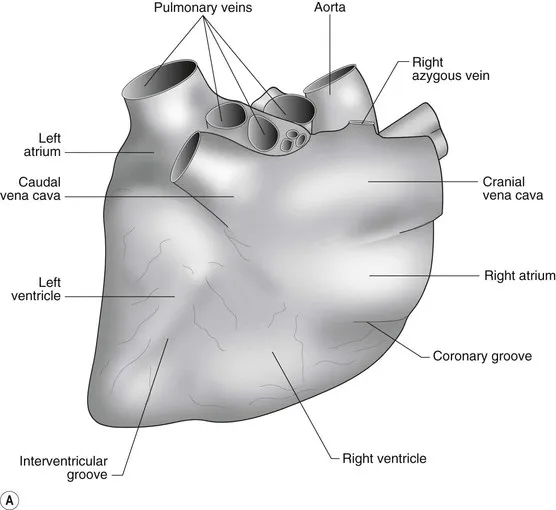
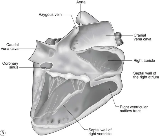
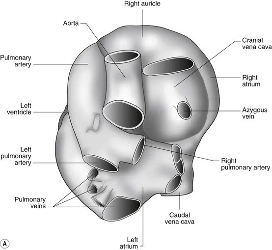
The heart can be regarded as a parallel pump system: deoxygenated blood returning from the body enters the right side from where it is directed via the pulmonary arterial system to the lungs for oxygenation. Oxygenated blood returns to the left side of the heart via the pulmonary veins and is then pumped to the body via the systemic arterial system. Deoxygenated blood returns via the systemic veins to the right side. The heart is located within the middle mediastinal space where its long axis is orientated at approximately 10° to vertical with its base lying dorsal and cranial to the apex. The apex is located above the last sternebra cranial to the sternal portion of the diaphragm. The heart consists of two atria and two ventricles, blood entering via the atria and leaving via the ventricles. The right atrium (RA) occupies the cranial part of the heart base and consists of two main parts, the larger part, the sinus venarum cavarum, into which the veins empty, and a conical out-pouching, the auricle. The auricle is triangular and broad-based and curves around the base of the heart towards the left ending cranial to the origin of the main pulmonary artery (Figs. 1.1 and 1.2). The cranial vena cava (draining structures of the head and neck) enters the most dorsal part of the RA, the caudal vena cava (draining abdominal structures) opens into the caudal part and the azygous vein (draining the caudal thorax) enters between the two cavae. The coronary sinus (draining the coronary circulation) opens into the RA ventral to the caudal vena cava. There are also several smaller veins that drain directly into the RA (see Figs. 1.1 and 1.2). On the internal surface of the RA there are pronounced ridges formed by extensive bands of pectinate muscles and dorsally these form the terminal crest at the base of the auricle (Fig. 1.3). The oval fossa is a diverticulum at the point of entrance of the caudal vena cava that is a remnant of the foramen ovale, the communication that exists between the two atria in the fetus.
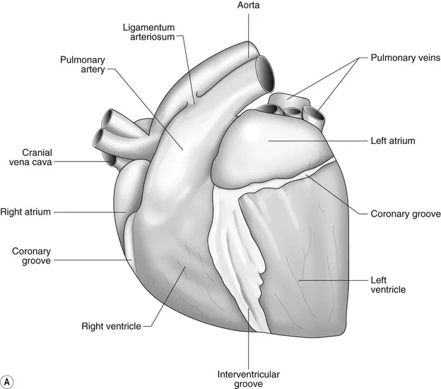
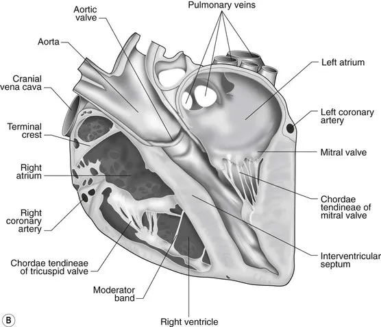
Figure 1.1(A) The intact heart viewed from the right side: note the boundaries of the right atrium and ventricle and the left and right ventricle are delineated by the coronary and interventricular grooves, respectively. (B) The right aspect of the heart following removal of the right wall.(C) A section through the centre of the heart viewed from the right side.Adapted with permission from Ghoshal NG. Equine heart and arteries. In: Getty R (ed) Sisson and Grossman's The Anatomy of the Domestic Animals, Vol 1, 5th ed. Philadelphia: WB Saunders, 1975:554–618.
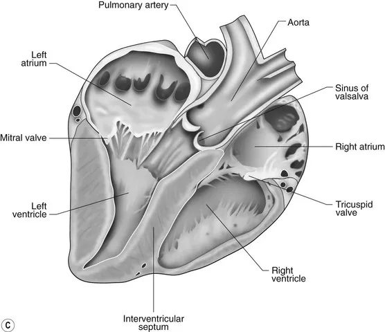
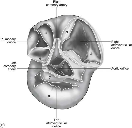
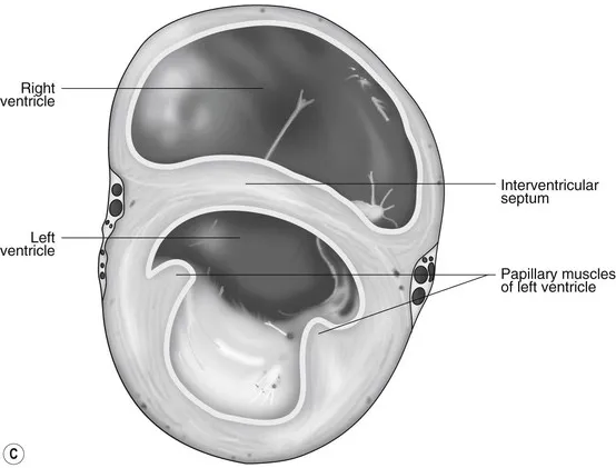
Figure 1.2(A) The heart base viewed from above.(B) A cross-section through the heart base illustrating the valve leaflets: Tricuspid valve 1 = septal, 2 = right, 3 = left, Pulmonary valve 4 = right, 5 = left, 6 = intermediate, Mitral valve 7 = septal, 8 = nonseptal, Aortic Valve 9 = right coronary, 10 = left coronary, 11 = noncoronary. (C) A cross-section through the ventricles.Adapted with permission from Ghoshal NG. Equine heart and arteries. In: Getty R (ed) Sisson and Grossman's The Anatomy of the Domestic Animals, Vol 1, 5th ed. Philadelphia: WB Saunders, 1975:554–618.
Figure 1.3(A) The intact heart viewed from the left side. Note the boundaries of the left atrium and ventricle and the left and right ventricle are delineated by the coronary and interventricular groves respectively.(B) A section through the centre of the heart viewed from the left side.Adapted with permission from Ghoshal NG. Equine heart and arteries. In: Getty R (ed) Sisson and Grossman's The Anatomy of the Domestic Animals, Vol 1, 5th ed. Philadelphia: WB Saunders, 1975:554–618.
The right atrioventricular (AV) or tricuspid valve forms the ventral floor of the RA and the entrance to the right ventricle (RV) (see Figs. 1.1 and 1.2). As its name suggests, the tricuspid valve is composed of three large leaflets: one is septal, one lies on the right margin (parietal) and the third lies between the AV opening and the right outflow tract (angular). The leaflets are anchored to the papillary muscles of the RV by a series of chordae tendineae. The RV is a crescent-shaped structure in cross-section and triangular when viewed from its inner aspect (see Fig. 1.2). It wraps around the cranial aspect of the heart and, in this respect, the convention derived from human anatomy ascribing the terms right and left to the heart, is rather misleading. In the horse, the heart would be better defined as having cranial and caudal components. The internal surface of the RV is trabeculated and moderator bands cross the lumen of the RV from the septum to the opposite wall carrying conduction tissue (see Fig. 1.3). These moderator bands vary in size greatly among individuals. Ventrally, the RV does not reach the heart's apex. It extends dorsally and to the left to form the right outflow tract leading to the main pulmonary artery (PA) via the pulmonary valve (right semilunar valve) valve (see Fig. 1.3). The pulmonary valve consists of three half-moon-shaped cusps, the right, left and intermediate, which occasionally have small fenestrations along their free edges and are attached to a fibrous ring at the base of the pulmonary artery. The PA arises from the left side of the RV and curves dorsally, caudally and medially to run under the descending aorta where it branches into left and right. The right PA passes over the cranial part of the left atrium and under the trachea while the left PA is in contact with the bulk of the dorsal surface of the left atrium (LA) (see Fig. 1.2).
The LA forms the caudal part of the base of the heart and also has an auricle, extending laterally and cranially on the left side. The left auricle is more pointed than the right auricle and lacks a terminal crest (see Fig. 1.2). The LA lacks the extensive pectinate muscles that characterize the RA. Seven or eight pulmonary veins enter the LA around its caudal and right aspects. A depression may be appreciated on the septal surface, corresponding with the site of the fetal foramen ovale. The ventral floor of the LA co...