Rostam Farhadieh, Neil Bulstrode, Babak J. Mehrara, Sabrina Cugno
This is a test
This is a test
Condividi libro
924 pagine
English
ePUB (disponibile sull'app)
Disponibile su iOS e Android
eBook - ePub
Plastic Surgery - Principles and Practice E-Book
Rostam Farhadieh, Neil Bulstrode, Babak J. Mehrara, Sabrina Cugno
Dettagli del libro
Anteprima del libro
Indice dei contenuti
Citazioni
Informazioni sul libro
Withdetailed, expert guidance on each essential topic, Plastic Surgery: Principles and Practiceoffers single-volume convenience without sacrificing complete coverage of this multi-faceted field.Written by global leading authorities, it provides concise, easy-to-follow instruction with the clinical details and supportive data needed to achieve optimal patient outcomes.
Offersthorough coverageof facelift procedures, rhinoplasty, otoplasty and more, along withclinical pearlsfrom masters in the field.
Featureshundreds of high-quality imagesincluding anatomical line art, case photos, and procedural operative photos.
IIncludes a superb selection of procedural videos of global experts performing key techniques within operating room and close-up clinical pearls.
An ideal resource for residents, fellows, and practitioners in plastic surgery, as well as those in otolaryngology, vascular surgery, and cosmetic dermatology.
Domande frequenti
Come faccio ad annullare l'abbonamento?
È semplicissimo: basta accedere alla sezione Account nelle Impostazioni e cliccare su "Annulla abbonamento". Dopo la cancellazione, l'abbonamento rimarrà attivo per il periodo rimanente già pagato. Per maggiori informazioni, clicca qui
È possibile scaricare libri? Se sì, come?
Al momento è possibile scaricare tramite l'app tutti i nostri libri ePub mobile-friendly. Anche la maggior parte dei nostri PDF è scaricabile e stiamo lavorando per rendere disponibile quanto prima il download di tutti gli altri file. Per maggiori informazioni, clicca qui
Che differenza c'è tra i piani?
Entrambi i piani ti danno accesso illimitato alla libreria e a tutte le funzionalità di Perlego. Le uniche differenze sono il prezzo e il periodo di abbonamento: con il piano annuale risparmierai circa il 30% rispetto a 12 rate con quello mensile.
Cos'è Perlego?
Perlego è un servizio di abbonamento a testi accademici, che ti permette di accedere a un'intera libreria online a un prezzo inferiore rispetto a quello che pagheresti per acquistare un singolo libro al mese. Con oltre 1 milione di testi suddivisi in più di 1.000 categorie, troverai sicuramente ciò che fa per te! Per maggiori informazioni, clicca qui.
Perlego supporta la sintesi vocale?
Cerca l'icona Sintesi vocale nel prossimo libro che leggerai per verificare se è possibile riprodurre l'audio. Questo strumento permette di leggere il testo a voce alta, evidenziandolo man mano che la lettura procede. Puoi aumentare o diminuire la velocità della sintesi vocale, oppure sospendere la riproduzione. Per maggiori informazioni, clicca qui.
Plastic Surgery - Principles and Practice E-Book è disponibile online in formato PDF/ePub?
Sì, puoi accedere a Plastic Surgery - Principles and Practice E-Book di Rostam Farhadieh, Neil Bulstrode, Babak J. Mehrara, Sabrina Cugno in formato PDF e/o ePub, così come ad altri libri molto apprezzati nelle sezioni relative a Medicine e Ear, Nose & Throat Medicine. Scopri oltre 1 milione di libri disponibili nel nostro catalogo.
1: Skin Structure and Function, Wound Healing and Scarring
Justine Victoria Sullivan, and Simon Myers
Skin Structure and Function
The integument comprises the skin together with its appendages (Figs. 1.1 and 1.2). These include hair and hair follicles, sebaceous and sweat glands, and nails. The skin covers the entire body and is the largest organ of the body. It covers a surface area of more than 1.7 m2, making up in total about 16% of normal body weight.1 It has an array of functions. These include acting as a barrier to physical, biological and chemical agents, as well as to ultraviolet (UV) radiation. Skin barrier function also acts to control body hydration. Other functions include: sensory and thermoregulatory roles, vitamin D synthesis, immune surveillance, excretion of wastes through sweat glands, socio-sexual communication and reproduction, by virtue of its appearance and smell (e.g. hormones and pheromones).
Skin is divided into glabrous (covering the palms of the hands and soles of the feet) and hairy skin. The skin comprises of two layers, the outermost epidermis and the innermost layer, the dermis. Embryologically, these two layers of skin are derived from the ectoderm and mesoderm, respectively. The epidermis and dermis are firmly attached to each other, and together vary in thickness from around 0.5 to 4 mm or more depending on body site. At the point where the epidermis meets the dermis, invaginations that project into the dermis are formed, known as “rete ridges” or “pegs.” Complementary projections of the dermis are called dermal papillae.
Epidermis
The epidermis is defined as a stratified squamous epithelium made up of several cell types. These include melanocytes (production of melanin pigment), Langerhans cells (immune function), Merkel cells (sensory function), and keratinocytes, of which the latter make up at least 80% of its cellular population.
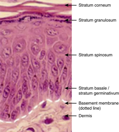
The epidermis is arranged into distinct layers, each showing a pattern of keratinocyte proliferation, differentiation, and maturation. Four main layers exist. These are strata basale (germinativum), spinosum, granulosum, and corneum.1 In thick skin, a fifth layer called the stratum lucidum is present and is found between the granular and the cornified layers. These layers reflect the sequential differentiation of keratinocytes as they migrate from the basal layer at the onset of terminal differentiation, having lost the ability to proliferate, to the outermost cornified layers where they are sloughed off. The process of terminal differentiation involves a series of biochemical and morphological changes, which result in the production of an anucleated cornified keratinocyte that forms the stratum corneum. Epidermal turnover takes on average 14–30 days.
The stratum basale is a single layer of cuboidal basal cells attached to the basement membrane by hemidesmosomes that contain integrins. Adjacent cells are attached by desmosomes that contain cadherins. The majority of the cells in this layer are mitotically active and are required for the continued renewal of the epidermis by upward displacement, replacing the cells of the outermost superficial layer that are lost during normal epidermal turnover.2 These mitotically active cells derive from a population of putative stem cells that are thought to reside in the deep rete ridges of glabrous skin and at the tips of the dermal papillae in interfollicular epidermis or in the bulge region of the outer root sheath (ORS) of adult human hair follicles in hairy skin. Also arising from stem cells in the basal layer are transit amplifying cells and post mitotic cells which are displaced into the suprabasal layers. Basal cells contain cytokeratins organized in bundles around the nucleus and insert into desmosomes peripherally.
Fig. 1.1 Diagrammatic representation of the skin. From Gawkrodger DJ, Ardern-Jones MR. Dermatology: An Illustrated Colour Text. 6th edn. Elsevier; 2017.
Fig. 1.2 Hematoxylin and eosin (H&E) histology of the skin.
Above this layer is the stratum spinosum, consisting of several layers of irregular, polyhedral shaped cells that display spiny projections. Cells from the previous layer lose contact with the basement membrane and are subsequently pushed up to form this one. Some cells in this layer are still mitotically active. Cells become progressively flattened as they move up towards succeeding layers. Cells contain lamellar granules (for the later provision of epidermal lipids responsible for the barrier properties of the stratified corneum) and more desmosomes for cell-to-cell adhesion.
The next layer, the stratum granulosum, comprises of three to five sheets of flattened cells. In this layer, as well as lamellar granules there are keratohyalin granules, which contain pro-filaggrin, a precursor of filaggrin that bundles the keratin filaments together.2,3
The stratum lucidum consists of several layers of flattened cells without nuclei and organelles, and a keratin-rich cytoplasm.
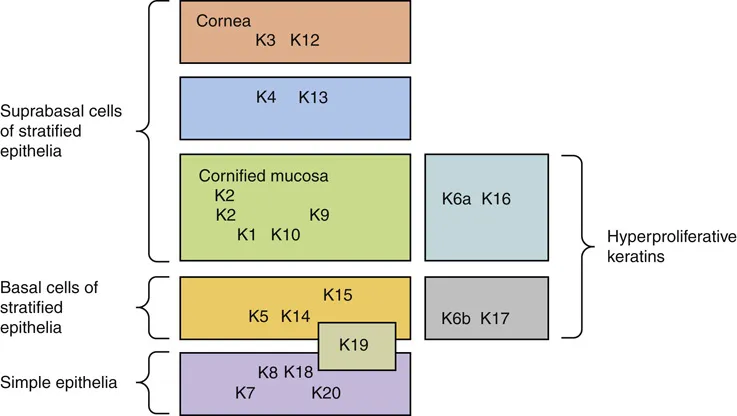
Fig. 1.3 Schematic representation of keratin expression in normal epithelia.
The cornified layer consists of dead, anucleated, highly keratinized cells called squames or corneocytes. Keratin filaments polymerize by forming strong disulphide bonds. Filaggrin, a protein component of the keratohyalin granule, is involved in this process. The cornified envelope forms due to the catalytic activity of the enzyme transglutaminase which crosslinks proteins such as involucrin (an insoluble 70–80 kDa cysteine rich protein) in the plasma membrane. Other proteins found as components of the cornified cell envelope include keratolinin, loricrin, small proline-rich proteins, the serine proteinase inhibitor elafin, filaggrin linker-segment peptide, and envoplakin. Lipids, discharged by lamellar granules, fill the intercellular spaces and contribute to the barrier properties of the epidermis. The lack of desmosomes in the cells that are closest to the outermost layer results in the loss or shedding of corneocytes from the skin.
Keratins
The cytoskeleton of all epithelial cells, including keratinocytes, is formed from the three groups of filaments, actin (microfilaments), tubulin (microtubules), and intermediate filaments. The keratins belong to the latter group, a multi-gene family of proteins that form filaments of 10 nm in diameter4 in which keratins form the two largest groups. These groups, which comprise more than 30 members of the keratin family, have been designated as type I keratins (acidic), numbered 9–20, and type II keratins (basic), numbered 1–8. Usually, type I and type II keratin subunits pair up and the heterodimers formed are expressed according to epithelial type and in a differentiation-state specific manner. For example, in simple epithelia, keratin K8 and K18 are expressed. Cells in the basal layer of stratified epithelia express K5 and K14.4 The differentiating suprabasal layers are characterized by the keratin pair K1 and K10. K6 and K16 are not found in normal epidermis except the outer root sheath of the hair follicle and junctional region. This keratin pair is also constitutively expressed in certain stratified squamous mucosal epithelia and the skin of the palm and sole. Expression of K6 and K16 is induced in wound healing epidermis, hyperproliferative epidermis such as psoriasis, squamous cell carcinomas, and hypertrophic scarring. Thus, keratin e...