![]()
Chapter 1
The history and importance of
venous disease in modern practice
Ian F Lane MA DM MCh FRCS
Consultant Vascular Surgeon, University Hospital of Wales, Cardiff, UK
Introduction
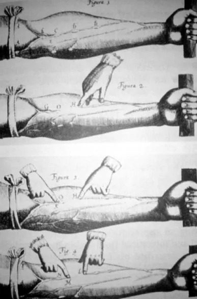
The modern management of venous disease is inextricably linked with advances in physiological measurement and imaging. Indeed, early accounts of disorders describe anomalies and only in the mid-20th century was there an attempt to merge ‘form and function’. Varicose veins have been reported for thousands of years with one of the earliest accounts of serpent-shaped dilatation of the lower limbs from about 1552BC in the Ebers papyrus (Figure 1). The first reliable description of intervention for extremity varices appears in a Turkish textbook, Imperial Surgery, written in 1465AD, although procedures appear to have taken place during the earlier Byzantine period. A published case history of venous thrombosis is recognised at the Bibliothèque Nationale in Paris, affecting a Normandy man aged 20 years in 1282AD. The patient sustained unilateral oedema with ulceration and exudation, and although able to walk only with crutches initially, the symptoms eventually settled leaving only mild long-term leg throbbing. Probably the most significant early work was by Fabricus in 1603 and his student Harvey in 1628, who described the structure and function of venous valves and the circulatory system respectively (Figure 2).
Figure 1 The Ebers papyrus was discovered in Egypt in the 1870s and contains 877 recipes for diseases or symptoms.
Figure 2 Illustration of venous valves from Harvey’s 1628 book De Moto Cordis.
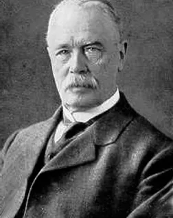
Paré in the 16th century underpinned surgical principles and these were developed by Trendelenburg (Figure 3) and others around the beginning of the 20th century, leading to Keller and Mayo in 1905/6 performing the first ablation of the great saphenous vein, thus providing a gateway to the modern era 1, 2.
Figure 3 Friedrick Trendelenburg (1844-1924) was Surgeon-in-Chief at Leipzig.
Courtesy of www.historiadelamedicina.org.
Historical landmarks
Investigation of venous disorders
Clinical examination with use of the Trendelenberg test and tourniquet has been the mainstay of diagnosis of superficial venous incompetence, even in recent years, despite the inaccuracies in differentiation of saphenopopliteal and perforator disease. The recognition of the physiological implications of deep venous incompetence remained elusive until the era of pulsed Doppler ultrasound imaging and flow measurements. Plethysmography was developed in the mid-20th century and is based upon measurment of venous refill times using strain gauge, electrical impedance or skin colour changes. Difficulties with reproducibility and standardisation has meant that plethysmography has limited use in clinical practice but is valuable in venous research, particularly where continual assessment of individual patients is required.
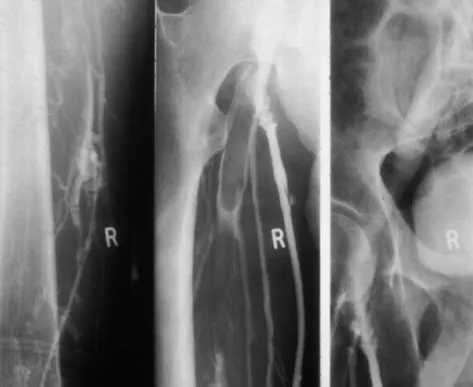
Figure 4 Ascending venogram with thrombus in the femoral vein.
Contrast venography was popularised in the 1960s and even now is regarded by many as the gold standard for the diagnosis of deep vein thrombosis (Figure 4). Descending femoral venography enabled diagnosis of deep venous incompetence based upon distal reflux of contrast, although the clinical implications of single or multiple valvular reflux are not clear. Venography or phlebography is still of value in planning surgery for pelvic venous obstruction and determining the origin of atypical lower limb varicosities, but its use has largely been superseded by duplex imaging.
Continuous-wave Doppler ultrasound has been used in fluid engineering since the 1950s and was further developed, initially in arterial disease where the velocity shifts are significant, by Satomura in the 1970s, when the importance of ankle/brachial systolic pressure indices was recognised. Strandness popularised the technique in both arterial and venous disease, but now modern duplex ultrasound systems combine flow measurement with imaging, enabling the function of individual valves to be identified whilst examining the quality of vein wall, as well as identifying deep vein thrombosis 3. The accurate assessment of perforator and pelvic vein incompetence has led to dilemmas in the significance, and need for treatment, of these conditions. Initial early concerns over the size of ultrasound equipment have been addressed by development of portable devices, although considerable operator training and experience is required. Hand-held Dopper devices, measuring flow alone, are useful as an adjunct to clinical examination but have limited specificity in determining saphenopopliteal and deep vein incompetence. Both duplex imaging and Doppler flow assessment have ensured that surgery for venous incompetence can be planned and is appropriate with venous surgeons recommending that all patients undergo pre-operative investigation.
Modern imaging techniques such as CT angiography have found a place in the diagnosis of pulmonary emboli, whilst magnetic resonance venography may be useful in the management of pelvic and thoracic outlet venous obstruction in the future.
Sclerotherapy and other local treatments
Sclerotherapy was commenced by Pravaz in approximately 1860 and promoted in Europe in 1947 with the foundation of Sociètè Francaise de Phlebologie. Fegan subsequently popularised and developed the technique that bears his name, although by the late 20th century enthusiasm, certainly in the UK, was reducing 4. The reasons for this were the recognition that sclerotherapy did not address the underlying valvular incompetence, leading to high recurrence rates reported as 90% at ten years, patient acceptability of prolonged compression and complications of the technique including ulceration and skin staining. Recently, the development of less toxic intravenous agents and the introduction of foam sclerotherapy under duplex guidance has led to a renaissance of the procedure. Foaming of the sclerosant enables the injection to remain within the targeted vein and can be accurately followed to the saphenofemoral junction. Trials of the technique are underway but if long-term recurrence rates are low then traditional surgery will be challenged. Within Europe the practice of phlebology is widespread leading to a new specialty for venous disease that is not found in the UK.
The rise in demand for cosmetic surgery has led to increasing requests for treatment of telangiectasia or ‘spider veins’. These are mainly present on the legs and can be unsightly, but normally are asymptomatic. A number of techniques are available including fine needle sclerotherapy using loupes and a 30-gauge needle, photodynamic therapy and laser. The result is not predictable on an individual basis and recurrence is usual as this is a progressive disease.
Surgery for varicose veins
Trendelenberg in 1891, described ligation of the great saphenous vein in the mid-thigh as a treatment for varicose veins, but the significance of flush saphenofemoral ligation was not appreciated until 1904 when Tavel indicated the advisability of this procedure in order to prevent recurrence. It was not until 1906 that Keller and Mayo described stripping with the operative techniques refined by Turner Warwick in the 1930s. Linton at same time published an account of the classic subfascial ligation of the perforating veins with a revival by Cockett in 1953 who applied a similar technique to the treatment of venous ulcers 5. The problems of operating through the infected ulcer base to ligate the perforators has been overcome with the recent development of Subfascial Endoscopic Perforator Ligation (SEPS) but even so, the procedure remains controversial with much debate over the significance of incompetence at this site (Figure 5).
Contrast imaging initially confirmed the importance of flush ligation of incompetent valves in order to prevent recurrence, but duplex ultrasound now ensures that visualisation is both sensitive and non-invasive. Latterly, not only is the need to treat Hunterian thigh perforators and the Giacomini vein recognised, but vulval varicosities, pelvic tributaries and other minor branch incompetencies have been identified, posing dilemmas as to the need for ligation which are still debated today. The 1980s were concerned with the prevention of recurrence after saphenofemoral ligation. Initially all recurrence was attributed to incorrect surgery and inability to ligate all branches of the saphenofemoral junction, particularly when the majority of varicose vein surgery at that time was performed by t...