
Radiology at a Glance
Rajat Chowdhury, Iain Wilson, Christopher Rofe, Graham Lloyd-Jones
- English
- ePUB (disponibile sull'app)
- Disponibile su iOS e Android
Radiology at a Glance
Rajat Chowdhury, Iain Wilson, Christopher Rofe, Graham Lloyd-Jones
Informazioni sul libro
Radiology at a Glance
The market-leading at a Glance series is popular among healthcare students, and newly qualified practitioners for its concise and simple approach and excellent illustrations.
Each bite-sized chapter is covered in a double-page spread with clear, easy-to-follow diagrams, supported by succinct explanatory text.
Covering a wide range of topics, books in the at a Glance series are ideal as introductory texts for teaching, learning and revision, and are useful throughout university and beyond.
Everything you need to know about Radiology… at a Glance!
Addressing the basic concepts of radiological physics and radiation protection, together with a structured approach to image interpretation, Radiology at a Glance is the perfect guide for medical students, junior doctors and radiologists.
Covering the radiology of plain films, fluoroscopy, CT, MRI, intervention, nuclear medicine and mammography, this edition has been fully updated to reflect advances in the field and now contains new spreads on cardiac, breast and bowel imaging, as well as further information on interventional radiology.
Radiology at a Glance:
- Assumes no prior knowledge of radiology
- Addresses both theory and clinical practice through theoretical and case-based chapters
- Provides structured help in assessing which radiological procedures are most appropriate for specific clinical problems
- Includes increased image clarity
Supported by 'classic cases' chapters in each section, and presented in a clear and concise format, Radiology at a Glance is easily accessible whether on the ward or as a quick revision guide.
For more information on the complete range of Wiley medical student and junior doctor publishing, please visit: www.wileymedicaleducation.com
To receive automatic updates on Wiley books and journals, join our email list. Sign up today at www.wiley.com/email
All content reviewed by students for students
Wiley Medical Education books are designed exactly for their intended audience. All of our books are developed in collaboration with students. This means that our books are always published with you, the student, in mind.
If you would like to be one of our student reviewers, go to www.reviewmedicalbooks.com to find out more.
This title is also available as an e-book. For more details, please see www.wiley.com/buy/9781118914779
Domande frequenti
Informazioni
Part 1
Radiology physics
Chapters
- Plain X-ray imaging
- Fluoroscopy
- Ultrasound
- Computed tomography
- Magnetic resonance imaging
1
Plain X-ray imaging
Plain X-ray physics
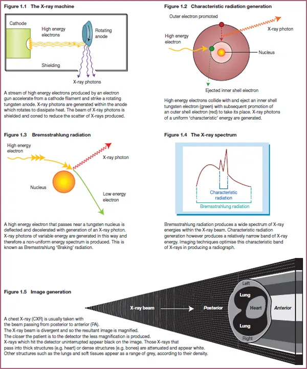
- An accelerated electron displaces an electron from within a shell of the atom. The vacant position left in the shell is filled by an electron from a higher level shell, which results in the release of X-ray photons of uniform energy. This is known as characteristic radiation.
- Accelerated electrons passing near the nucleus of the atom may be deviated from their original course by nuclear forces and thereby transfer some energy into X-ray photons of varying energies. This is known as Bremsstrahlung radiation.
- Absorption – this prevents the X-rays reaching the X-ray detector plate. Absorption contributes to patient dose and therefore increases the risk of potential harm to the patient.
- Scatter – scattering of X-rays is the commonest source of radiation exposure for radiological staff and patients. It also reduces the sharpness of the image.
- Transmitted – transmitted X-rays penetrate completely through the body and contribute to the image obtained by causing a uniform blackening of the image.
- Attenuation – an X-ray image is composed of transmitted X-rays (black) and X-rays which are attenuated to varying degrees (white to grey). Attenuation can be thought of as a sum of absorption and scatter and is determined by the thickness and density of a structure. In the chest, structures such as the lungs are relatively thick but contain air, making them low in density. The lungs therefore transmit X-rays easily and appear black on the X-ray image. Conversely, bones are not thick but are very dense and therefore appear white. Attenuation can be controlled by varying the power or ‘hardness’ of the X-ray beam.
The X-ray machine (tube)
Applying physics to practice
- If the subject to be imaged is placed further from the detector, the image created will be magnified. This is based on the principle that X-ray beams travel in diverging straight lines.
- Scatter from the patient and other objects degrades the resolution. This will cause the image to be blurred.
- Beams of lower energy are absorbed more than beams of higher energy. This affects the difference in clarity between the soft tissue detail and artefact.
Image quality
- Inherent unsharpness – this is caused by the structures involved not having sharp, well-defined edges.
- Movement unsharpness – this can be reduced by using short exposures, as with light photography.
- Photographic unsharpness – this is dependent on the quality and type of imaging equipment and the method of capturing the image.
Contrast
Plain X-ray densities
| Air/gas Fat So... |